DEPARTAMENTO DE BIOLOGIA ANIMAL
Role of the proto‐oncogene Ret during
haematopoietic differentiation
Mariana Rodrigues Campos
MESTRADO EM BIOLOGIA EVOLUTIVA E DO DESENVOLVIMENTO
2009
UNIVERSIDADE DE LISBOA
FACULDADE DE CIÊNCIAS
DEPARTAMENTO DE BIOLOGIA ANIMAL
Role of the proto‐oncogene Ret during
haematopoietic differentiation
Mariana Rodrigues Campos
Dissertação orientada por Doutor Henrique Veiga‐Fernandes
Orientador Interno: Prof. Doutor Élio Sucena
MESTRADO EM BIOLOGIA EVOLUTIVA E DO DESENVOLVIMENTO
2009
“Men love to wonder, and that is the seed of science” Ralph Waldo Emerson
A todos aqueles que contribuíram para esta dissertação
RESUMO
O desenvolvimento embrionário é um período crítico da vida do indivíduo durante o qual são formados os sistemas biológicos essenciais à sua sobrevivência. Exemplo disso é o sistema hematopoiético, responsável pela produção das células sanguíneas. Este sistema, crucial no transporte de oxigénio, na defesa contra microorganismos patogénicos e no processo da coagulação, é constituído por eritrócitos, granulócitos, linfócitos, plaquetas e células NK. Nos mamíferos, a hematopoiése inicia‐se nos ilhéus sanguíneos localizados no saco vitelino sendo esta função assegurada mais tarde pelo fígado fetal, o principal órgão hematopoiético na embriogénese. No caso do adulto, este processo ocorre maioritariamente na medula óssea, exceptuando‐se no entanto o desenvolvimento dos linfócitos T que ocorre no timo.
Na base desta complexa cascata encontram‐se as células estaminais hematopoiéticas. Estas células são multipotentes e, como tal, capazes de originar todas as linhagens do sistema hematopoiético. Por outro lado, por terem a capacidade de se dividir dando origem a uma célula igual a si mesma permitem a manutenção constante de uma fracção de células estaminais hematopoiéticas multipotentes.
Nas últimas décadas vários grupos têm concentrado os seus esforços em identificar moléculas responsáveis pela função das células estaminais hematopoiéticas, nomeadamente no que respeita aos sinais responsáveis pela cascata de diferenciação e manutenção destas células. Entre as moléculas identificadas até à data, a família das tirosina cinases está altamente representada. Um exemplo clássico dessas moléculas é cKit, um receptor de membrana que é, actualmente, um dos marcadores utilizados na identificação de células estaminais hematopoiéticas. Nesta família de tirosina cinases inclui‐se também a cinase RET, cuja expressão foi descrita em órgãos linfóides primários, locais onde o essencial da diferenciação hematopoiética decorre.
Ret foi inicialmente identificado por Takahashi e colaboradores como um proto‐oncogene
tendo a sua expressão sido inicialmente identificada em células derivadas da crista neural e em células urogenitais. Este receptor está descrito como crucial no desenvolvimento do sistema nervoso periférico, na morfogénese dos rins e na espermatogénese. RET é o receptor de um complexo ao qual se ligam factores neurotróficos da família do GDNF. Sendo uma tirosina cinase este receptor é incapaz de realizar transdução de sinal per se e, como tal, necessita da associação a um de quatro co‐receptores: GFRα1, GFRα2, GFRα3 e GFRα4. Estão descritas
quatro moléculas capazes de sinalizar através desta via e que se associam preferencialmente a um dos co‐receptores; assim, GDNF liga‐se preferencialmente a GFRα1, NRTN a GFRα2, ARTN a GFRα3 e PSPN a GFRα4.
Apesar de estas moléculas estarem classicamente associadas ao sistema nervoso, são várias as evidências da sua expressão no sistema hematopoiético. Se por um lado a expressão de Gfra1 e Gfra2 foi descrita em células do estroma da medula óssea por outro existem também evidências de expressão de Ret em células de fígado fetal. Para além disso, foi descrita a expressão de intervenientes desta via em precursores de células B na medula, em células B e T maduras e em timócitos adultos e embrionários.
Relevante é também o facto de GDNF e GFRα1 terem sido implicados na sobrevivência de timócitos em ensaios in vitro. Para além disso, se por um lado na ausência de Ret o desenvolvimento das placas de Peyer, estruturas linfóides secundárias, é altamente afectado, por outro, a adição de ARTN exógena leva à formação ectópica destas estruturas.
Todas estas evidências sugerem um envolvimento desta via de sinalização na hematopoiése. De modo a compreender este envolvimento, a expressão de Ret e seus co‐receptores foi avaliada em precursores hematopoiéticos tanto a nível do fígado fetal como na medula óssea adulta. Análise por RT‐PCR revelou elevada expressão de Ret e dos co‐receptores Gfra1 e
Gfra2 em células estaminais hematopoiéticas embrionárias, reafirmando o possível
envolvimento desta via de sinalização no processo hematopoiético.
Com este pressuposto em mente, foram analisados embriões homozigóticos para uma deficiência em Ret. A análise do fígado destes embriões revelou uma redução no número de células estaminais hematopoiéticas, repercutindo‐se também nos números totais de células do fígado fetal. É importante focar ainda que, no compartimento das células estaminais hematopoiéticas, existe uma redução significativa do número de células capazes de reconstituir a longo prazo hospedeiros irradiados.
A realização de ensaios in vitro, quer por co‐cultura com uma linhagem de estroma quer por cultura em metilcelulose, revelou que as células originárias de animais Ret‐/‐ são capazes de se
diferenciar em linhagem mielóide, eritróide e B com a mesma eficiência que as células de embriões selvagem. Estes resultados sugerem então que Ret não estará envolvido na diferenciação de uma linhagem específica.
A análise de ciclo celular por incorporação de BrdU não revelou quaisquer diferenças entre embriões Ret‐/‐ e embriões selvagem. Por outro lado a marcação com Ki‐67, que nos permite
G0, ou seja células quiescentes. Esta redução tem sido frequentemente associada na literatura a dificuldades na reconstituição do sistema hematopoiético de animais irradiados.
De modo a testar se esta hipótese se adequava aos nossos resultados, foram realizadas experiências de reconstituição clássicas. Nestas experiências os animais foram irradiados de modo a eliminar o seu sistema hematopoiético e, posteriormente, injectados com baixos números de precursores hematopiéticos. Após injecção, estas células migram para o baço onde formam colónias macroscopicamente visíveis. Estas experiências revelaram que os animais injectados com células Ret‐/‐ possuem um menor número de colónias,
comparativamente a animais injectados com células de animais selvagem.
Também com o objectivo de testar esta hipótese foram realizados, em paralelo, ensaios de competição. Nestes ensaios os animais irradiados foram injectados com uma mistura de células de fígado fetal, Ret‐/‐ ou selvagem, e células de medula de animais selvagem. De modo
a poder distinguir as células do fígado e as células de medula adulta foram utilizados animais com um marcador halotípico distinto, respectivamente CD45.2 e CD45.1. Os resultados destes ensaios sugerem que as células de origem Ret‐/‐ são pouco eficazes na reconstituição do
sistema imunitários dos indivíduos injectados. Dois meses após injecção, nos animais que foram reconstituídos com células homozigóticas para o alelo nulo, menos de 50% do sistema hematopoiético era originário do dador, em comparação com mais de 80% no caso de animais injectados com células de origem selvagem. Em suma, estes resultados apontam claramente para um papel de Ret no desenvolvimento do sistema hematopoiético suportando a ideia da utilização de uma mesma via de sinalização no funcionamento de sistemas com origem em diferentes tecidos germinativos.
Palavras‐chave: Hematopoiése, proto‐oncogene Ret, células estaminais hematopoiéticas,
reconstituição hematopoiética, diferenciação.
ABSTRACT
Haematopoiesis is a tightly regulated developmental cascade responsible for the generation of all haematopoietic cell types. Many molecules have been involved in this process, namely tyrosine kinase family members. This family includes the tyrosine kinase receptor RET, encoded by a proto‐oncogene and classically allocated to nervous system function.
Herein we show that Ret and its co‐receptors Gfra1 and Gfra2 are highly expressed in haematopoietic stem cells (HSC). Using mice homozygous for a null mutation of Ret we show that ablation of this gene has a major impact in the foetal liver cellularity. Accordingly, the HSC compartment of Ret deficient embryos shows a 2.2 fold reduction when compared to their WT littermate controls. Despite reduced foetal liver cellularity, HSC from Ret null mice display normal differentiation into B lymphocyte, erythroid and myeloid lineages. Conversely, Ret null HSCs have a reduction of quiescent cells, which also correlated with a reduction in long‐term reconstitution haematopoietic stem cells.
These findings suggest defects in haematopoietic activity and reconstitution potential. Accordingly, HSCs from Ret‐/‐ origin have a reduction in CFU‐S potential and poorly reconstitute
lethally irradiated hosts. In fact, two months after competitive transplantation, cells from Ret null origin represented less than half of the haematopoietic cells in recipient mice, while WT cells already accounted for more than 80% of the haematopoietic pool of the host.
Thus RET emerges as a novel molecule in haematopoiesis, supporting the concept that molecular mechanisms historically ascribed to specific tissues can be more widely used in order to orchestrate the function of systems derived from different germ layers. Key words: Haematopoiesis, proto‐oncogene Ret, haematopoietic stem cells, haematopoietic reconstitution, differentiation.
TABLE OF CONTENTS
I. INTRODUCTION
12
1. GENERAL ASPECTS
13
1.1. Functions of the haematopoietic system
13
1.2. Haematopoietic cell lineages
14
1.3. Haematopoietic organs
16
2. HAEMATOPOIESIS
17
2.1. Haematopoietic stem cells
17
2.2. Characterisation of haematopoietic stem cell function
18
2.3. Foetal Haematopoiesis
20
3. THE PROTO‐ONCOGENE RET
22
3.1. GDNF family ligands and signalling axes
23
3.2. Impact of deregulated expression of Ret
23
3.3. Ret expression in the haematopoietic system
24
4. AIMS AND EXPERIMENTAL MODELS
25
II. MATERIAL AND METHODS
26
1. MICE
27
2. MOUSE ANALYSIS
27
3. CELL STAINING AND FLOW CYTOMETRY
27
4. GENE EXPRESSION ANALYSIS
29
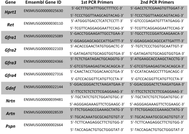
4.1. First PCR Amplification
29
4.2. Real‐time quantitative PCR
29
5. GENOTYPING PCR
30
6. COCULTURE ASSAYS
30
7. CFU ASSAYS
31
7.1. Methylcellulose assays
31
7.2. CFU‐S assays
31
8. CELL CYCLE ANALYSIS
32
8.1. BrdU analysis
32
8.2. Ki‐67 analysis
32
9. ADOPTIVE TRANSFERS
32
10. STATISTICS
33
III. RESULTS
34
1.
Expression of Ret and its signalling partners in non‐committed progenitor cells 35
2. Expression of Ret and its signalling partners in committed cell precursors
37
3. Impact of Ret ablation in haematopoietic stem cells
37
4. Impact of Ret ablation in lineage committed cell populations
39
5. Analysis of precursor frequency in Ret deficient embryos
40
6. Cell cycle analysis
41
7. Short‐term reconstitution of Ret deficient HSCs
42
8. Long‐term reconstitution of Ret deficient HSCs
43
IV. DISCUSSION
45
V. REFERENCES
50
VI. ANNEXES
55
i. Used solutions
56
ii. Multiplex PCR
57
iii. Genotyping primers
58
iv. Expression of Ret and its signalling partners in adult bone marrow
59
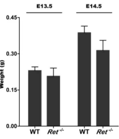
v. Embryos’ Weight
60
vi. Units and Abbreviations
61
I. INTRODUCTION
1.
G
ENERAL
A
SPECTS
1.1. Functions of the haematopoietic system
The haematopoietic system is composed of a myriad of cell types and tissues that ensure vital physiological functions. The main roles of this system are i. oxygen transportation; ii. defense against pathogenic microorganisms; and iii. contribution to blood clotting1.
Oxygen transportation is critical for survival and thus required from very early phases of embryonic development. This function is ensured by erythrocytes, or red blood cells, which are a specialised enucleated cell type that contains haemoglobin. Erythocytes are the first cells to differentiate in the embryo, being detectable at yolk sac’s blood islands at embryonic day 7.5 (E7.5). However, the final enucleated stage of development, characteristic of adult erythrocytes, is only achieved by E12.52.
Protection against microbes is ensured by the production of immune cell types, which can make rapid, specific, and protective responses against a limitless variety of potentially pathogenic microorganisms. Furthermore, immune cells can also mediate the rejection of tumours and may even exert important effects in regulating other systems1.
Elimination of microbial agents can be exerted either through nonspecific mechanisms (via the innate immune system), or by a highly specific recognition and elimination of foreign antigens (via the adaptive immune system). Importantly, while innate immune responses are not modified by repeated exposure to a given infection, adaptive responses are improved with each successive encounter with the same pathogen. Hence the two main key features of the adaptive immune system are specificity and immunological memory1.
Clotting is an important part of haemostasis, the cessation of blood loss from a damaged vessel, and this function is partly ensured by the haematopoietic system. When a blood vessel is damaged its wall is immediately covered by platelets that form a plug at the site of injury (primary haemostasis). Secondary haemostasis occurs simultaneously when clotting factors present in the blood plasma respond in a complex cascade to form fibrin strands, which strengthen the platelet plug3.
1.2. Haematopoietic cell lineages
The haematopoietic system is composed of a wide range of distinct cell types that differentiate and develop through a process named haematopoiesis. Haematopietic cells can be divided into three different lineages: i.erythroid; ii. lymphoid and iii. myeloid, each of them mediating specialised functions1. Figure 1. Conventional view of haematopoiesis. In this process, multipotent stem cells are self‐generating and also produce precursor cells with progressive restriction of their lineage potential (adapted from Metcalf, 20074). The erythroid lineage differentiates early in development, as it plays a critical role in carrying oxygen. In mice, between E7.0 and E8.0, blood‐islands’ peripheral cells assume a spindle shape as they differentiate into endothelial cells. At this stage, their counter‐parts inner cells progressively lose their intercellular attachments differentiating into primitive erythroblasts. Soon after, the latter enter the newly formed vasculature of the embryo where they divide for several days. Thus, primitive erythroblasts differentiate within the bloodstream, gradually accumulating increasing amounts of haemoglobin and reaching the definitive enucleated stage by E12.55.
The lymphoid lineage, derived from common lymphoid progenitors (CLP), includes the cells that ensure specificity and immunological memory. There are two broad classes of lymphocytes: B‐lymphocytes (or B cells) and T‐lymphocytes (or T cells).
B cells are precursors of antibody‐secreting cells. Upon antigen stimulation, specific B cells proliferate and differentiate into plasma cells, which produce large amounts of soluble
receptor molecules known as antibodies. The latter are found in the blood and tissue fluids and bind the antigen that initially activated B cells6. T‐lymphocytes express a cell surface
receptor known as the T cell receptor (TCR). These receptors are composed of protein heterodimmers αβ or γδ, defining different T cell populations. T cells bearing a αβ TCR can be further subdivided in two distinct populations: the subset that expresses the CD4 marker, also known as T helper cells, and cells carrying the CD8 marker, the cytotoxic T cells. CD4 T cells aid in the development of specific immune responses, including the production of antibody by B‐ cells and the increase in anti‐microbial activity by macrophages. Furthermore, specialised CD4 T cells, named regulatory T cells, have the capacity to suppress specific immune responses. On the other hand, CD8 T cells are involved in direct killer functions, such as the lysis of virus‐ infected and neoplastic cells7,8.
Haematopoietic cells from the myeloid lineage are derived from a common myeloid progenitor (CMP). Myeloid cells interact with lymphocytes playing a crucial role both in antigen presentation and mediation of the immune responses. These cells are classified into two main families: mononuclear and polymorphic cells. The mononuclear family is composed by monocytes, which circulate in the blood, and macrophages, which are found in peripheral tissues. During haematopoiesis, granulocyte‐monocyte progenitor cells differentiate and enter the blood, where they further differentiate into monocytes. The latter can then differentiate into macrophages and dendritic cells upon their exit from the blood stream to peripheral tissues. Polymorphonuclear cells include neutrophils, eosinophils, basophils and mast cells. Mast cells and basophils play important roles in the induction of allergic inflammatory responses; neuthrophils are critical in clearing infections by extracellular bacteria and eosinophils have been implicated in protective responses to helminths and in allergic inflammation1.
Finally, it is worth notice that specialised epithelial and stromal cells from non‐haematopoietic origin provide the anatomic environment in which haematopoiesis and immune responses occur. These cells secrete critical factors, such as citokines and chemokines that regulate migration, growth, and/or gene activation of haematopoietic cells. As an example, in haematopoiesis they provide the physical support and the chemical clues essential to create the correct niche where this process occurs9.
1.3. Haematopoietic organs
In mammalian embryos, haematopoiesis begins in the yolk sac, where primitive erythroid cells are formed10. Consecutively, haematopoietic stem cells (HSCs) formed in the aorta‐gonad‐
mesonephros region migrate to the foetal liver, which assumes most of the foetal haematopoietic activity. At later stages of gestation, differentiation and maintenance of HSCs starts to occur in the bone marrow (BM), and by birth there’s almost no haematopoietic activity in the liver11,12.
During adulthood haematopoiesis occurs mainly in the so called primary lymphoid organs. The BM represents the major site of haematopoietic activity, although late B‐cell maturation stages occur in the spleen and T‐cell differentiation in the thymus.
A major challenge for the immune system is to allow the encounter of pathogens with rare pathogen‐specific B and T cells soon after the breakage of the protective epithelial and/or mucosal barriers. Secondary lymphoid tissues have seemingly evolved to meet this goal. These secondary lymphoid tissues include the spleen, lymph nodes and organized lymphoid tissues associated with mucosal surfaces (Peyer’s patches, tonsils, bronchial associated lymphoid tissues and other gut associated lymphoid tissues)9.
Each of these secondary lymphoid organs consists of a supporting stromal matrix that allows the interaction between T,B and antigen‐presenting cells. These structures are interconnected by a network of lymphatic vessels and drain strategic regions where foreign antigens and pathogenic microbes may enter the body, either by penetrating the skin, by crossing a mucosal barrier or by injection into the bloodstream9.
2.
H
AEMATOPOIESIS
Haematopoiesis is a tightly regulated developmental cascade responsible for the formation of all blood cell types via the expansion and differentiation of haematopoietic precursors. This process has been highly conserved during evolution. However, in avian models, a function similar to that of the FL seems to be carried out by a structure called the para‐aortic foci and in adulthood the bursa of Fabricius is the primary lymphoid organ responsible for the production of B cells13.
Haematopoietic differentiation depends on cellular and molecular signals that lead to cell division and progressive restriction of lineage potential. Importantly, the integrity of the haematopoietic process is strictly dependent on the existence and persistence of rare long‐ lived haematopoietic stem cells that ensure long‐term haematopoietic cell production14.
2.1. Haematopoietic Stem Cells
Haematopoietic stem cells (HSCs) are multipotent cells that, under a permissive environment, are capable of self‐renewal, thus maintaining a constant pool (probably through asymmetric cell division15). Importantly, in certain conditions they can also give rise to a differentiated
progeny that comprises different haematopoietic cell types14. The hallmark of haematopoietic
stem cell function is therefore their capacity to provide long‐term haematopoietic reconstitution after transplantation.
Phenotypically, HSCs can be identified by the absence of expression of lineage markers. Furthermore, HSCs are characterised by the expression of both cKit and Sca1, which became canonical markers used to identify these cells. Even though, phenotype changes can occur, which correlates with cell activation, differentiation and cell cycle status16.
HSCs are mostly quiescent in adulthood but they can be mobilized from their niche to proliferate and differentiate into lineages of the innate and adaptive immune system, as well as into red blood cells and platelets. Cell‐fate decisions are initiated and maintained by specific combinations of transcription factors, the activity of which can be orchestrated by extrinsic and intrinsic signals17. During the last decade a great effort has been put into identifying these
mediate critical events in the biology of HSCs. A classical example of a tyrosine kinase is cKit. The expression of cKit on haematopoietic progenitor cells was first identified by Ogawa et al. in 199118. When Ogawa and colleagues injected mice with an anti‐cKit monoclonal antibody most
haematopoietic progenitor cells disappeared from the bone marrow, leading to the absence of myeloid and erythroid cells. These results were the first direct evidence that cKit is an essential molecule for haematopoiesis, especially for the self‐renewal of haematopoietic progenitor cells. Another member of this family is the tyrosine kinase receptor RET, which is described in detail in following sections of this thesis. From a clinical perspective, haematopoietic stem cells continuously replenish blood cells that are lost by attrition or tissue damage and are currently the only adult stem‐cell type routinely used to replace lost cells, namely after chemotherapy treatment19.
2.2. Characterisation of haematopoietic stem cell function
There are different methods for characterising haematopoietic stem cells, which range from immunophenotypical analysis to in vitro and in vivo functional assays. In the past two decades different methods have been used to distinguish HSCs and progenitor cells by immunophenotypical analysis; all these techniques are fluorescence activated cell sorting (FACS)‐based. The purity of the populations achieved using these methods has increased within recent years, such that approximately 50%‐96% of single cells in certain purified populations can give rise to long‐term reconstitution after transplantation.
One of the most commonly used FACS‐purified populations of HSC/progenitor cells is characterised by the absence of lineage markers and by the expression of both cKit and Sca1 (linnegcKitposSca1pos, usually referred as LKS cells)20,21.
In 1992 Suda’s laboratory reported the isolation of HSCs from C57BL/6 mice within the linnegcKitposSca1pos population21. However, this population is heterogeneous and consists
predominantly of progenitor cells with less than 10% of representing HSCs. More recently, the expression of CD34 and Flt3 (CD135) has been used to further purify long‐term repopulating HSCs (LT‐HSCs) (LKSposCD34negFlt3neg) from short‐term repopulating HSCs (ST‐HSCs)
(LKSposCD34posFlt3neg) and multipotent progenitors (LKSposCD34posFlt3pos)22. LT‐HSCs are capable
into irradiated hosts, while ST‐HSCs only show multi‐lineage repopulation for a few weeks after transplantation23. Besides from CD34 and Flt3 expression also the levels of CD38 have
been used to distinguish between long‐term and short‐term reconstitution haematopoietic stem cells. linnegcKitposSca1pos cells showing low or absent levels of CD38 contain most of day‐12
colony‐forming‐units‐spleen (CFU‐S) but are not long‐term reconstituting cells. On the other hand, their counterparts, linnegcKitposSca1pos CD38pos/high contain virtually all the haematopoietic
stem cells capable of long‐term reconstitution24.
The colony forming cell assays measure progenitor cells in a given population, using semisolid agar or, more commonly, a well‐defined methylcellulose‐based culture media in vitro. The majority of colony forming cells (CFCs) consist of lineage‐restricted colonies: erythroid‐ restricted burst‐forming units‐erythroid (BFU‐E), which are more immature than the colony‐ forming units‐eythroid (CFU‐E); megakaryocyte‐restricted (CFU‐Mk); colony‐forming units‐ granulocytes (CFU‐G); colony‐forming units‐monocyte/macrophages (CFU‐M); and colony‐ forming units‐granulocyte/macrophages (CFU‐GM). The most immature CFC measurable originates granulocytes, erythrocytes, macrophages, and often megakaryocytes (CFU‐GEMM). This type of assay is informative about the frequency of progenitor cells in a given population but does not measure numbers of HSCs25.
Analysis of B and T potential is difficult to assess by this method requiring specialised co‐ culture systems. For instance, the OP9 BM stromal cell line26 has been shown to support the
differentiation of HSCs into multiple lineages, including B cells, in vitro27. As Notch signalling is
critical for the earliest stages of T cell commitment28,29 stromal cells expressing Delta‐like‐1
(Dll1) have been used to support T cell lineage commitment30.
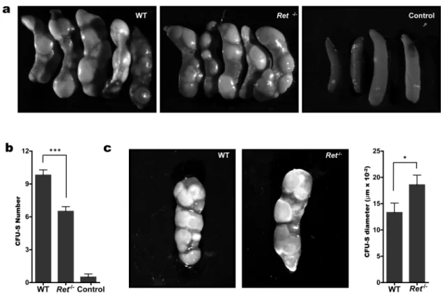
Short‐Term in vivo assays are also classically used to analyse HSC function. Colony‐forming unit‐spleen (CFU‐S) cells are cells that, once injected into irradiated recipient, home to the spleen and form macroscopic colonies that provide very short‐term (usually 1 to 3 weeks) in
vivo repopulation of the mouse. The CFU‐S are therefore early engrafting cells, providing
radioprotection to the mouse and allowing it to survive more readily in the first 2‐3 weeks
post‐transplantation when pancytopenia usually occurs31.
Another type of assay is the long‐term repopulating assays used to measure HSC numbers and to analyse their functional potential. The competitive repopulating assay is the most common. In these experiments is measured the functional potential of an unknown source of HSCs against a set known number of HSCs (usually whole bone marrow cells from congenic wild‐
type mice). In these experiments the halotype markers CD45.1/CD45.2 are used in order to detect donor versus radio‐resistant host cells or to distinguish between competitor cells32.
2.3. Foetal Haematopoiesis
Foetal haematopoiesis is a temporal and spatial complex developmental process. During this period successive sites achieve the production of differentiated haematopoietic cells until adult haematopoietic organs are fully developed and ready to take over.
The first detected haematopoietic activity occurs in the yolk sac’s (YS) blood islands, between E7.0 and E12.0. During this period, the vast majority of produced cells are nucleated erythrocytes expressing embryonic haemoglobin. These erythrocytes resemble erythrocytes precursors in the BM and erythrocytes in lower vertebrate groups, such as birds, fish and amphibians. These cells never reach the final enucleated stage and for this reason they have been called primitive erythrocytes2.
Although the YS is the primitive site of haematopoiesis, precursors here formed proved unable to reconstitute all blood cell types, as they can’t give rise to lymphoid cells. This ability is a trait of hematopoietic stem cells (HSC), which are originated in the aorta‐gonad‐mesonephros (AGM) region, between E9.0‐E12.033.
Although HSCs are originated in the AGM region, haematopoiesis does not occur at this site but in the foetal liver (FL), which then becomes the major haematopoietic organ during mammals’ embryonic development34. The liver primordium is evident by E9.0 and colonization
by AGM and YS derived haematopoietic precursors occurs between E10.0 and E12.0. The majority of cells that initially colonize the FL are progenitors of YS origin that rapidly differentiate to enucleated erythrocytes and myeloid cells. Thus, within 24 hours, between E10.0 and E11.0, the frequency of primitive erythrocytes decreases because these cells reach terminal differentiation in the FL by exposure to erythropoietin35.
The foetal liver provides a niche with a unique set of environmental conditions that allow the expansion of HSCs. Until E15.0, HSCs expand in numbers and acquire the final characteristic surface markers that define adult HSCs. In addition to the expansion of HSCs, the FL also supports the differentiation of erythrocytes, myeloid cells, and lymphocytes36.
The foetal liver remains an active site of haematopoiesis until after birth, by which time the bone marrow (BM), after being colonized by HSC migrating from FL, takes over as the major organ where haematopoiesis occurs18.
The foetal spleen is also a transient haematopoietic organ, which activity starts between E13.0‐14.0 until the first weeks of postnatal life. HSCs, circulating from FL, home to the spleen but in contrast to the FL, they do not expand significantly in the foetal spleen. Thus, in the absence of a robust maintenance of an HSC pool at the embryonic spleen, haematopoietic differentiation relies on the presence of intermediate precursors. These precursors either derive from differentiation of HSCs or home directly from the FL to the foetal spleen37.
3.
T
HE PROTO
‐
ONCOGENE
R
ET
Ret encodes a receptor tyrosine kinase initially described on neural crest‐derived and
urogenital cells. This receptor is a key regulator in different processes that encompass the peripheral nervous system development, kidney morphogenesis and spermatogenesis38‐40.
Figure 2. RET tyrosine kinase receptor, co‐ receptors and specific ligands. GDNF and three
related GFL proteins (NRTN, ARTN and PSPN) signal through RET and the specificity of the RET/GFL axis is determined by one of the four GDNF family alpha co‐receptors (GFRα): GFRα1 binds preferentially to GDNF, GFRα2 to NRTN, GFRα3 to ARTN and GFRα4 to PSPN (adapted from Airaksinen, 200241).
The Ret gene was first identified by Takahashi et al.42, who reported a novel gene
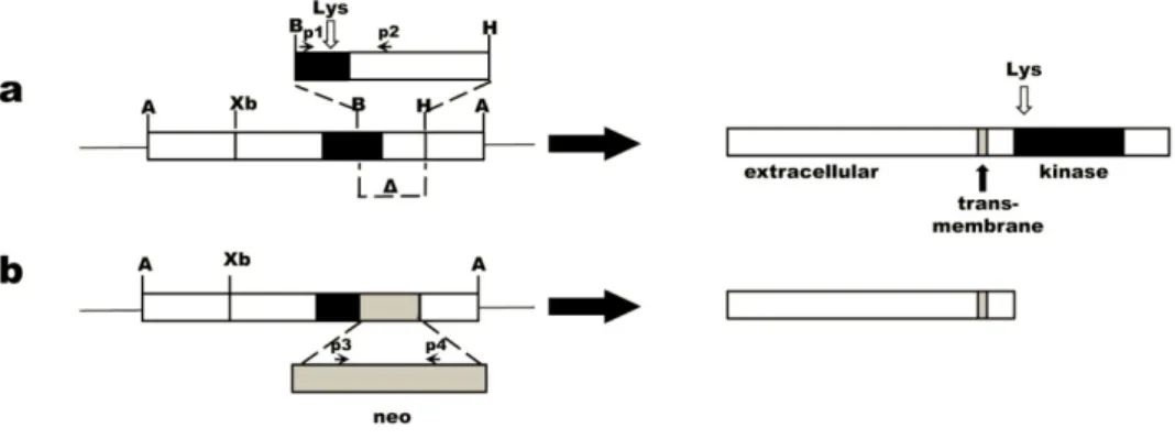
rearrangement with transforming activity in NIH 3T3 cells transfected with human lymphoma DNA. The transforming gene resulted from a recombination event between two unrelated DNA sequences that occurred during the transfection process. Hence, the name RET stems for “rearranged during transfection”. The resulting chimeric gene encodes for a fusion protein comprising an N‐terminal region with a dimerising motif fused to a tyrosine kinase (TK) domain.
The proto‐oncogene Ret encodes for a transmembrane protein of the tyrosine kinase (TK) family, which is composed of three domains: at the extracellular portion there is a ligand‐ binding domain with four cadherine‐like repeats and a cysteine‐rich region; at the cytoplasmic level the protein is composed by a TK domain interrupted by an insertion of 27 amino acids and, linking these two, there is a hydrophobic transmembrane domain43,44.
3.1. GDNF family ligands and signalling axes
RET is the signalling receptor of a multi‐molecular complex that binds growth factors of the glial cell line‐derived neurotrophic factor (GDNF) family45. GDNF‐family ligands (GFLs) bind to
and activate RET when bound to a GDNF‐family receptor‐alpha (GFRα) protein. GFRα are ligand‐binding co‐receptors, anchored to the cell surface by glycosylphosphatidyl inositol (GPI)‐ linkage, which lack intercellular or transmembrane domains46.
GDNF and three related GFL proteins, i.e. Neurturin (NRTN), Artemin (ARTN) and Persephin (PSPN) signal through RET and the specificity of the RET/GFL axis is determined by one of the four GFRα: GFRα1 binds preferentially to GDNF, GFRα2 to NRTN, GFRα3 to ARTN and GFRα4 PSPN47. Although this preferential binding is likely to be the dominant signalling mechanism, signs of cross talk between ligands and co‐receptors have been described in vitro48. The downstream RET signalling pathways are thought to be mostly common for all four GFLs, since all GFRαs bind to and activate RET with similar kinetics49, inducing the phosphorilation of the same four key tyrosine residues (Tyr905, Tyr1015, Tyr 1062, Tyr 1096). Nevertheless, each of these co‐receptors has a unique, tissue‐specific and developmentally regulated expression pattern suggesting they are likely to have distinct cell type‐specific roles in RET‐activation50.
3.2. Impact of deregulated expression of Ret
Over expression and expression of aberrant forms of RET are characteristic of human oncogenic diseases including leukaemia. Somatic chromosomal rearrangements involving the
Ret gene represent the most frequent genetic alteration in papillary thyroid carcinoma (PTC),
the most common thyroid malignancy. These rearrangements lead to the fusion of the Ret tyrosine kinase with the 5’‐terminal regions of heterologous genes, generating chimeric oncogenes. The fusion between tyrosine kinase domain and activating genes lead to ligand‐ independent dimerization constitutively activating these chimeric proteins51.
Moreover, activating germline point mutations of Ret are also responsible for multiple endocrine neoplasia type 2 (MEN 2A and 2B) and familial medullary thyroid carcinoma (FMTC). These Ret mutations fall into two major groups: those affecting the extracellular and those affecting the tyrosine kinase domain. All these mutations have a gain‐of‐function effect as they lead to constitutive dimerization of the transmembranar receptor52.
Loss‐of‐function caused by hypomorphic mutations of RET cause Hirschsprung’s disease (HSCR) or colonic aganglionosis. In this disease, mutations in the extracellular domain impair Ret cell surface expression leading to unresponsiveness to GDNF, which prevent chemotatic migration of enteric neurons and consequent hind‐gut enervation53.
In mice, homozygosity for null mutations of Ret results in peri‐natal lethality, hindering analysis of post‐natal biological events. Nonetheless, Ret null embryos exhibit a wide range of developmental abnormalities affecting the nervous system (enteric aganglionosis38), the
lymphoid system (impaired Peyer’s Patch development54) and the kidney (kidney
hypodysplasia or aplasia38).
3.3. Ret expression in the haematopoietic system
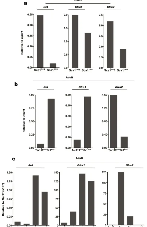
Although neurotrophic factors and the associated signalling cascade are classically allocated to nervous system function, various research groups showed Ret expression in the haematopoietic system. Expression of Gfra1 and Gfra2 in Bone Marrow (BM) stromal cells and
Ret expression in foetal liver (FL) have been previously reported55‐58. Furthermore, Ret has
been shown to be widely expressed by Bone Marrow B cell precursors, from pre‐pro B to immature B cells59. Ret, Gfra1 and GDNF have also been shown to be expressed in embryonic
and adult thymocytes60. There is also clear evidence that RET and its partners are expressed in
mature T and B cells61.
Interestingly, Kondo et. al.60 showed that both GDNF and GFRα1 are involved in the survival of
thymocytes as addition of rGDNF to in vitro cultures induced increased thymocyte cell survival. Furthermore, Veiga‐Fernandes et. al54 recently showed that in the absence of Ret Peyer’s
Patch development is severely compromised and the RET ligand ARTN functions as a strong attractant of gut haematopoietic cells, inducing the formation of ectopic Peyer’s patch‐like structures.
Taken together these observations suggest that the RET signalling pathway is likely to play a role in the molecular mechanisms that orchestrate haematopoiesis.
4.
A
IMS AND EXPERIMENTAL MODELS
The GDNF ligands (GFLs), which signal through the RET tyrosine kinase receptor, have recently emerged as key players during enteric lymphoid organogenesis and although RET expression has been reported during haematopoiesis, its functional significance remains elusive. In this thesis project we used combined genetic, cellular, and molecular approaches in order to determine, quantify and manipulate the function of RET during foetal haematopoiesis. In order to achieve this, we first analysed the patterns of Ret expression during different stages of haematopoiesis and in different haematopoietic cell lineages. By using this strategy we identified differentiation stages where the proto‐oncogene Ret was likely to exert its role. We then assessed the functions of RET by studying the impact of Ret gene ablation. To achieve this, we studied haematopoietic cells at different differentiation stages from mice homozygous for a null mutation of Ret and determined the impact that this molecule plays in key cellular and molecular events during foetal haematopoiesis. Thus, we investigated the differentiation and reconstitution potential of Ret deficient HSCs using in vivo, and in vitro approaches.
We foresee the haematopoietic differentiation as a valuable model for the assessment of new roles for the proto‐oncogene Ret, allocating its functions to defined in vivo differentiation processes that form the basis of immune fitness.
II. MATERIAL AND METHODS
1.
M
ICE
C57Bl/6J mice were purchased from Instituto Gulbenkian de Ciência (IGC) and from Harlan. C56Bl/6J CD45.1 and Rag2 deficient mice62 were purchased from IGC and The Jackson
Laboratory. Ret‐/‐ 38 mice were bred and maintained at the IMM animal facility.
2.
M
OUSE ANALYSIS
In order to obtain embryos at different developmental stages, mice were naturally timed‐ mated and the presence of a vaginal plug was evaluated daily. The day of vaginal plug observation was designated as 0.5 day of gestation. To obtain Ret‐/‐ embryos, Ret+/‐ females
were crossed with Ret+/‐ males. Pregnant females, at gestational day 13.5 or 14.5, were
sacrificed and dissected. Embryos were then collected and the foetal liver was removed. Foetal livers were then ressuspended in GIBCO® Dulbecco's Modified Eagle Medium (DMEM) (Invitrogen), supplemented with 2% Foetal Bovine Serum (Invitrogen), 1% Penicillin and Streptomycin (Invitrogen) and 1% Glutamine (Invitrogen), and passed through 70 µM cell strainers (Becton Dickinson). Viable cells were counted in a Neubaeur hemocytometer using Trypan Blue exclusion to assess cell viability.
For adult haematopoietic cell analysis, 6‐8 weeks‐old C57Bl/6J mice were dissected, bone marrow was extracted from both forelimbs and hind limbs and cells were processed as previously described for the foetal liver. Red blood cells were then lysed using Erythrocyte Lyses Buffer (Annex I) during 5 minutes at 4ºC and then ressuspended in FACS buffer (PBS containing 2% FBS and 0,05% azide, Annex I).
All animal experiments were done in accordance to institutional and national guidelines.
3.
C
ELL STAINING AND FLOW CYTOMETRY
Antibodies were purchased from BD Pharmingen, e‐Bioscience or Biolegend. The following antibodies were used: Ter119‐biotin/PE (Ter119), B220‐APC‐Cy7/PE (RA3‐6B2), CD3‐biotin/PE (145‐2C11), DX5‐biotin/PE (DX5), Gr‐1‐biotin/PE (RB6‐8C5), cKit‐APC/PE‐Cy7/PE (2B8), Sca1‐ FITC/APC (D7), CD19‐biotin/PE (1D3), NK1.1‐biotin/PE/FITC (PK136), Ly6C‐biotin (HK1.4), CD11b‐biotin/PE/APC (M1/70), CD38‐APC (90), CD45‐APC (30‐F11), CD93‐APC (AA4.1), CD43‐ FITC (S7), BP1‐biotin (6C3), CD24‐PE‐Cy7 (M1/69), IgM‐FITC (II/41), IgD‐PerCP (11‐26c), BrdU‐
FITC (B44), Ki‐67‐FITC (B56), CD34‐PerCP (HM34), CD16/32‐PE (93), CD45.2‐APC‐Cy7 (104), CD45.1‐PE‐Cy7 (A20) and Streptavidin‐PerCP/APC‐Cy7/PE‐Cy7.
In order to purify haematopoietic sub‐populations, cells either from E14.5 C57Bl/6J embryos or from C57Bl/6J adult bone marrow were FACS sorted. Prior to FACS sorting, Ter119pos cells
(erythroid cells) were depleted by negative selection using Dynabeads Biotin Binder (Invitrogen), according to manufacturer’s instructions. Briefly, Dynabeads were added to cell suspensions and incubated for 25’ at 4ºC in a rotating device. Magnetically labelled Ter119pos
cells were eliminated after 5’ incubation in a magnetic field.
Ter119neg cells were then incubated with a lineage antibody cocktail (B220, CD3, CD19, DX5,
Gr1 and Ter119), cKit and Sca1. Stainings were performed for 20 minutes on wet ice after which the cells suspensions were washed with FACS buffer (Annex I). Stained cells were sorted using BD FACSAria Cell Sorting System (Becton Dickinson) into linnegcKitposSca1neg and
linnegcKitposSca1pos. The purity of the sorted populations was then analysed (Figure 3a).
In order to purify B cell developmental stages, cells from C57Bl/6J adult bone marrow were lineage depleted (using CD3, Gr1, Nk1.1 and Ter119 mAb) as previously described and FACS sorted. Cell suspension was sorted into Pre‐pro‐B cells (B220posCD93posCD43posBP1negCD24low ),
Pro‐B cells (B220posCD93posCD43posCD24pos), Pre‐B cells (B220posCD93posCD43negIgMnegIgDneg) and
Immature‐B cells (B220posCD93posCD43negIgMposIgDneg).
Purification of Ter119pos and Gr1pos cells both from FL (E14.5) and adult BM was performed in a
two‐round protocol. An initial step of negative selection was done using Dynabeads Biotin Binder (Invitrogen), as previously described, after which Magnetic Activated Cell Sorting (MACS) was performed in order to enrich cell suspension in desired cells. Briefly, cells were incubated with anti‐PE MicroBeads (Miltenyi Biotec) for 15’ on ice. After incubation, magnetically labelled cells were flushed through an MACS column. The purity of the enriched populations was then analysed by FACS.
Cell staining for flow cytometry analysis was performed as previously described for sorting, using appropriate antibody cocktails. Results were acquired using BD FACSCanto (Bencton Dickinson) and analysed using FlowJo (Tree Star Inc., version 8.8.4).
4.
G
ENE EXPRESSION ANALYSIS
Total RNA was extracted from sorted cells using RNAeasy Mini Kit (Qiagen), according to manufacturer’s instructions. RNA was then maintained at ‐80ºC.
cDNA synthesis and PCR amplifications were performed on a Veriti 96‐Well Thermal Cycler (Applied Biosystems).
cDNA synthesis was performed as previously described63. Briefly, RNA was specifically
retrotranscribed for 1h at 37ºC by adding a mix containing 0.13 µM reverse primer (Annex II), 50mM KCL and 10 mM Tris‐HCl at pH 8.3 (Applied Biosystems), 3.3 mM MgCl2 (Applied
Biosystems), 1mM dNTPs (Applied Biosystems), 39 units of RNAse Block (Stratagene) and 11.5 units of MuLV Reverse Transcriptase (Applied Biosystems) in a 15 µL reaction. The reaction was stopped by 3 min incubation at 95ºC.
4.1. First PCR Amplification
The cDNAs resulting from the reverse transcription were then amplified. The first round of amplification consisted of a initial step of denaturation at 95ºC for 10 min, followed by 15 cycles of amplification (45 sec at 95ºC, 1 min at 60ºC and 1 min and 30 sec at 72ºC) with 50 mM KCl and 10 mM Tris‐HCl ph 8.3 (Applied Biosystems), 2.0 mM MgCl2 and 0.2 mM dNTPs
(Applied Biosystems), 3 units of AmpliTaq Gold DNA Polymerase (Applied Biosystems) and 0.015 µM specific primers (Annex II).