REVIEW
Personalized medicine: caught between hope, hype
and the real world
Marc Dammann, Frank Weber
University Hospital Essen, Endocrine Surgery, Department of General, Visceral and Transplantation Surgery, Hufelandstrasse/Essen, Germany.
Genomic and personalized medicine have become buzz phrases that pervade all fields of medicine. Rapid advances in ‘‘-omics’’ fields of research (chief of which are genomics, proteinomics, and epigenomics) over the last few years have allowed us to dissect the molecular signatures and functional pathways that underlie disease initiation and progression and to identify molecular profiles that help the classification of tumor subtypes and determine their natural course, prognosis, and responsiveness to therapies. Genomic medicine implements the use of traditional genetic information, as well as modern pangenomic information, with the aim of individualizing risk assessment, prevention, diagnosis, and treatment of cancers and other diseases. It is of note that personalizing medical treatment based on genetic information is not the revolution of the 21st century. Indeed, the use of genetic information, such as human leukocyte antigen-matching for solid organ transplantation or blood transfusion based on ABO blood group antigens, has been standard of care for several decades. However, in recent years rapid technical advances have allowed us to perform high-throughput, high-density molecular analyses to depict the genomic, proteinomic, and epigenomic make-up of an individual at a reasonable cost. Hence, the so-called genomic revolution is more or less the logical evolution from years of bench-based research and bench-to-bedside translational medicine.
KEYWORDS: Inherited Endocrine Neoplasia; Genomic Medicine; Tumor Syndromes.
Dammann M, Weber F. Personalized medicine: caught between hope, hype and the real world. Clinics. 2012;67(S1):91-97.
E-mail: [email protected] Tel.: 49 201 7231469
INTRODUCTION
Genomic medicine and personalized medicine have become buzz phrases that pervade all fields of medicine. The rapid advances in ‘‘–omics’’ research (chief of which are genomics, proteinomics, and epigenomics) over the last few years have allowed us to dissect the molecular signatures and functional pathways that underlie disease initiation and progression and to identify molecular profiles that help the classification of tumor subtypes and determine their natural course, prognosis, and responsiveness to therapies. Genomic medicine implements the use of traditional genetic information, as well as modern pangenomic information, with the aim of individualizing risk assessment, prevention, diagnosis, and treatment of cancers and other diseases.
It is of note that personalizing medical treatment based on genetic information is not a revolution of the 21st century. Indeed, the use of genetic information, such as human leukocyte antigen-matching for solid organ transplantation or blood transfusion based on ABO blood group antigens, has been standard of care for several decades. However, in recent years rapid technical advances have allowed us to
perform high-throughput, high-density molecular analyses to describe the genomic, proteinomic, and epigenomic make-up of an individual at a reasonable cost. Hence, the so-called genomic revolution is more or less the logical evolution from years of bench-based research and bench-to-bedside translational medicine. The challenge is to deter-mine how the structural changes, both within the germline and soma, affect how genes globally interact within the germline, how they interact locally between the germline and tumor and, finally, how the environment and lifestyle choices can alter genes at a functional level in order to modulate thyroid cancer susceptibility, aggressiveness, and the likelihood of cure.
Molecular testing for non-inherited genetic profiles (i.e., the somatic alteration of genes such as KIT, BRAF, and EGFR) to tailor and optimize treatment approaches has become implemented in the routine armament for the diagnosis and treatment of various tumors (i.e., lung, colon, breast, gastrointestinal stromal, thyroid, etc.) and will not be discussed in this review (1–4). The impact on practicing personalized medicine will be tremendous. For instance, it will not be long before we can tailor our surgical approa-ches and surveillance of patients with papillary micro-carcinomas based on theBRAF(V600E) mutation status (5). On the other hand, human disease susceptibility can be seen as the result of either rare germline genetic variations of high penetrance or as common genomic variants of low penetrance. The latter path on the road to practicing genomic medicine is using the technical advances of high-throughput,
Copyrightß2012CLINICS– This is an Open Access article distributed under
the terms of the Creative Commons Attribution Non-Commercial License (http:// creativecommons.org/licenses/by-nc/3.0/) which permits unrestricted non-commercial use, distribution, and reproduction in any medium, provided the original work is properly cited.
high-density analyses (e.g., whole-genome linked analysis) to correlate common genetic variations (e.g., single nucleotide polymorphisms) to common diseases (6). In this way, a vast variety of allelic alterations have been identified that correlate with the risk of cancer, other common diseases (e.g., diabetes, hypertension, autism, obesity, etc.) or even a patient’s ability to respond to severe sepsis (7–10). There is tremendous hype to push forward on this path in order to implement ‘‘–omics’’-based analyses into clinical practice (11,12). However, there are some obstacles to clinical implementation that must not be underestimated and the impact on public health must be critically discussed (12–14). Beyond ethical and legal issues, numerous studies have shown that susceptibility variants for common diseases only marginally contribute to the disease risk with odd ratios of around 1.5 (6). For instance, in coronary heart syndrome, other medical conditions and lifestyle factors, such as blood pressure, diabetes, or smoking, are better predictors of cardiovas-cular events than are genomic variants (15). Furthermore, easy access to affordable, high-volume sequencing/geno-typing techniques (i.e., the ability to sequence thousands of genes or millions of single nucleotide polymorphisms) resulted in a flood of studies reporting the association of one or another genetic variant to disease or disease outcome (6). This harbors a profound statistical dilemma. Among the major limitations of many association studies is that they are profoundly underpowered. For instance, to detect an association with an odds ratio of 1.5 (i.e., a patient with the variant is 1.5 times more likely to have the disease or outcome) more than 1,000 people need to be tested when the suspected susceptibility variant occurs with a frequency of 0.2 (20% of the population). Furthermore, none of the technologies used today achieves 100% accuracy. Even if we propose a test accuracy of 99% — to be more realistic, we should estimate it between 80% and 95% — at least one of 100 patients would receive an erroneous result. Hence, there is the widely accepted opinion that clinical implementation should be scrutinized under the framework of analytic validity, clinical validity, clinical utility, and ethical, legal, and social implications. The last of these implies that for each test there has to be a medical treatment available to prevent the outbreak or influence the course of the disorder in those patients who are identified as being at risk.
However, one can envision that using population-wide ‘‘–omics’’ screening methods should help to establish which genetic changes, whether static (structural genomic) or dynamic (expression profiling), predispose under which environmental conditions to disrupt the usual cellular balances of apoptosis, survival, and maintenance of differ-entiation. In the future, this will provide us with unique genetic signatures for the prediction of outcomes and choices of therapy or prevention, or, in short, personalized
genomic healthcare. Furthermore, the field of pharmacoge-nomics allows us to anticipate what the future can hold. For instance theHCP5 single nucleotide polymorphism serves as a predictor for a hypersensitivity to antiviral drug medications, and theCYP2C19 variant is associated with a diminished clopidogrel response (16,17). Hence, patients at risk of adverse events or with limited response to medica-tion can be identified using virtually ‘‘bed-side’’ genetic testing methods and the treatment regimen can be tailored accordingly.
PERSONALIZED MEDICINE FOR HERITABLE ENDOCRINE NEOPLASIAS
This review will focus on the implementation of validated personalized healthcare practices and the utilization of evidence-based genetic tests for the individualized treat-ment of inherited endocrine, especially thyroid, cancers. Looking back in history, one finds that endocrine neoplasias and those involved in their treatment have pioneered the field of clinical cancer genetics and personalized medicine. There is an array of heritable endocrine neoplasias for which genes that connote a high degree of penetrance have been identified and validated. In these situations, molecular diagnostic and predictive testing leads to tailored surveil-lance and clear therapeutic implications that, at best, allow for preventive surgery.
MEN 2 and RET: the paradigm for the practice of genomic medicine
The inherited tumor syndrome multiple endocrine neo-plasia type 2 (MEN 2) can be considered the role model for the practice of genomic medicine (4,18–20). MEN 2 is an autosomal-dominant, transmitted tumor syndrome caused by germline mutation in the RET (REarrangend during Transfection) proto-oncogene and is comprised of the key endocrine neoplasia components of pheochromocytoma, hyperparathyroidism, and, importantly, medullary thyroid carcinoma (MTC) as the life-limiting alteration (21,22). About 25% of all MTCs occur as part of the autosomal-dominant MEN 2 syndrome, hence all patients treated for MTC should be tested for this mutation regardless of age or
known family history. The presence of the RET MTC
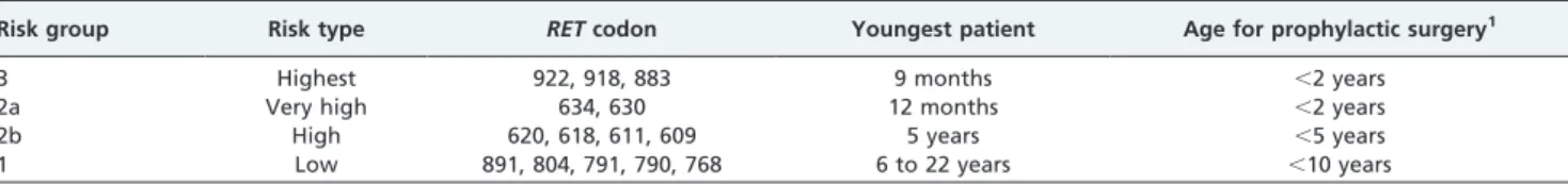
susceptibility gene enabled us to implement a powerful molecular diagnostic test to identify mutation carriers at premorbid stages (i.e., predictive testing) (Table 1). In this situation, family members who are found to carry the family-specific mutation but who are as yet unaffected can be subjected to life-saving prophylactic surgery and surveillance (18). Importantly, the delineation of a geno-type-phenotype correlation allows us to tailor surgical techniques based on the exact amino acids that are altered (23). Furthermore, family members not harboring family-specific disease-associated mutations can be definitively
Table 1 -Risk stratification in MEN 2.
Risk group Risk type RETcodon Youngest patient Age for prophylactic surgery1
3 Highest 922, 918, 883 9 months ,2 years
2a Very high 634, 630 12 months ,2 years
2b High 620, 618, 611, 609 5 years ,5 years
1 Low 891, 804, 791, 790, 768 6 to 22 years ,10 years
advised that they only have the same risk as the general population of developing the component neoplasias of MEN 2, so could be excluded from lifelong clinical surveillance and prophylactic surgeries.
There are three forms of MEN 2. First, MEN 2A, the most common form, is characterized by the components MTC: primary hyperparathyroidism and pheochromocytoma. Because MTC can show a high penetrance at a young age, prophylactic surgery should be offered depending on the risk genotype before the age of 5–10 years. MEN 2B is the most aggressive form caused by germline mutations in codon 918 (98%) or codon 883 (2%) and is associated with an age of onset for MTC in the first year of life. Therefore, genetic testing should be offered shortly after birth in order to identify those infants who require early preventive surgery. Of note, in MEN 2B MTC occurs together with pheochromocytoma (50%) and marfanoid habitus (100%), intestinal ganglioneuromatosis, mucosal neuromas, and thickening of the corneal nerve (all up to 90%), but not primary hyperparathyroidism. Familial MTC is often considered a separate entity giving rise to MTC as the sole clinical manifestation. Whether this holds true today has to be critically discussed. Most likely, familial MTC is just the result of variability in penetrance, thus surveillance for hyperparathyroidism and pheochromocytoma should be offered to these patients as well. Importantly, for the most common exon 11 mutation (RET C634G), a penetrance of 21% by age 50 for hyperparathyroidism and 52% by age 30 and 83% by age 50 for pheochromocytoma have been reported. However, it is not possible to predict precisely if and when an individual patient might suffer from para-thyroid hyperplasia or pheochromocytoma (24). Hence, all patients should undergo lifelong surveillance. Furthermore, there are other clinical aspects that cannot be entirely explained by the traditional mutations inRET. For instance, within some MEN 2 families, the age of onset and severity of disease vary considerably among the affected family members despite carrying identical germline RET muta-tions. For example, for the most common exon 11 mutation (Cys634Trp) the age of onset for MTC varies between
families from 3 years to .60 years. To what extent allelic variants play a role is a matter of continued investigation (25). For instance, the variant IVS1-126G . T in a large family with the G533C mutation was associated with an early onset of disease (26). One might envision that the integration of classic Mendelian cancer predisposition genes with genomic alterations of low penetrance might bring the practice of personalized genomic medicine to the next level (26,27).
Heritable adrenal neoplasias
Heritable adrenal neoplasias can affect the medulla (pheochromocytoma) as well as the adrenal cortex. The genetic differential diagnoses for adrenocortical neoplasia are summarized in Table 2. Diagnostic work-up, manage-ment, and surveillance of patients with hereditary diseases can differ significantly from their sporadic counterparts, so it is important to identify those patients. For instance, in MEN 1, adrenocortical lesions occur in as many as 40% of patients. Most manifest as non-functional, hyperplastic lesions and present an indolent course. Although the incidence of adrenocortical carcinoma has been reported to be between 2% and 6%, there is no a consensus regarding the management of MEN 1-associated adrenocortical lesions. Even though there is no genotype-phenotype correlation, it is a widely accepted protocol to surgically remove lesions exceeding a cutoff size of 3 cm in diameter. A considerable proportion of pheochromocytomas are heritable and form part of tumor syndromes other than MEN 2. In addition, studies have shown that pheochromo-cytomas can precede the development of MTC, so knowl-edge about differential diagnosis is important. Pheochromo-cytomas can occur in the adrenal medulla but also extra-adrenally in the head-and-neck region as well as being abdominal and thoracic, where they are termed paragan-glioma (PGL). It used to be thought that about 10% of all pheochromocytomas occur in a familial setting and are caused by germline genetic alterations. Today, there is growing evidence that about 25–30% of all patients with pheochromocytoma and paraganglioma harbor a germline
Table 2 -Genetic differential diagnosis for adrenocortical neoplasias.
Location Heritable tumor syndrome and phenotype Gene
Adrenocortical neoplasia
+ Multiple endocrine neoplasia type 1 (MEN 1)
Adrenocortical adenoma, HPT, pituitary and neuroendocrine tumors
+ Beckwith–Wiedemann syndrome (BWS)
ACC, Wilms’ tumor, hepatoblastoma, rhabdomyosarcoma
+ McCune–Albright syndrome
Cushing syndrome, pituitary tumors, polyostotic fibrous dysplasia, patchy skin pigmentation, thyrotoxicosis, gigantism
+ Li–Fraumeni Syndrome (LFS)
ACC, breast cancer, sarcomas, leukemia,
+ Adrenocortical hyperplasia
Nodular hyperplasia
+ Gardner syndrome
ACC, polyps of the colon, osteomas, fibromas, thyroid cancer
MEN1
CDKN1C (KIP2)
GNAS1
TP53
CYP21
APC
Adrenal medulla/
Pheochromozytoma + Multiple endocrine neoplasia type 2 (MEN 2)
+ Von Hippel–Lindau disease (VHL)
+ Neurofibromatosis type 1 (NF1)
+ Pheochromocytoma–paraganglioma syndrome (PC-PGL syndrome)
RET VHL NF1 SDHB, SDHC, SDHD
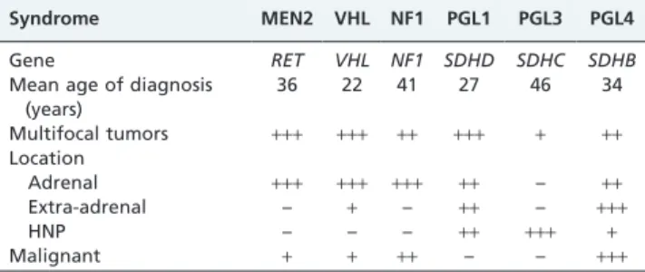
mutation. Six tumor syndromes, including MEN 2, Von Hippel–Lindau disease, neurofibromatosis (NF1) and pheo-chromocytoma-paraganglioma syndrome (PGL1, PGL2 and PGL3), and their predisposing genes have been identified (Table 3). The predominant signs of neurofibromatosis type 1 include characteristic cafe´-au-lait spots, neurofibromas, and other lesions such as freckling of the skin. Although penetrance of pheochromocytoma in NF1 is low (,5%), malignant pheochromocytomas are not infrequent. In patients with NF1 and pheochromocytoma there are no mutation hot spots, instead the alterations are spread all over the gene and include large deletions or rearrangements in about 10% of patients. As outlined above, pheochromo-cytoma in MEN 2 shows a variable penetrance that can be as high as 50%. Most notably, mutations in codons 634 and 918 harbor a very high risk of developing this disease. Pheochromocytomas in MEN 2 are commonly benign and located bilaterally in the adrenal medulla. Von Hippel-Lindau syndrome is an autosomal-dominant disorder that predisposes individuals to pheochromocytoma and other important neoplasias such as clear cell renal carcinomas, pancreatic islet cell tumors, and cystic disease of the kidney and pancreas. Interestingly, although the overall penetrance of pheochromocytoma ranges between 20% and 30% of patients with Von Hippel-Lindau syndrome, in certain families the prevalence of pheochromocytoma can be more than 90%. As a rule, all tumorous components of Von Hippel-Lindau syndrome can be effectively treated if diagnosed in time. Three paraganglioma syndrome predis-position genes have been identified so far, namely, SDHB the predisposition gene for PGL4,SDHC(PGL3), andSDHD (PGL1). Patients with PGL1 nearly always display benign but multiple adrenal pheochromocytomas and paraganglio-mas of the head and neck. Patients with PGL4 often display extra-adrenal, thoracic, or head-and-neck paragangliomas. In contrast to PGL1, they are frequently malignant and about one-third of patients develop metastases. PGL3 is not associated with malignant pheochromocytoma/paragan-glioma and patients display the characteristics of age, manifestation, and tumor number similar to those with sporadic HNPs. One has to be aware of the clinical symptoms and signs that allow for an operational division, which in turn is the cornerstone of prioritized genetic testing and personalized management of patients and their relatives (28). Hereditary pheochromocytoma and paragan-glioma syndromes should be recognized as early as possible in order to tailor medical management and follow-up.
Whereas the clinical and pathologic call of malignancy remains a diagnostic challenge and preventive therapy cannot be advised, it is key to conduct predictive testing of as-yet-unaffected relatives, so that mutation carriers can received a tailored screening and surveillance program (29).
Heritable non-medullary thyroid cancers
In contrast to heritable MTC, for which the genetic differential diagnosis is straightforward and results in only MEN 2, heritable non-MTC (NMTC) is more complex (4,20,24,30). In this setting we have to differentiate between NMTCs that are part of a heritable syndrome and those that occur non-syndromically as familial NMTC. There are three well-characterized syndromic forms of heritable NMTC—Cowden syndrome, Gardner syndrome (familial adenomatous polyposis syndrome), and Werner syndrome — all of which are transmitted in an autosomal-dominant fashion (31,32). Cowden syndrome is caused by germline mutations of the PTEN tumor suppressor gene. Patients with Cowden syndrome present with characteristic multi-ple hamartomas and mucocutaneous lesions (e.g., oral papillomatous papules, trichilemmoma, and acral kerato-sis). In addition, affected patients are at high risk of developing various benign and malignant neoplasias, especially of the breast (up to 50% lifetime risk in affected females) and thyroid gland. While benign thyroid nodules are observed in up to 75% of patients, NMTC commonly presents as a follicular thyroid carcinoma and is associated with a 10-fold increased lifetime risk compared with the general population.
Gardner syndrome is caused by germline mutations in the APC gene and is characterized by gastrointestinal adeno-matous polyps and a high risk of developing gastrointest-inal and other cancers. It is estimated that the lifetime risk of developing NMTC ranges between 2% and 10% in those affected with familial adenomatous polyposis syndrome. In contrast, the gastrointestinal manifestation will progress to colorectal cancer in 100% of affected patients and preventive surgery is advised for these patients. Interestingly, NMTC will present in this setting as a unique pathologic variant known as cribriform morular variant of papillary thyroid carcinoma (33). Therefore, any patients with this diagnosis who are operated on for thyroid disease should be screened for familial adenomatous polyposis syndrome as well.
Werner syndrome is a rare autosomal recessive disease that results in premature aging (34). Besides their elderly appearance, affected patients develop various age-related disorders such as diabetes, osteoporosis, heart disease, and other malignancies. The latter two are responsible for a diminished life expectancy of around 50 years. The incidence of thyroid cancer (e.g., papillary, follicular, and anaplastic thyroid cancer) is about 20%. Because of the characteristic physical appearance, genetic testing is com-monly used to confirm the clinical diagnosis.
Molecular diagnosis and predictive testing for these syndromes are available and highly accurate, and the results change medical management with regard to pro-phylaxis and surveillance. What cannot be done currently is accurately predict who will develop thyroid cancer and what the age of maximal risk is. However, the high incidence of malignant thyroid disease warrants regular thyroid screening and a low threshold for recommending thyroidectomy.
Table 3 -Comparison of pheochromocytoma-associated and paraganglioma-associated syndromes.
Syndrome MEN2 VHL NF1 PGL1 PGL3 PGL4
Gene RET VHL NF1 SDHD SDHC SDHB
Mean age of diagnosis (years)
36 22 41 27 46 34
Multifocal tumors +++ +++ ++ +++ + ++
Location
Adrenal +++ +++ +++ ++ – ++
Extra-adrenal – + – ++ – +++
HNP – – – ++ +++ +
Malignant + + ++ – – +++
+++: very high.++: high.+: low. –: absent. HNP: head-and-neck paraganglioma.
It has been suggested that there is an association between non-MTC and two other familial cancer syndromes, Carney complex (PRKAR1a) and Pendred syndrome. Based on a small series of 15 kindreds with Carney complex the prevalence of thyroid cancer is estimated 4% in affected family members (35). For Pendred Syndrome the estimated 1% prevalence for thyroid cancer is deduced from a single family report in which two of the three affected family members developed follicular thyroid cancer (36). Whether these are true associations due to the germline alterations that are involved, or if additional modifying events participate, is a matter of debate. However, patients with Carney complex typically develop nodular thyroid disease in childhood (about 60%) and in Pendred syndrome there is a high frequency of multinodular goiters and hypothyroidism. Hence, while a preventive thyroidectomy cannot be advised for any of these syndromes, a regular thyroid work-up should be part of the routine surveillance for patients identified as being at risk.
Non-syndromic familial non-MTC
For non-syndromic familial NMTC (FNMTC), epidemio-logic data indicate a very high likelihood of familial aggregation and, hence, a strong genetic component (4,37). Indeed, it is estimated that about 10% of all cases of NMTC are hereditary, putting affected first-degree relatives at up to a 10-fold increased risk compared with the general popula-tion (38,39). In fact, differentiated epithelial thyroid carcino-mas have one of the highest familial risks among all cancer sites. FNMTC is characterized by two or more first-degree relatives who are affected by thyroid carcinomas (NMTC) but lack the signs of other hereditary syndromes or exposure to other risk factors (e.g., radiation). This operational definition is based on the assumption that the chance of clustering of three or more relatives with NMTC in one family by chance is less than 6% (40). In contrast, there is a.60% likelihood that two affected first-degree relatives will harbor sporadic disease (40). The standardized incidence ratio is an index that estimates the familial risk of developing a malignancy based on epidemiologic data, and as a result may be viewed as a measure of cancer risk for the children of parents with a specific malignancy. For NMTC, the standardized incidence ratio exceeds an impressive value of 3.8 (38,41). One might argue that the familial aggregation might be the result of environmental factors shared by the family members because environmental factors have been implicated in thyroid carcinogenesis. However, studies comparing cancer risk between spouses showed that the standardized incidence ratio does not exceed 1.4. Therefore, for NMTC, it has been argued that heritability is most likely the main contributor to the high standardized incidence ratios and that familial aggregations of NMTC are most likely the result of germline mutations in susceptibility genes.
Despite the impressive standardized incidence ratio for FNMTC and tremendous efforts over the last 10 years to identify the cancer-associated gene, no susceptibility genes have been identified so far (4,42). Nevertheless, at least five putative susceptibility loci have been identified. The caveat remains that for each locus evidence both in favor and opposing association with FNMTC has been published (42). This can be explained by the fact that the operational definition for FNMTC varies between studies (some already include families with two affected first-degree relatives) and some include benign thyroid diseases as well. Of the five loci on 1q21 (PRN), 2q21 (NMTC1), 8p23.1-p22 (FTEN), 14q31 (MNG1), and
19p13.2 (TCO), only TCO and NMTC1 have been replicated in independent family sets, but the susceptibly genes mapped to these loci have not been identified (42–46).
The NMTC1 locus has been replicated in 10 kindreds and linkage to FNMTC is proposed with a logarithm of the odds (LOD) score of 2.85 (where a LOD score.3 is accepted to confirm linkage) is suggested (42). However, it has been suggested that NMTC1 has a more significant association with the follicular variant of papillary thyroid carcinoma. A linkage between the TCO (thyroid tumors with cell oxyphilia) locus and NMTC has been identified and validated in four studies comprised of 21 families (LOD score of 3.01) (42,43). In contrast, studies with a total of 65 relatives failed to show a linkage, so TCO might contribute only slightly to FNMTC and be associated with cell oxyphilia.
The MNG1 locus has only shown linkage (LOD score of 4.88) in one kindred with two family members who were affected by papillary thyroid carcinoma. Interestingly, 18 family members showed multinodular goiters (44). Given that six other studies that included.100 kindreds failed to reproduce this association, MNG1 might rather account for a familial form of benign nodular disease rather than FNMTC. Linkage of the PRN locus (LOD score of 3.58) was identified in one large kindred with five cases of papillary thyroid carcinoma and two cases of papillary renal neoplasm (45). However, no additional families have been identified so far. Thus, the 1q21 locus might be associated with rare phenotypes of papillary thyroid carcinoma and papillary renal neoplasm. Recently, another association was identified in one kindred with five cases of thyroid cancer and 11 cases of benign nodular disease (47). The putative susceptibility locus was mapped to 8q23.1-p22 with a LOD score of 4.41. However, a follow-up study by the same group included six families but failed to validate the linkage (48).
Thus, clinical genetic counseling or advice for families segregating NMTC is imprecise. Notably, there is ongoing debate as to whether or not FNMTC displays a more aggressive phenotype compared with its sporadic counter-part (49). Indeed, several studies have found that FNMTC presents at a younger age and displays more aggressive features, such as multifocality, local and lymph node invasion, and diminished tumor-free and overall survival rates (50–53). In contrast, other studies have failed to show a difference between FNMTC and sporadic cancer (49,54,55). A recent study that included 67 patients from 46 families with FNMTC and 375 controls was not able to identify a more aggressive phenotype (49,54). Still, there is a tendency for a lower age of onset (42 vs. 47 years) in the FNMTC group compared with the control group. Others have proposed prophylactic thyroidectomy for FNMTC (42). However, from a surgeon’s standpoint, the current data are too weak and inconsistent for such a general demand. First and foremost ‘‘primum non nocere’’has to prevail. Thus, in the setting of two or more family members with NMTC, a thyroid work up should be offered to all first-degree relatives and the presence of thyroid nodular disease should have a lower threshold for recommending thyroidectomy to these patients.
KNOWING HERITABLE CANCER PHENOMICS: THE FIRST STEP TO PRACTICE PERSONALIZED MEDICINE
personalized medicine is snowballing out of control when direct-to-consumer genomic testing is becoming increasingly available (56). Indeed, since 2007 the interested consumer is able to receive a whole-genome scan for less than US$500, without ever having to see a doctor or receive medical advice. This kind of self-administered genetic testing is problematic, as shown by Bloss et al. (56) who found a correlation between test-related distress and increased lifetime risk. Various professional organizations, including the American Medical Association and the American College of Medical Genetics, state that such genetic testing should not be performed without the supervision of healthcare providers. However, centers for genomic medicine, and even clinical cancer genetics, are far from being ubiquitously established even in high-level medical centers. It is estimated that only 1% of patients that would benefit from this 21st century approach to medical treatment will be referred to professional genetic counseling (31). The implementation of state-of-the-art clinical genetic counseling within the constraints of budget and limited human resources is one of the important tasks that have to be tackled.
Eventually, one has to keep in mind that, notwithstanding all of these technological advances, several tumor syn-dromes can be identified by rigorous clinical analysis (57). Especially regarding endocrine-related neoplasia, the med-ical practitioner has to be aware of the phenomics, or phenotypic profiles, that comes along with the array of inheritable endocrine tumor syndromes. It is of no surprise that several of the inherited tumor syndromes were first described and named long before genetic causes had been identified. In fact, it should be paramount to the medical professional to use meticulous observation to ‘‘personalize’’ the decision for treatment options and, much more importantly, to prioritize medical genetic testing.
ACKNOWLEDGMENTS
Frank Weber is deeply grateful to his mentor, Charis Eng, who always encouraged and inspired him. Thank you for your friendship and advice.
AUTHOR CONTRIBUTIONS
Weber F and Dammann M were responsible for the selection of relevant literature as well as for writing and revising the manuscript.
REFERENCES
1. Weber F, Fukino K, Sawada T, Williams N, Sweet K, Brena RM, et al. Variability in organ-specific EGFR mutational spectra in tumour epithelium and stroma may be the biological basis for differential responses to tyrosine kinase inhibitors. Br J Cancer. 2005;92(10):1922-6, http://dx.doi.org/10.1038/sj.bjc.6602557.
2. Hou YY, Grabellus F, Weber F, Zhou Y, Tan YS, Li J, et al. Impact of KIT and PDGFRA gene mutations on prognosis of patients with gastrointest-inal stromal tumors after complete primary tumor resection. J Gastrointest Surg. 2009;13(9):1583-92, http://dx.doi.org/10.1007/s11605-009-0842-6. 3. Landa I, Montero-Conde C, Malanga D, De Gisi S, Pita G,
Leandro-Garcia LJ, et al. Allelic variant at -79 (C.T) in CDKN1B (p27Kip1) confers an increased risk of thyroid cancer and alters mRNA levels. Endocr Relat Cancer. 2010;17(2):317-28.
4. Weber F, Eng C. Gene-expression profiling in differentiated thyroid cancer: a viable strategy for the practice of genomic medicine? Future Oncol. 2005;1(4):497-510.
5. Niemeier LA, Kuffner Akatsu H, Song C, Carty SE, Hodak SP, Yip L, et al. A combined molecular-pathologic score improves risk stratification of thyroid papillary microcarcinoma. Cancer. Aug 31 [epub ahead of print]. 6. Manolio TA. Genomewide association studies and assessment of the risk
of disease. N Engl J Med. 2010;363(2):166-76.
7. Adamzik M, Frey UH, Mohlenkamp S, Scherag A, Waydhas C, Marggraf G, et al. Aquaporin 5 gene promoter: 1364A/C polymorphism associated with 30-day survival in severe sepsis. Anesthesiology. 2011;114(4):912-17.
8. Anney R, Klei L, Pinto D, Regan R, Conroy J, Magalhaes TR, et al. A genome-wide scan for common alleles affecting risk for autism. Hum Mol Genet. 2010;19(20):4072-82.
9. McCarthy MI, Hirschhorn JN. Genome-wide association studies: potential next steps on a genetic journey. Hum Mol Genet. 2008;17(R2):R156-65, http://dx.doi.org/10.1093/hmg/ddn289. 10. Dorn GW, 2nd. The genomic architecture of sporadic heart failure. Circ
Res. 2011;108(10):1270-83.
11. Park SJ, Jung EH, Ryu RS, Kang HW, Ko JM, Kim HJ, et al. Clinical implementation of whole-genome array CGH as a first-tier test in 5080 pre and postnatal cases. Mol Cytogenet. 2011;4:12.
12. Guttmacher AE, McGuire AL, Ponder B, Stefansson K. Personalized genomic information: preparing for the future of genetic medicine. Nat Rev Genet. 2010;11(2):161-5.
13. Hall WD, Mathews R, Morley KI. Being more realistic about the public health impact of genomic medicine. PLoS Med. 2010;7(10):e1000347, http://dx.doi.org/10.1371/journal.pmed.1000347
14. Feero WG, Guttmacher AE, Collins FS. Genomic medicine: an updated primer. N Engl J Med. 2010;362(21):2001-11, http://dx.doi.org/10.1056/ NEJMra0907175.
15. Paynter NP, Chasman DI, Buring JE, Shiffman D, Cook NR, Ridker PM. Cardiovascular disease risk prediction with and without knowledge of genetic variation at chromosome 9p21.3. Ann Intern Med. 2009;150(2):65-72.
16. Colombo S, Rauch A, Rotger M, Fellay J, Martinez R, Fux C, et al. The HCP5 single-nucleotide polymorphism: a simple screening tool for prediction of hypersensitivity reaction to abacavir. J Infect Dis. 2008;198(6):864-7, http://dx.doi.org/10.1086/591184.
17. Buchan BW, Peterson JF, Cogbill CH, Anderson DK, Ledford JS, White MN, et al. Evaluation of a Microarray-Based Genotyping Assay for the Rapid Detection of Cytochrome P450 2C19 *2 and *3 Polymorphisms From Whole Blood Using Nanoparticle Probes. Am J Clin Pathol. 2011;136(4):604-8.
18. Frilling A, Weber F, Tecklenborg C, Broelsch CE. Prophylactic thyroidectomy in multiple endocrine neoplasia: the impact of molecular mechanisms of RET proto-oncogene. Langenbecks Arch Surg. 2003;388(1):17-26.
19. Frilling A, Weber F. Prophylactic thyroid surgery. Chirurg. 2006;77(1):6-14, http://dx.doi.org/10.1007/s00104-005-1118-7.
20. Weber F, Eng C. Update on the molecular diagnosis of endocrine tumors: toward -omics-based personalized healthcare? J Clin Endocrinol Metab. 2008;93(4):1097-104, http://dx.doi.org/10.1210/jc.2008-0212.
21. Eng C. RET proto-oncogene in the development of human cancer. J Clin Oncol. 1999;17(1):380-93.
22. Frilling A, Dralle H, Eng C, Raue F, Broelsch CE. Presymptomatic DNA screening in families with multiple endocrine neoplasia type 2 and familial medullary thyroid carcinoma. Surgery. 1995;118(6):1099-103; discussion 1103-4, http://dx.doi.org/10.1016/S0039-6060(05)80120-5 23. Eng C, Clayton D, Schuffenecker I, Lenoir G, Cote G, Gagel RF, et al. The
relationship between specific RET proto-oncogene mutations and disease phenotype in multiple endocrine neoplasia type 2. International RET mutation consortium analysis. JAMA. 1996;276(19):1575-9.
24. Milos IN, Frank-Raue K, Wohllk N, Maia AL, Pusiol E, Patocs A, et al. Age-related neoplastic risk profiles and penetrance estimations in multi-ple endocrine neoplasia type 2A caused by germ line RET Cys634Trp (TGC.TGG) mutation. Endocr Relat Cancer. 2008;15(4):1035-41, http:// dx.doi.org/10.1677/ERC-08-0105.
25. Weber F, Eng C. Editorial: germline variants within RET: clinical utility or scientific playtoy? J Clin Endocrinol Metab. 2005;90(11):6334-6, http:// dx.doi.org/10.1210/jc.2005-2030.
26. Tamanaha R, Camacho CP, Pereira AC, da Silva AM, Maciel RM, Cerutti JM. Evaluation of RET polymorphisms in a six-generation family with G533C RET mutation: specific RET variants may modulate age at onset and clinical presentation. Clin Endocrinol (Oxf). 2009;71(1):56-64, http:// dx.doi.org/10.1111/j.1365-2265.2008.03491.x.
27. Jiang Q, Ho YY, Hao L, Nichols Berrios C, Chakravarti A. Copy number variants in candidate genes are genetic modifiers of Hirschsprung disease. PLoS One. 2011;6(6):e21219.
28. Erlic Z, Rybicki L, Peczkowska M, Golcher H, Kann PH, Brauckhoff M, et al. Clinical predictors and algorithm for the genetic diagnosis of pheochromocytoma patients. Clin Cancer Res. 2009;15(20):6378-85, http://dx.doi.org/10.1158/1078-0432.CCR-09-1237.
29. Eisenhofer G, Bornstein SR, Brouwers FM, Cheung NK, Dahia PL, de Krijger RR, et al. Malignant pheochromocytoma: current status and initiatives for future progress. Endocr Relat Cancer. 2004;11(3):423-36, http://dx.doi.org/10.1677/erc.1.00829.
30. Eng C. Familial papillary thyroid cancer: many syndromes, too many genes? J Clin Endocrinol Metab. 2000;85(5):1755-7, http://dx.doi.org/ 10.1210/jc.85.5.1755.
31. Eng C. Mendelian genetics of rare—and not so rare—cancers. Ann N Y Acad Sci. 2010;1214:70-82.
33. Tomoda C, Miyauchi A, Uruno T, Takamura Y, Ito Y, Miya A, et al. Cribriform-morular variant of papillary thyroid carcinoma: clue to early detection of familial adenomatous polyposis-associated colon cancer. World J Surg. 2004;28(9):886-9, http://dx.doi.org/10.1007/s00268-004-7475-4.
34. Muftuoglu M, Oshima J, von Kobbe C, Cheng WH, Leistritz DF, Bohr VA. The clinical characteristics of Werner syndrome: molecular and biochemical diagnosis. Hum Genet. 2008;124(4):369-77, http:// dx.doi.org/10.1007/s00439-008-0562-0.
35. Stratakis CA, Courcoutsakis NA, Abati A, Filie A, Doppman JL, Carney JA, et al. Thyroid gland abnormalities in patients with the syndrome of spotty skin pigmentation, myxomas, endocrine overactivity, and schwan-nomas (Carney complex). J Clin Endocrinol Metab. 1997;82(7):2037-43, http://dx.doi.org/10.1210/jc.82.7.2037.
36. Snabboon T, Plengpanich W, Saengpanich S, Sirisalipoch S, Keelawat S, Sunthornyothin S, et al. Two common and three novel PDS mutations in Thai patients with Pendred syndrome. J Endocrinol Invest. 2007;30(11):907-13.
37. Malchoff CD, Sarfarazi M, Tendler B, Forouhar F, Whalen G, Malchoff DM. Familial papillary thyroid carcinoma is genetically distinct from familial adenomatous polyposis coli. Thyroid. 1999;9(3):247-52, http:// dx.doi.org/10.1089/thy.1999.9.247.
38. Hemminki K, Eng C, Chen B. Familial risks for nonmedullary thyroid cancer. J Clin Endocrinol Metab. 2005;90(10):5747-53, http://dx.doi.org/ 10.1210/jc.2005-0935.
39. Goldgar DE, Easton DF, Cannon-Albright LA, Skolnick MH. Systematic population-based assessment of cancer risk in first-degree relatives of cancer probands. J Natl Cancer Inst. 1994;86(21):1600-8.
40. Charkes ND. On the prevalence of familial nonmedullary thyroid cancer in multiply affected kindreds. Thyroid. 2006;16(2):181-6, http:// dx.doi.org/10.1089/thy.2006.16.181.
41. Hemminki K, Eng C. Clinical genetic counselling for familial cancers requires reliable data on familial cancer risks and general action plans. J Med Genet. 2004;41(11):801-7, http://dx.doi.org/10.1136/ jmg.2004.022731.
42. Khan A, Smellie J, Nutting C, Harrington K, Newbold K. Familial nonmedullary thyroid cancer: a review of the genetics. Thyroid. 2010; 20(7):795-801.
43. Lesueur F, Stark M, Tocco T, Ayadi H, Delisle MJ, Goldgar DE, et al. Genetic heterogeneity in familial nonmedullary thyroid carcinoma: exclusion of linkage to RET, MNG1, and TCO in 56 families. NMTC Consortium. J Clin Endocrinol Metab. 1999;84(6):2157-62, http://dx.doi.org/10.1210/ jc.84.6.2157.
44. Bignell GR, Canzian F, Shayeghi M, Stark M, Shugart YY, Biggs P, et al. Familial nontoxic multinodular thyroid goiter locus maps to chromo-some 14q but does not account for familial nonmedullary thyroid cancer. Am J Hum Genet. 1997;61(5):1123-30, http://dx.doi.org/10.1086/301610. 45. Malchoff CD, Sarfarazi M, Tendler B, Forouhar F, Whalen G, Joshi V, et al. Papillary thyroid carcinoma associated with papillary renal neoplasia: genetic linkage analysis of a distinct heritable tumor
syndrome. J Clin Endocrinol Metab. 2000;85(5):1758-64, http:// dx.doi.org/10.1210/jc.85.5.1758.
46. McKay JD, Lesueur F, Jonard L, Pastore A, Williamson J, Hoffman L, et al. Localization of a susceptibility gene for familial nonmedullary thyroid carcinoma to chromosome 2q21. Am J Hum Genet. 2001;69(2):440-6, http://dx.doi.org/10.1086/321979.
47. Cavaco BM, Batista PF, Sobrinho LG, Leite V. Mapping a new familial thyroid epithelial neoplasia susceptibility locus to chromosome 8p23.1-p22 by high-density single-nucleotide polymorphism genome-wide linkage analysis. J Clin Endocrinol Metab. 2008;93(11):4426-30, http:// dx.doi.org/10.1210/jc.2008-0449.
48. Cavaco BM, Batista PF, Martins C, Banito A, do Rosario F, Limbert E, et al. Familial non-medullary thyroid carcinoma (FNMTC): analysis of fPTC/PRN, NMTC1, MNG1 and TCO susceptibility loci and identifica-tion of somatic BRAF and RAS mutaidentifica-tions. Endocr Relat Cancer. 2008;15(1):207-15, http://dx.doi.org/10.1677/ERC-07-0214.
49. Loh KC. Familial nonmedullary thyroid carcinoma: a meta-review of case series. Thyroid. 1997;7(1):107-13, http://dx.doi.org/10.1089/ thy.1997.7.107.
50. Burgess JR, Duffield A, Wilkinson SJ, Ware R, Greenaway TM, Percival J, et al. Two families with an autosomal dominant inheritance pattern for papillary carcinoma of the thyroid. J Clin Endocrinol Metab. 1997;82(2):345-8, http://dx.doi.org/10.1210/jc.82.2.345.
51. Capezzone M, Marchisotta S, Cantara S, Busonero G, Brilli L, Pazaitou-Panayiotou K, et al. Familial non-medullary thyroid carcinoma displays the features of clinical anticipation suggestive of a distinct biological entity. Endocr Relat Cancer. 2008;15(4):1075-81, http://dx.doi.org/ 10.1677/ERC-08-0080.
52. Alsanea O, Wada N, Ain K, Wong M, Taylor K, Ituarte PH, et al. Is familial non-medullary thyroid carcinoma more aggressive than spora-dic thyroid cancer? A multicenter series. Surgery. 2000;128(6):1043-50; discussion 50-1, http://dx.doi.org/10.1067/msy.2000.110848
53. Uchino S, Noguchi S, Kawamoto H, Yamashita H, Watanabe S, Shuto S. Familial nonmedullary thyroid carcinoma characterized by multifocality and a high recurrence rate in a large study population. World J Surg. 2002;26(8):897-902, http://dx.doi.org/10.1007/s00268-002-6615-y. 54. Robenshtok E, Tzvetov G, Grozinsky-Glasberg S, Shraga-Slutzky I,
Weinstein R, Lazar L, et al. Clinical characteristics and outcome of familial nonmedullary thyroid cancer: a retrospective controlled study. Thyroid. 2011;21(1):43-8.
55. Ito Y, Kakudo K, Hirokawa M, Fukushima M, Yabuta T, Tomoda C, et al. Biological behavior and prognosis of familial papillary thyroid carci-noma. Surgery. 2009;145(1):100-5, http://dx.doi.org/10.1016/ j.surg.2008.08.004.
56. Bloss CS, Schork NJ, Topol EJ. Effect of direct-to-consumer genomewide profiling to assess disease risk. N Engl J Med. 2011;364(6):524-34. 57. Zbuk KM, Eng C. Cancer phenomics: RET and PTEN as illustrative