The Value of a Routine Histopathological
Examination of Uterine Specimens in Dogs
and Cats
ABSTRACT
Uterine lesions are common in small animals. The most frequent one is pyometra, which gross features included distended uterine walls by a mucopurulent fluid and a change in uterine wall thickness. However, pyometra is a secondary lesion that may mask several other pathologies of the endometrium in both the bitch and queen. In here we present some notes related to the submission of uterine specimens for histopathology. This chapter further discusses the role of histopathology in the diagnosis of various uterine pathologies sharing common gross appearance or hidden behind pyometra, such as the endometrial adenocarcinoma or the squamous endometrial metaplasia. It also briefly describes the differences between those lesions. Although the surgeons often question how important may histopathology be when excised material so obvious fit pyometra gross pathological findings, in fact most professionals are often unreliable to perceive the co-existing pathology. Thus, the histopatological analysis of uterine specimens will support a
Rita Payan-Carreira* and Maria dos Anjos Pires
CECAV- Animal and Veterinary Research Center, Universidade de Trás-os-Montes e Alto Douro, Portugal
*Corresponding author: Payan-Carreira R, Animal and Veterinary Research Center, Univer-sidade de Trás-os-Montes e Alto Douro, Portugal, Tel: +351 259350425; Fax: +351 259350480; Email: [email protected]
Published Date: June 25, 2016
Gr up
patient care and establish the need for additional treatment. Also, the histopathological exam is of utmost importance when conducting medical research, in particular if addressing diagnostic decision making in pathology or on the identification of diagnostic or prognostic markers.
Keywords: Histopathology; Gross morphology; Histological exam; Uterine lesions; Dog; Cat Abbreviations: FEA-Feline Endometrial Adenocarcinoma; CEH-Cystic Endometrial
Hyperplasia; PolCEH-Polypoid Cystic Endometrial Hyperplasia; SM-Squamous Metaplasia; v/v-volume/volume
INTRODUCTION
Lesions of the uterus are common in dogs and cats, in particular, pyometra. Frequently, acute inflammatory conditions of the uterus, usually cursing as pyometra, lead the animal to surgery for ovariohysterectomy. However, seldom the excised uterus is submitted for analysis since its gross appearance fits the changes typically described in pyometra: a uterus distended by fluid, which is often mucopurulent, and changes in the uterine wall thickness [1,2]. And practitioners often question how important may histopathology be when excised material so obvious fit the gross pathological findings associated with pyometra. Most clinics only submit excised uteri for histopathology when the post-operative findings are suspect, or a notorious mass is observed. However, in either dogs or cats, pyometra may hide/mask more serious lesions, as the uterus in these species is prone to react to an irritator stimulus with inflammation [3,4]. In cats, for example, lesions of endometrial adenocarcinoma are frequently found masked by pyometra features [5]. In either case, progesterone seems to play a major role in the pathogenesis of reactive uterine inflammatory conditions and consequently, most situations are diagnosed in diestrus [3,4,6]. Notwithstanding, it is important to the practitioners to retain that pyometra is usually a complication of a previous lesion arising in the uterus, which may deserve to be explored.
Still, there are other lesions, such as masses or chronic inflammatory conditions, driving the need for a thorough histopathological examination of the entire excised organ or a uterine surgical biopsy, the later often requested during infertility assessment.
The histopathological exam of surgical uterine specimens may have an impact on patient management or outcome in situations that were not suspected on macroscopic examination at the time of surgery. The histopatological analysis of surgical material serves to support a proper diagnosis of the situation, to grade the severity of a condition and therefore to guide patient care, to establish the need for additional treatment, and to estimate a prognosis for situations representing uterine neoplasia. Moreover, the histopathological exam will further provide additional insights into the etiology of a process or the characterization and grading of a lesion, which are useful in medical research aiming to establish the best therapeutic approach for each variant or to identify a molecular marker for a particular uterine disease, like pyometra.
Below, the importance of histopathology in the diagnosis of the uterine lesions will be addressed in more detail, supported by our experience. Lesions will be grouped according to the changes in the gross morphology of the uterine specimen submitted to analysis.
A NOTE ON THE SUBMISSION OF A UTERINE SPECIMEN FOR
HISTOPATHOLOGY
For a good histopathological analysis, the excised material or biopsy specimens must be properly handled and transported into the lab. Several fixatives may be used for transport and preservation of the specimens, but 4% buffered formalin is the mostly used and all routine histopathological and molecular techniques are standardized for this fixative. The organ or a fragment must be immersed in the fixative, in a proportion of 1/10 to 1/15 (v/v), immediately after its collection. If particular exams are requested (e.g., histochemistry technique, immunohistochemistry, fluorescent microscopy or molecular biology), contact previously the laboratory to check the existence of additional concerns on sample preservation, and transport samples in appropriate sealed containers [7,8]. The size of the container is an important issue because fixed tissues are more rigid than the fresh´s; also, it should have a large enough opening. If space is insufficient, it may be difficult to remove the specimens or it may compromise its fixation [7-9]. To allow a proper fixation, gently mix the content of the container, as the fixative must surround the specimen on all sides [8,10].
An important step in the histopathological exam respects the adequate evaluation and gross description of lesions. The lesions will be oriented, measured and described regarding the consistency, tissue composition or disposition, its anatomical relations and its content annotated. This description requires the whole specimen to be sent to the lab [9]. However, due to the large size of uterine specimens, it is not always feasible to submit the whole organ to the lab. Then, only a sample of the specimen is commonly sent for analysis. Still, one should remember that this is not always the best option for a proper diagnosis. In such cases, it could be important to take several pictures (don´t forget to include a ruler scale in it, to create a frame of reference) [10] showing the total morphological appearance of the specimen (external and internal or the cut surface appearance, after the longitudinal section of the uterus). An annotation can be included regarding the area where the sample was collected. The surgical specimen should also be measured. In the case of the uterus, each uterine horn should be measured, longitudinally (i.e., the length from the base up to the apex) and transversally (the width); if irregularly shaped, the largest transversal measurement should be taken. Fat compromises the fixation; therefore, the fat located in the broad ligament should be carefully removed, except when it contains any lesion. When existing a mass, a net transversal cut following its major axis is often needed for proper fixation, and should be performed with caution, not to destroy the anatomical relationship with the surrounding tissues or to alter its features [1,9].
Important uterine lesions evolve during the luteal stage, so it is also important that the ovaries are sent along with the uterus. In the dog, extraction from the ovarian bursa is vital to ensure proper fixation. If it is not possible to include the ovaries, the stage of the estrous cycle and information on any contraceptive treatment should be conveyed to the pathologist.
On the other hand, biopsy specimens are often too small, and can therefore easily suffer from artifacts [10]. Biopsy samples may be obtained transcervical or by abdominal laparoscopy or laparotomy [11,12]. A higher risk for uncontrolled hemorrhage has been reported for transcervical collection of biopsy specimens [13], as well as an increased risk for secondary contamination of the uterus by vaginal biota [14], for which the transmural collection of material is preferred [12]. In the later, the obtained specimens contain the representation of all the uterine layers. These samples are easily orientated in the lab and give complete information on processes occurring in the organ. Contrasting, in transcervical-collected biopsies, only the endometrium is represented [13]. The precise orientation of the samples is more difficult in these cases, particularly when the endometrium is thicker than the normal, and the myometrium is not represented in the tissue collected [12,13]. Biopsy samples are usually small and frequently float in the fixative. To avoid losing the specimen, cover it with a gauze tissue. Whenever more than one sample was collected, the proper identification of the collection area is needed. As it is often difficult to proper mark each specimen, each one should be conveyed to the histopathology lab in a different container, properly labeled and containing information on the place of collection [8]. In the requisition form, the description related to the specimen in each container should be acknowledged.
For submission of a specimen for histopathological analysis, a proper requisition form must be filled. Besides the animal-related information (species, breed, age and reproductive status), it is of utmost importance to include a proper clinical history, information on the results from particular complementary exams, the description of the lesions (such as site, size, shape, dimensions) and also the suspicions of the clinician [8]. Such information is relevant to the pathologist, and will support him to establish a final diagnosis.
WHAT MAY HISTOPATHOLOGY REVEAL IN A DISTENDED
UTERUS?
Distension due to the accumulation of fluid often accompanies diverse pathological conditions originating in the uterus. The degree of distension of the uterus is variable, depending on the amount of fluid retained in the uterine cavity, which in turn depends on the cervix patency [15,16]. Fluid retention is higher when the cervical canal is closed; thereby the changes in the uterus are more pronounced, fluid accumulation increasing intra-uterine pressure and also provoking compression on the endometrium. In parallel, the endometrial inflammation and the cysts rupture lead to a destruction of a large portion of the endometrium [17]. When this happens, the uterine walls are thinner than the normal.
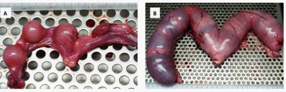
When assessing a distended uterus, one should first exclude the existence of unknown pregnancy, or any interruption of pregnancy leading to the accumulation of content in the uterus. In normal pregnancies, the uterus presents multiple or solitary swellings, usually similar in size and appearance, which in the last third of pregnancy let show evidence of the placentation areas (Figure 1). Uterus displaying regular pregnancy features does not usually impose any difficulty to the clinician or the pathologist.
Figure 1: Gross morphology of the uterus in a 4-week feline pregnancy (A) and an 8-week
canine pregnancy (B).
Some of those features are also present when pregnancy was interrupted. The death of the fetus may lead to maceration or mummification. Depending on the age of the process, the uterine swellings loss their tension and with time the fetal fluids are resorbed and fetal tissues may be compacted and maintained within the uterine cavity, but lose their normal disposition (Figure 2). These changes give the uterus an irregular contour and increased consistence; crepitation may also be presented. In cross-sections of the uterus, the walls are distended by the accumulation of heterogeneous material, containing different tissues of various consistencies and sometimes also bone and hair. The histological exam in such cases may reveal a neutrophilic infiltrate in the endometrium and necrotized fetal remnants surrounded by purulent material (fetal maceration) or dehydrated/mummified fetus surrounded by a viscous mucous secretion (fetal mummification) [2].
Figure 2: Specimen of a feline uterus representing a case of fetal maceration. The uterine horn is
notoriously distended (A) by a heterogeneous content (B) that corresponds to the remnants of 3 fetuses that died in the uterus.
Cases of uterine distension in the absence of pregnancy may be however more challenging to the pathologist. An important aspect to remember is that in small animal species, the uterus may present different co-existing lesions in the same region or adjoining areas. Thus, when submitting only one fragment of the uterus for histopathological analysis, or in the case of a biopsy, the results must be interpreted as respecting only to the fragment submitted [14]; any extrapolations for the whole organ cannot be attributed to the pathologist.
The amount of retained fluid (and therefore the degree of uterine distension), as well as the thickness of uterine walls, varies with the existence of a patent cervix and the extension of the primary lesion and the process originating the distension. Therefore, the volume of the excised uterus may vary from a subtle increase in size to a severe enlargement of the uterine horns [2,5]. Pyometra is the most common clinical syndrome that course with a distended uterus. In the majority of cases, particularly in dogs, it occurs in older animals. Pyometra is triggered by a pre-existing process – the cystic endometrial hyperplasia – that in a progesterone-primed endometrium originates the accumulation of mucinous secretion in the lumen that later suffer from the ascending contamination of vagina [2,18]. In open pyometra, the uterus usually presents thickened uterine walls and lower amount of fluid, while in close pyometra it typically shows thin walls and larger amount of fluid. The increased thickness of the uterine wall in dogs commonly accompanies a CEH process while, in cats, it is frequently associated with feline endometrial adenocarcinoma. On a recent survey, Pires & Carreira [19] refer that pyometra was diagnosed by histopathology in 29% of the inflammatory processes of the canine uterus, from which 25% represented processes secondary to CEH. In cats, another survey by Pires et al. [20] points a pyometra prevalence of 36%, and further refers that pyometra often co-exists with endometrial adenocarcinoma (45%). When pyometra exists, the main signs evidenced on the surgical specimen relates with inflammation and the accumulation of a mucopurulent fluid, which may mask any underlying, less evidenced lesion.
However, a fluid without purulent characteristics can also distend the uterus, such as in the case of hydrometra or mucometra, which are more frequent in dogs than in cats. In contrast to pyometra, hydrometra and mucometra evolve silently and are often a casual finding during the clinical exam or ultrasound or radiographic exploitation of abdomen [21]. Also, hydrometra and mucometra generally do not induce changes in the blood hematology [21], a useful sign when distinguishing these clinical situations. In a large number of cases diagnosed in older dogs, mucometra co-exists with CEH. However, not always the motif for the accumulation of non-inflammatory fluid is determined upon the macroscopic inspection of the uterus, particularly in hydrometra cases. In hydrometra, a disturbance in the levels of estrogen may trigger the pathogenesis of the process. In 2006, Payan-Carreira et al. described a case of hydrometra associated with granulosa cell tumour in a female dog [22]. Only the histopathological exam allowed reaching the diagnosis, as the cause of the lesion remained unapparent during the clinical exam. Chronic low stimulation of (endogenous or exogenous) estrogens may stimulate the secretion of low viscosity fluid that accumulates and distends the uterus.
Squamous metaplasia is another estrogen-induced lesion of the uterus that is often accompanied by a variable degree of pyometra. Pathogenesis of uterine squamous metaplasia in dogs remains unclear. In large animals, squamous metaplasia of the endometrium has been associated with zearalenone mycotoxicosis or prolonged follicular ovarian cysts [23]. In dogs, few cases of squamous endometrial metaplasia (SM) of the uterus have been described. McEntee [23] refers the occurrence of SM following depot estradiol treatment, while Zanghi et al [24] describe a uterine SM associated with granulosa cell tumor. These findings suggest that chronic estrogen stimulation may play an important role in the pathogenesis of the uterine squamous metaplasia. The chronic estrogenic impregnation might be achieved by increased or prolonged estrogen levels of endogenous or exogenous origin, or by a prolonged stimulation of estrogen receptors. The squamous metaplasia lesions may be focal or generalized. In the former, it is easy to lose track on the lesion, or it may appear as an incidental finding in the histological exam of hyperplastic lesions of the endometrium [24]. However, in generalized lesions, the overall uterus is distended by a mucopurulent fluid, the uterine walls are thinner, and the internal uterine relief is changed due to a loss of the expression of endometrial glands and to the proliferation of the surface epithelia, which changed into a keratinized stratified squamous epithelium.
In cats, FEA often presents distended uterus in the absence of a pyometra or fluid accumulation (Figure 3). The main feature is the increased thickness of the uterine walls, due to proliferation of the endometrium that contributes to a slight distension of the uterine cavity.
Figure 3: Uterine specimen from a female cat with endometrial adenocarcinoma not
accompanied by pyometra. The inspection of the uterus shows the distension of the organ (A) while on the sagittal cut surface (B) the increased thickness of the endometrium is observed.
When assessing the gross appearance of a distended uterus it may be important to remark: - The amount and the physical characteristics of the intrauterine fluid (viscosity, color, smell or existence of purulent material) – this may allow a gross distinction between pyometra and hydrometra/mucometra conditions. If necessary, cytology of the mucous or a cytospin, or even the apposition cytology of the endometrial surface can be quickly performed to check on the presence of neutrophils in the fluid or the surface of the organ.
- The thickness of the uterine walls and its regularity (related to the retention of the fluid within the uterus) may also give some information on the severity of the process and may be useful for emission of a prognosis in particular cases.
- The spraying of a lesion to surrounding areas of the organ or other tissues, or the appearance of a solitary mass, as well as the changes in the normal arrangement of the layers in the organ.
WHAT MAY HISTOPATHOLOGY REVEAL FROM FOCAL
DISTENSION OF THE UTERUS?
Focal distension of the uterus in dogs and cats is usually associated with polypoid cystic endometrial hyperplasia [2,17,18], mesenchymal neoplasia [25], focal endometrial adenocarcinoma lesions [25,26] or, less frequently, by fluid accumulation upstream an area of uterine aplasia [27]. In the later lesions, upstream to the obstruction point, a segmental distension of the uterine horn (Figure 4) resembles a mucometra or pyometra lesions, while downstream the uterus may be normal, or present any age-related degenerative change [27].
Figure 4: Uterine specimen from a female dog with aplasia of one uterine horn, showing
distension of a segment cranial to the obstruction point (arrow).
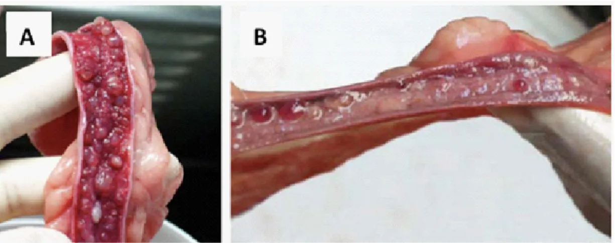
In cats and dogs, a polypoid or segmental variant of cystic endometrial hyperplasia may be found [2,17]. These polyps are localized overgrowths that, in some cases, reach considerable dimensions leading to a segmental distension of the uterine horn; these are commonly benign pedunculated lesions that may be found as solitary cystic masses containing connective tissue stroma with dilated endometrial glands, or as multiple masses arising in any portion of the uterine horns. They often co-exist with a local thinning of the uterine wall and accumulation of fluid of variable viscosity (Figure 5). With the progression of the disease, polypoid lesions tend to protrude into the uterine lumen and assume a fusiform shape [2,23,28]. Sporadic reports exist on the prolapse of such uterine polyps into the vagina, in either dogs or cats, which may originate inversion/invagination of a segment of the uterine horn [23].
Figure 5: Sections of the uterus with a polypoid cystic endometrial hyperplasia lesion in
a female cat (A –in a sagittal cut) and a female dog (B - in a transversal cut). It is observed concurrent severe atrophy of the uterine wall in the area surrounding the polyps and the accumulation of the fluid within the uterus, which is less viscous in the feline specimen,
contrasting to the mucous aspect of the fluid contained in the canine sample.
Although in cats endometrial adenocarcinoma often occurs in the form of diffuse or multiple lesions in the endometrium, this pathology may also sporadically develop in dogs and cats as a solitary lesion that which gross appearance is a focal dilatation of a uterine horn [29]. The longitudinal cut surface of the lesions may give some indications on the nature of the process although, in this case, the histopathological exam is crucial to distinguish these lesions from a focal lesion of endometrial adenocarcinoma whether in dogs or cats (Figure 6).
Figure 6: The overall appearance of the focal distension of the uterine horn (arrow) in a queen
associated with a lesion of feline endometrial adenocarcinoma (A) is rather similar to the one linked to a solitary lesion of polypoid cystic endometrial hyperplasia (arrow) in this dog specimen (B). Sometimes, these lesions may be confused with a solitary embryonic vesicle in a 3
Mesenchymal tumours of the uterus (e.g., fibroma and leiomyoma) may also present focal dilatation of the uterine horn and appear occasionally in female dogs and cats [1,31]. In most cases, similar lesions may be concurrently found in the vagina, particularly the leiomyoma lesions in dogs. They correspond to solid, firm, broad-based masses developing from the uterine wall (Figure 7). On the transversal cut surface, it may be observed the deviation of the uterine lumen by white masses [1,2]. Fibromas appear as hard spherical nodules composed of collagenous fibrous tissue while the leiomyoma is firm, opalescent to tan masses [1], composed of interwoven smooth muscle bundles [1,2]. Unless they obliterate the uterine wall and promote the accumulation of fluids, fibroma lesions are asymptomatic. In the dog, leiomyoma represents the most common tumours of the tubular genital tract, and are supposedly hormone-dependant [2]. Also in this species, they are usually multiple. Larger lesions may present a harder consistency because of the increased participation of fibrous connective tissue [1]. Leiomyoma lesion extends either inward, filling the uterine lumen, or outward, projecting at the external surface of the organ [2].
Figure 7: Focal distension (asterisk) within the walls of a uterine horn in female dog
associated to a mesenchymal tumour that seems to grow outward and to distort the normal alignment of the uterine horn (arrow) (A). A transversal cut of the lesion (B) showed that the lesion contained two nodules (asterisks), one of them growing into the uterine cavity, dislocating the uterine lumen (arrow), and another projecting towards the broad ligament, both
composed of tissues disposed in a storiform pattern.
WHAT MAY HISTOPATHOLOGY REVEAL FROM A
NON-DISTENDED UTERUS?
In situations of chronic uterine inflammation, often underlying infertility in bitches and queens, the size of the uterus remains frequently unchanged. Also, in most situations occurring in breeding females, the specimen is submitted for histopathology as a biopsy [11,14].
Not all dogs with pyometra have cystic endometrial hyperplasia, and not all the inflammatory process in the uterus co-exist with pyometra or mucometra. The histopathological exam has a major role in identifying those processes. Chronic endometritis is often found in breeding bitches
lymphocytic infiltration of the endometrium were frequently observed in routine histological observation of these lesions [11,14].
In acute or sub-acute endometritis not associated with pyometra, the endometrium is infiltrated with neutrophils or sporadically with also eosinophils, and only a small to moderate amount of fibrosis is occasionally observed [12].
In the initial steps of a process such as CEH (pre-clinical grades I and II) [16,17] or FEA [30], the changes in the uterine dimensions may only be fairly noticed, particularly when the existing lesion represents a relatively homogenous increased in the thickness of the uterine wall in the absence of fluid accumulation. The visual inspection may raise the suspicion of a rounder contour of the uterine horns. However, the proper diagnosis of the disease is only possible if the specimens are examined microscopically.
In the CEH grade I and II, the morphological changes in the endometrium are represented mainly by dilatations of the endometrial glands (Figure 8) [16,17,31], and subtle mononuclear cells infiltration of the endometrium and cysts [16]. Therefore the endometrial thickness is close to the normal. As usually these two grades evolve in the absence of fluid accumulation, discrete changes in the volume of the uterus are seldom reported [16,17]. Larger endometrial cysts may be perceived in the endometrial surface on the cut surface, or by palpating the specimen; otherwise, they are only perceived upon the histological exam in tissue sections or biopsies.
Figure 8: Internal morphology of a uterine specimen presenting canine CEH lesions. A- Cystic
endometrial hyperplasia grade I. B- Cystic endometrial hyperplasia grade II.
FEA present different morphological patterns – papillary, in situ and clear cell adenocarcinomas [30] - as well as different biological behavior and invasiveness ability. The pathogenesis and the mechanism underlying the malignant transformation observed in some cases are still unclear. Some FEA lesions do not co-exist with pyometra, and the volume of the organ is therefore closer to the normal. These mainly represent the clear cell and the in situ variants of FEA. The age of the queen seems not to affect the malignant or benign behavior of FEA [6,30], and metastatic
spread of FEA lesions into the uterine wall and abdominal cavity have been reported in female cats as young as one-year-old [6]. Whether or not associated with pyometra, a focal or disperse thickness of the uterine wall in cats should raise the suspicion of FEA, which can only be correctly diagnosed upon histopathological examination. This exam may disclose other pathologies not evident on the uterus gross examination. Additionally, the characterization of the variant of FEA and its invasiveness is exclusively possible under microscopic examination.
CONCLUSION
This work reinforces the need to submit to histopathological exam the uterine specimens excised at surgery or the biopsies in case of infertility in breeding females. In here we discuss several situations sharing a common gross external appearance of the uterus but that course as distinct diseases with different prognostic value, which when remaining undiagnosed may negatively impact on patient management or the outcome.
ACKNOWLEDGEMENT
The authors gratefully acknowledge the financial support of “Fundação para a Ciência e Tecnologia” (FCT – Portugal), through the project UID/CVT/00772/2013.
References
1. Kennedy PC, Cullen JM, Edwards JM, Goldshmidt MH, Larsen S, et al. Histological Classification of Tumors of the Genital System of Domestic Animals. Washington, DC: Armed Forces Institute of Pathology in cooperation with the American Registry of Pathology and The World Health Organization Collaborating Center for Worldwide Reference on Comparative Oncology. 1998; 79. 2. Schlafer DH, Miller RB. Female genital system. In: Maxie MG (Editor) Jubb, Kennedy and Palmer’s Pathology of Domestic Animals,
volume 3 (5th edn). Philadelphia: Saunders. 2007; 429-564
3. Nomura K. Canine pyometra with cystic endometrial hyperplasia experimentally induced by E. coli inoculation. Nihon Juigaku Zasshi. 1983; 45: 237-240.
4. Nomura K. Histological evaluation of canine deciduoma induced by silk suture. J Vet Med Sc. 1995; 57: 9-16.
5. Pires MA, Vilhena H, Miranda S, Tavares Pereira M, Seixas F, et al. Proliferative Endometrial Lesions Hidden behind the Feline Pyometra, Insights from Animal Reproduction, Payan-Carreira, R (Editor), In Tech. 2016.
6. Payan-Carreira R, Saraiva AL, Santos T, Vilhena H, Sousa A, et al. Feline endometrial adenocarcinoma in females< 1 year old: a description of four cases. Reprod Domest Anim. 2013; 48, e70-e77.
7. Pires MA, Seixas F, Gama A, Payan-Carreira R. Basic Guidelines for the Collection and Submission of Necropsy Samples. In: Advances in Medicine and Biology. Leon V. Berhardt. NOVA Science Publishers, Hauppauge, USA. 2012; 46: 83-104.
8. Vala H, Pires MA. Recolha e Envio de Material para o Laboratório de Anatomia Patológica. Descrição Anatomopatológica em Medicina Veterinária. Payan-Carreira R, Pires MA (Editor) UTAD, Vila Real, Portugal. 2016; 65-76.
9. Pires MA, Payan-Carreira R. Procedimentos para Análise Histopatológica do Aparelho Genital Feminino. Descrição Anatomopatológica em Medicina Veterinária. Payan-Carreira R, Pires MA (Editor) UTAD, Vila Real, Portugal. 2016; 223-234. 10. Jain N. Essentials Before Sending Biopsy Specimens: A Surgeon’s Prespective and Pathologists Concern. J Maxillofac Oral Surg.
2011; 10: 361-364.
11. Mir F, Fontaine E, Albaric O, GreerM, Vannier F, et al. Findings in uterine biopsies obtained by laparotomy from bitches with unexplained infertility or pregnancy loss: an observational study. Theriogenology 2013; 79: 312-322.
12. Gifford AT, Scarlett JM, Schlafer DH. Histopathologic findings in uterine biopsy samples from subfertile bitches: 399 cases (1990-2005). J Am Vet Med Assoc. 2014; 244: 180-186.
14. Christensen B, Schlafer D, Agnew D, Wang C, Kozlowski C, et al. Diagnostic Value of Transcervical Endometrial Biopsies in Domestic Dogs Compared with Full-Thickness Uterine Sections. Reprod Domest Anim. 2012; 47: 342-346.
15. Johnston SD, Root Kustritz MV, Olson PS. Disorders of the canine uterus and uterine tubes (oviducts). In: Johnston SD, Root Kustritz MV, Olson PS (editors), Canine and Feline Theriogenology, 1stedn. Saunders, Philadelphia. 2001; 206-224.
16. Payan Carreira R, Pires MA. Hiperplasia qui´stica do endome´trio em candelas. Rev. Port. de Cie^ncias Veterina´rias. 2005; 100: 5-16.
17. Dow C. The cystic hyperplasia-pyometra complex in the bitch. J Comp Pathol. 1959; 69: 237-250.
18. Agudelo CF. Cystic endometrial hyperplasia-pyometra complex in cats. A review. Vet Quart. 2005; 27: 173-82.
19. Pires MA, Carreira R. Inflammatory diseases of the canine uterus: a retrospective study. Proceedings of the Gdansk-International Conference on Biology and Pathology of Reproduction in Domestic Animals- Endometritis as a cause of infertility in domestic animals. 2015.
20. Pires MA, Saraiva, AL, Vilhena H, Miranda S, Tavares Pereira M, et al. Inflammatory conditions in feline uterus. Proceedings of the Gdansk-International Conference on Biology and Pathology of Reproduction in Domestic Animals- Endometritis as a cause of infertility in domestic animals. 2015.
21. Pretzer SD. Clinical presentation of canine pyometra and mucometra: a review. Theriogenology. 2008; 70: 359-363.
22. Payan-Carreira R, Pina J, Costa M, Seixas F, Pires MA. Oestrogen receptors in a case of hydrometra in a bitch. Vet Rec. 2006; 158: 487-489.
23. McEntee K. The uterus: atrophic, metaplastic, and proliferative lesions. In: K McEntee (Editor), Reproductive pathology of domestic mammals. Academic Press Inc, New York. 1990; 170-176.
24. Zanghì A, Catone G, Marino G, Quartuccio M, Nicòtina PA. Endometrial polypoid adenomyomatosis in a bitch with ovarian granulosa cell tumour and pyometra. J Comp Pathol. 2007; 136: 83-86.
25. Baba AI, Câtoi C. Female Genital Tract Tumors. In: Comparative Oncology. Bucharest: The Publishing House of the Romanian Academy. 2007.
26. Miller MA, Ramos-Vara JA, Dickerson MF, Johnson GC, Pace LW. Uterine neoplasia in 13 cats. J Vet Diagn Invest. 2003; 15: 515-522.
27. Colaço B, Pires MA, Payan-Carreira R. Congenital aplasia of the uterine-vaginal segment in dogs. In: A Bird’s-Eye View of Veterinary Medicine, C.C. Perez-Marin (Editor), In Tech. 2012; 165-178.
28. Gelberg HB, McEntee K. Hyperplastic endometrial polyps in the dog and cat. Vet Pathol. 1984; 21: 570-573.
29. Pires MA, Seixas F, Palmeira C, Payan-Carreira R. Histopathologic and immunohistochemical exam in one case of canine endometrial adenocarcinoma. Reprod Domest Anim. 2010; 45: 545-549.
30. Saraiva AL, Payan-Carreira R, Gärtner F, Pires, MA. Feline endometrial adenocarcinomas. In: Longoria MA, Alcalá JI, editors. Adenocarcinoma: Pathogenesis Treatment and Prognosis. New York: Nova. 2012; 175-189.
31. De Bosschere H, Ducatelle R, Vermeirsch H, Van Den Broeck W, Coryn M, et al. Cystic endometrial hyperplasia-pyometra complex in the bitch: should the two entities be disconnected? Theriogenology. 2001; 55: 1509-1519.