ACADEMIE DE PARIS
ACADEMIE DE PARIS
ACADEMIE DE PARIS
ACADEMIE DE PARIS
UNIVERSITÉ RENÉ DESCARTES
UNIVERSITÉ RENÉ DESCARTES
UNIVERSITÉ RENÉ DESCARTES
UNIVERSITÉ RENÉ DESCARTES –
–
–
– PARIS V
PARIS V
PARIS V
PARIS V
_______________________
_______________________
_______________________
_______________________
FACULTÉ DE MÉDECINE RENÉ DESCARTES FACULTÉ DE MÉDECINE RENÉ DESCARTES FACULTÉ DE MÉDECINE RENÉ DESCARTES
FACULTÉ DE MÉDECINE RENÉ DESCARTES ––– PARIS 5– PARIS 5 PARIS 5 PARIS 5 –
––
– site Necker site Necker site Necker site Necker ––––
THÈSE THÈSE THÈSE THÈSE
Pour obtenir le grade de Pour obtenir le grade de Pour obtenir le grade de Pour obtenir le grade de
DOCTEUR DE L’UNIVERSITÉ DE PARIS V DOCTEUR DE L’UNIVERSITÉ DE PARIS VDOCTEUR DE L’UNIVERSITÉ DE PARIS V DOCTEUR DE L’UNIVERSITÉ DE PARIS V
Spécialité Spécialité Spécialité
Spécialité: : : : ImmunologieImmunologieImmunologieImmunologie Présentée par Présentée parPrésentée par Présentée par
Marta Ferreira Monteiro
Présentée et soutenue publiquement le 18 juillet 2006 Présentée et soutenue publiquement le 18 juillet 2006 Présentée et soutenue publiquement le 18 juillet 2006 Présentée et soutenue publiquement le 18 juillet 2006
C
HARACTERIZATION OF
CD8
+T-
CELL POPULATIONS
OF THE HUMAN PERIPHERAL BLOOD
Professeur Christian BOITARD Professeur Christian BOITARDProfesseur Christian BOITARD
Professeur Christian BOITARD PrésidentPrésident PrésidentPrésident Docteur
DocteurDocteur
Docteur Nathalie R Nathalie R Nathalie R Nathalie RUFERUFERUFER UFER RapporteurRapporteur RapporteurRapporteur Docteur
DocteurDocteur
Docteur António BANDEIRA António BANDEIRA António BANDEIRA António BANDEIRA RapporteurRapporteur RapporteurRapporteur Professeur
ProfesseurProfesseur
Professeur Eric TARTOUREric TARTOUREric TARTOUR Eric TARTOUR ExaminateurExaminateur ExaminateurExaminateur Docteur Victor APPAY
Docteur Victor APPAYDocteur Victor APPAY
Docteur Victor APPAY ExaminateurExaminateur ExaminateurExaminateur Docteur Benedita ROCHA
Docteur Benedita ROCHADocteur Benedita ROCHA
‘Make not your thoughts your prisons’
ACKNOWLEDGEMENTS
Doing a PhD is may be one of the most difficult challenges one can face in life. Not only because of the responsibility, the amount of work, the disappointments, the pressure, etc., but probably because of the combination of all these factors. It is quite difficult to put in words and everyone I know who has a PhD felt the same way. Hence, the support I had from some people was fundamental for me to reach the day when I wrote the last page of this manuscript. I own them these words… and much more.
First, I would like to thank Benedita Rocha, the first person to believe I could do this thesis. When I first met Benedita, I dared to confess to her that I did not know anything about immunology. Even though, she accepted me as a PhD student in her lab and I must thank her for also having dared to agree me this wonderful and challenging opportunity. Being integrated in such a competitive and proficient team was very stimulating and helped me to develop important skills, throughout an enriching process from an absolute “naïve” state to the “acquisition of multiple competences”. I would also like to thank her for the availability, the stimulating discussions, the sharp constructive critics and all the freedom to run my work and trace my path. Especially, I won’t forget the trust she always demonstrated to have on me, which together with the constant good mood made of this PhD a very pleasant way of learning how to do science.
I am very grateful to Martine Papiernik as well, for having accepted me in the 345 Unit and also for being kindly available every time I needed.
I have also to thank the many people that have been part of the team at our lab across these years, from 345 to 591 Unit, with whom I shared benches, protocols, scientific discussions and beers, and whose friendship and talent transformed my PhD period into an extremely rich and pleasant experience. I therefore thank to Adelaide, Agnès Le Bon, Amine, Armelle, Benoît, Bruno, Chantal, Christiane, Christophe, Diego, Emilie, Evelyne, Florence Vasseur, Laetitia Rapetti, Marie-Christine, Sylvain, Renée, Valérie and Vanessa, it has been really nice to work with you! I have a special “merci beaucoup” to Anne-Marie, for her extraordinary effort to get all my orders in time for the experiments. I am extremely grateful to Sophie and Corinne as well, who have always been available for discuss or to help in all the unexpected problems arising during these years. I shall also mention how great it was after Agnès Legrand arrived to the lab, not only because she was a great help, but mainly because for the first time I had someone working directly with me and it was very motivating that this person was Agnès, since she is extremely interested in and
critic with the work she performs. A very special and kind thank you, Agnès! In terms of technical assistance and performance, I would also like to remind and thank Corinne Cordier and Jerôme, who were infinitely patient and careful with my cell sortings.
I will never forget how exceptional, helpful, generous and supportive you were, my colleagues and friends Alf, Alice, César, Florence Lambolez, Henrique, Ivana, Laetitia Peaudecerf and Titi! Your friendship was precious during this period, your support encouraging and your words very wise in critical moments. With you I grew up as a scientist, between discussions in the corridors, never-ending experiments and cocktails of publishing commemoration. Thank you for everything.
A very special thanks to César, my closest colleague who never got tired of discussing my work, reinventing new theories or revisiting all the immunology concepts to improve our knowledge and save experiments. Doing science next to you is compulsorily exciting! Thank you for being always available to help in everything, even in experiments that forced you to miss the games of the European Championship.
It was very motivating to collaborate with Pina, Toni and Emilie Groyer. I am extremely grateful for our hallway exhausting discussions. It is a pleasure to work along with Emilie, finally someone more organized than me, but still amusing! And all my gratitude to Toni, who dedicated a couple of days of his work to develop the means of saving months of mine!
I must also acknowledge Mafalda for some exciting discussions, for being always available to help and for all the effort that she put on my work in a certain period.
Still a deep acknowledgment thought to César, Corinne and Sheila, for their trouble and sharp criticism while reading this manuscript.
The strength I needed to accomplish this PhD thesis up to the very end came from the wonderful force team I joined in Paris: Ana Maria, Pata, Guillaume, Viegas, César, Kakes, Jenny, Sheila, Carlos, Agnès, Pedro Bordalo and Tremoço. I own them not only countless and unforgettable moments of joy, but also an everlasting support and encouragement. Thank you for everything! Paulo and Bubba, even if you were not in Paris, it seemed sometimes you were. Thank you for all your interest and friendship! The Cochin-Necker-Pasteur Triangle was the proof that science moves forward much faster when people collaborate openly. I thank all of you for the spontaneous discussions, the initiative to help, the reagents, your expertise to save experiments… and for the fun!
The four pillars: Ana, your bravery was one of the most inspiring examples to me; Jenny, you were my escape to the outside world, my gateway to the non-immunological world where we can just relax and be ourselves; Sheila, you were always there, ready for anything – you were such an important support! Patrícia, there are no words to thank your friendship along these years (nor to thank you for being the only audience of my lab meeting rehearsals!!!). It was great to having all of you around, thanks for being always present, to celebrate cheerful events or to help turning the difficult pages…
Away from the sight, but not from the heart... My dearest friends from Portugal that always claimed my return, but saved wise words for the difficult moments: Jo, Susana, Marta Maria, Kika, Tiago, Tomé, Hugo and all the folks from the “Tuna”, who always make me feel so welcome that I hold the feeling of belonging to that place… and distance becomes insignificant.
A special thanks to Luís, for believing that we could climb this mountain… together. I caught my dream tightly as you told me and surprisingly here I am in the heights! This thesis became incredibly more important because of you. Thanks for the courage of waiting all this time…
Finally, I have no words to thank my family for all the encouragement, especially in the tough first years. In particular, mummy and Filipa, you were incredible. Thank you for the endless support, your courage and your love. You were my undying fans! And this thesis is for you.
INDEX
ABBREVIATIONS __________________________________________________________ 9 LIST OF FIGURES ________________________________________________________10 RESUME ________________________________________________________________11 SUMMARY ______________________________________________________________12 INTRODUCTION ________________________________________________ 13 PART I. General aspects _______________________________________ 141. The immune system: an overview _______________________________ 14 2. Lymphocyte ontogeny and diversity ______________________________ 16 2.1. A glance on T-cell development _________________________________ 17 2.2. Diversity of T-cell repertoire_____________________________________ 18 3. T lymphocytes: key players on adaptive immunity ___________________ 20 3.1. Structure and function of the TCR ________________________________ 20 3.2. The CD3 complex ____________________________________________ 21 3.3. The T cell co-receptors CD4 and CD8 ____________________________ 23 3.3. T-cell receptor signaling _______________________________________ 25 3.3.1. Signaling events initiated upon antigen recognition _________________ 25 3.3.2. TCR signaling final destination: the nucleus_______________________ 28
Part II. T-cell immune responses_________________________________ 31
1.1. APCs interact with naïve T cells in the secondary lymphoid organs ______ 32 1.2. Requirements for T-cell activation ________________________________ 35 1.3. Co-stimulation may be also required for full activation of T cells _________ 37 1.3.1 CD28 _____________________________________________________ 37 1.3.2. CD27 ____________________________________________________ 40 1.4. Strength of TCR signaling encompasses multiple components and can
1.5. Signal strength determines T-cell fate _____________________________ 45 1.6. Lymphocyte differentiation and the pivotal role of cytokines ____________ 47 1.6.1. CD4+ T lymphocyte commitment into TH1 or TH2 cell types ___________ 48
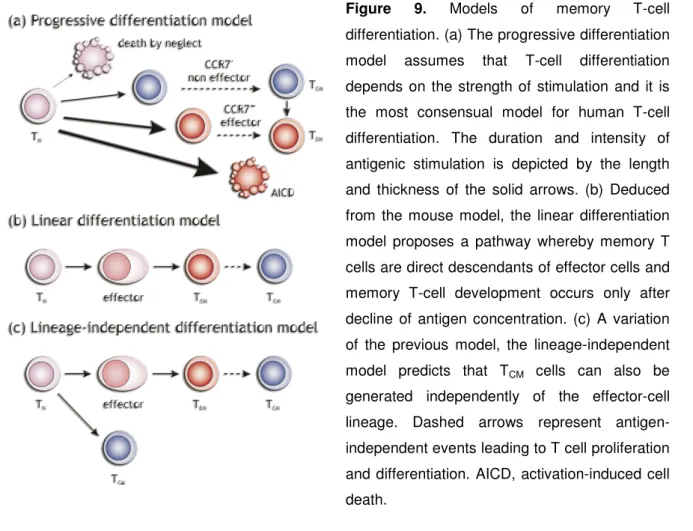
1.6.2. CD8+ T lymphocytes differentiate into cytotoxic T cells ______________ 51 2.1. FADD pathway ______________________________________________ 57 2.2. Perforin/granzymes pathway ____________________________________ 59 2.2.1. Components of the granules __________________________________ 59 2.2.2. The universe of granzymes ___________________________________ 62 2.3. Other molecules expressed by CD8+ T cells ________________________ 65 2.3.1. TGF-β ____________________________________________________ 65 2.3.2. Chemokines: a matter of attraction______________________________ 67 3.1 Memory T-cell responses are different from primary responses__________ 72 3.1.1. Memory cell subsets have particular turnover and activation properties _ 72 3.1.2. Memory cells provide quantitatively and qualitatively enhanced protection 74 3.1.3. Memory cells have distinct survival requirements __________________ 75 3.1.4. Cell surface molecules associated to memory phenotypes ___________ 78 3.2 Immunological memory generation and lineage relationships ___________ 82
PART III. AIMS AND METHODOLOGICAL APPROCHES ______________ 87 RESULTS______________________________________________________ 90
MANUSCRIPT #1________________________________________________ 91 MANUSCRIPT #2_______________________________________________ 103
DISCUSSION __________________________________________________ 134 Part I. Single-cell multiplex RT-PCR _____________________________ 135 Part II. Characterization of human CD8+ T-cell subpopulations _______ 138 Rarely detected genes: IL-10, IL-2 and MIP-1α _________________________________138 Expression of cytokines by CD8+ T-cell subsets _________________________________142 Expression of several types of receptors by CD8+ T-cell subsets____________________143 Gene expression profiles within the CD8+ T-cell subsets __________________________149
Expression of CD62L within the CCR7– CD8+ T-cell subsets _______________________160 Reversion of phenotype from CD45R0+ to CD45RA+_____________________________162 Loss of expression of CCR7, CD27 and CD28 – the way to differentiation ____________166
CONCLUDING REMARKS AND PERSPECTIVES_____________________ 172 REFERENCES_________________________________________________ 176 ANNEXES ____________________________________________________ 194
ABBREVIATIONS
APC Antigen Presenting Cell BCL-2 B-Cell Lymphoma-2 CD Cluster of Differentiation
CMV Cytomegalovirus
CTL Cytotoxic T-Lymphocyte DC Dendritic Cell
EBV Epstein Barr Virus HCV Hepatitis C Virus
HEV High Endothelial Venules
HIV Human Immunodeficiency Virus
IFN Interferon
IL Interleukin
LFA Lymphocyte Function-Associated Antigen
LN Lymph Nodes
LTA Lymphotoxin α
MIP Macrophage Inflammatory Protein MHC Major Histocompatibility Complex PCR Polymerase Chain Reaction PTK Protein Tyrosine Kinase
RANTES Regulated Upon Activation, Normally T-Expressed, and Presumably Secreted
RT Reverse Transcription TCM Central Memory T Cells
TCR T-Cell Receptor
TEM Effector Memory T Cells
TEMRA Effector Memory CD45RA+ T Cells
TGF Transforming Growth Factor
TH T Helper Cells
TN Naïve T Cells
TNF Tumor Necrosis Factor
LIST OF FIGURES
Figure 1. Structure of the T-cell receptor
Figure 2. Simplified structure of the TCR-CD3 complex Figure 3. Structure of CD4 and CD8 molecules
Figure 4. Cleavage of PIP2 into DAG and IP3, an intermediate step in the
signaling cascade initiated upon TCR stimulation
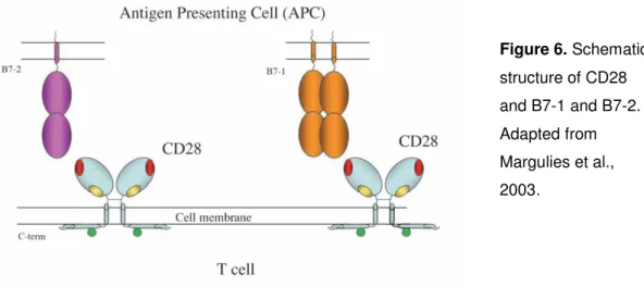
Figure 5. Trafficking of cells of the immune system through the lymph node Figure 6. Schematic structure of CD28 and B7-1 and B7-2
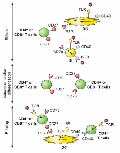
Figure 7. CD27-CD70 interactions in different phases of the immune
response
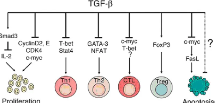
Figure 8. TGF-β regulation of immune responses Figure 9. Models of memory T-cell differentiation
Figure 10. Number of mRNA molecules per cell coding for IFN-γR2 in naïve
and central memory cells
Figure 11. Sequence of gene expression events after T-cell activation by
antigen
Figure 12. Hierarchy of activation stages and putative lineage relationships
RESUME
Après stimulation antigénique, les lymphocytes T CD8+ naïves subissent plusieurs modifications, comme l’expression des molécules de surface. Chez l’homme, l’association du CCR7, CD45RA, CD27 et CD28 est fréquemment utilisée pour discriminer de façon reproductible des sous-populations de cellules T CD8+ fonctionnellement différentes. Néanmoins, la description de ces populations reste incomplète, puisque plusieurs études ont utilisé des associations différentes et limitées de molécules de surface. En conséquence, certaines sous-populations de cellules T CD8+ n’ont pas encore été établies, en particulier dans les compartiments CCR7–CD45RA+ et CCR7–CD45R0+. De plus, les voies de différenciation de ces sous-populations ainsi que leurs rôles respectifs ne sont pas encore définis.
L’objet de ce travail était de définir une corrélation précise et prévisible entre un phénotype de surface donné et des propriétés fonctionnelles des cellules T CD8+. Nous avons associé les niveaux d’expression de CCR7, CD45RA, CD27 et CD28 pour subdiviser les cellules T CD8+ en quatorze sous-types cellulaires différents. Ces populations ont été isolées et l’expression génique de 18 gènes a été étudiée simultanément sur des cellules uniques, par une nouvelle méthode de RT-PCR multiplex que nous avons développée. Nos résultats démontrent que les différentes populations présentent des profils d’expression génique caractéristiques et distincts, reproductibles entre différents donneurs. L’expression de CD45RA est nécessaire pour identifier les cellules naïves, mais ne discrimine pas les différentes populations de cellules qui ont déjà rencontré l’antigène. Par contre, les profils d’expression génique des cellules T CD8+ CCR7– montrent une importante corrélation avec les niveaux d’expression de CD27 ainsi qu’avec la co-expression CD27/CD28. Une hiérarchie d’activation a été établie de la façon suivante : naïve<CD27high<CD27+CD28+<CD28+CD27–<CD27+CD28–<CD27–CD28–. De plus, nous montrons que les cellules CD45RA+ et CD45RA– appartenant à ces sous-populations ont des profiles d’expression génique identiques, au niveau qualitative et quantitative. Par ailleurs, nous avons identifié des sous-populations mineures avec des caractéristiques d’activation récente parmi les compartiments CD45RA+ et CD45RA–. Ces résultats suggèrent que la différentiation des cellules naïves T CD8+ en cellules effectrices n’oblige pas à une perte d’expression de CD45RA. Nous avons, donc, décrit des nouvelles populations T CD8+ et établis une corrélation entre le phénotype de surface et les fonctions cellulaires, ce qui permet d’identifier des populations cellulaires homogènes.
SUMMARY
Following antigenic challenge, naïve CD8+ T lymphocytes undergo several changes, including the expression of cell-surface molecules. In humans, the association of CCR7, CD45RA, CD27 and CD28 is widely used to discriminate a reproducible set of functionally different subpopulations of CD8+ T cells. However, the prevailing data concerning the description of these subsets remains fragmentary, since a multitude of studies used a different and limited set of surface markers. Hence, some CD8+ T-cell subsets are still not clearly established, especially within the CCR7–CD45RA+ and CCR7–CD45R0+ compartments, and the correspondent differential roles and lineage relationships remain undisclosed.
The present study aims to define a predictable and precise correlation between particular cell surface markers and CD8+ T-cell functional properties. We associated CCR7, CD45RA, CD27 and CD28 expression levels to subdivide CD8+ T cells into fourteen different cell types. These populations were further isolated and gene expression of 18 genes was assessed, simultaneously, in single-cells by a novel multiplex RT-PCR method we developed. Our results demonstrate that the different subpopulations display distinct and characteristic gene co-expression patterns, reproducible between donors. CD45RA expression is required to define the naïve subset, but does not discriminate functionally different populations of primed cells. In contrast, gene expression profiles of CCR7-CD8+ T cells correlate significantly to CD27 expression levels and CD27/CD28 co-expression, and a hierarchy of activation stages could be established as follows: naïve < CD27high < CD27+CD28+ < CD28+CD27– < CD27+CD28– < CD27–CD28–. Surprisingly, we found that CD45RA+ and CD45RA– cells of each of these subsets had the same gene expression patterns at both qualitative and quantitative level. Importantly, we identified minor subsets displaying characteristics of recent activation that could be found in both CD45RA+ and CD45RA– compartments. These findings strongly suggest that differentiation of naïve CD8+ T cells into effectors does not necessarily imply CD45RA downregulation. Furthermore, they describe novel CD8+ T cell subsets and establish a correlation between surface phenotype and cell function, which helped to identify homogeneous populations.
PART I. General aspects
1. The immune system: an overview
Every living being is continuously threatened by environmental aggressions, including other organisms. These threats have acted as selective pressures among evolution driving species to evolve several defensive mechanisms that allow them to escape and survive such dangers. In response to invading pathogens, eukaryotes have evolved an elaborate protective apparatus generally known as the immune system. The word “immune” comes from the Latin immunis, meaning “exempt”. In this way, an organism that is immune to a specific infecting agent is able to remain free from infection by that agent.
The immune system functions in two distinct lines of defense: the innate immunity and the adaptive (or acquired) immunity. Types of immune response classified as “innate” are present already in almost all metazoans and depend on germ line-encoded receptors that recognize highly conserved pathogen-associated molecular patterns. These responses are extremely fast and constitute the earlier line of action of the immune system. In addition to innate immunity, jawed vertebrates have evolved an adaptive immune system that provides highly specific immune responses to particular pathogens. In contrast to the immediate induction of defense mechanisms from innate immunity, adaptive immune responses can take several days to mount. However, acquired immunity can confer in many cases lifelong protection to re-infection with the same pathogen, a phenomenon also known as immunological memory (Pancer and Cooper, 2006).
Innate immunity action is based on the recognition and killing of invading agents by phagocytes, a class of white blood cells (or leukocytes) specialized in the ingestion and digestion of unwanted materials. This cell type includes neutrophils and macrophages, which are the major players of the innate immunity. Other cells, called dendritic cells, are highly specialized in capturing antigen and present it to cells of the adaptive immune system. Cells of the innate immunity have a relatively short life-span and are thought to proliferate only in the generative hematopoietic tissues. Phagocytes express a large panel of surface receptors, such as integrins, scavenger receptors, Toll-like receptors, and others,
that recognize microbial conserved components and through which they can be activated to become effector cells.
If the innate immune defenses are by-passed, evaded or overwhelmed, an adaptive immune response normally ensues. Adaptive immunity is mediated by a very important class of leukocytes with potential for self-renewal and clonal expansion that carries on the surface specific recognition molecules, the antigen receptors. Such cells are called lymphocytes and fall into two major categories: B lymphocytes (or B cells) and T lymphocytes (or T cells). B lymphocytes are the major players of humoral immunity, and T cells are responsible for cell-mediated immunity, but often they work in concert, and also with other types of cells, to accomplish an effective immune response.
2. Lymphocyte ontogeny and diversity
All the cellular components of immune system develop from a population of multipotent and self-renewing hematopoietic progenitors, the hematopoietic stem cells (HSCs), which can be found in the adult bone marrow. HSCs differentiate into two main lineages: myeloid, which originates erythrocytes, megakaryocytes, granulocytes and monocytes, and lymphoid, which generates B, T and natural killer (NK) cells (Laiosa et al., 2006).
Adult mammalian B lymphocytes develop in the bone marrow in a sequence of discrete stages that can be identified by the differential expression of several surface markers and are associated to the progressive rearrangement of the immunoglobulin (Ig) loci. These events lead to the formation of a highly specific B cell receptor (BCR) which is expressed on the surface of mature B cells. Once activated, B lymphocytes differentiate into plasma cells, which are highly specialized in antibody production and secretion. The antibodies, which indeed correspond to a secreted form of the BCR, specifically bind the antigen that led to B cell activation. This can result in neutralization or opsonization that leads to pathogen destruction by phagocytes or complement.
T-cell precursors also originate from adult bone marrow-derived cells. Although it is not yet clearly established where T-cell commitment occurs, T-cell precursors eventually migrate at a very early stage to the thymus, where the receptor gene rearrangements and T-cell maturation can occur. Similarly to B lymphocytes, T-cell differentiation is a stepwise process towards the generation of T-cell receptor (TCR) expressing cells.
Unlike B cells, T lymphocytes recognize non-soluble antigens presented by highly polymorphic cell surface molecules generally known as major histocompatibility complex (MHC). T cells are said to be MHC restricted because they specifically recognize antigenic peptides presented by particular MHC alleles. MHC restriction is achieved during thymic T-cell development by positive selection of T lymphocytes recognizing self MHC-peptide complexes. In addition, a process of negative selection also takes place during T-cell maturation, leading to the deletion of lymphocytes with strong affinity to self peptides bound to self MHC molecules. Together, these two selection processes confer T lymphocytes the capacity of discrimination between “nonself” and “self”.
NK cells are also generated from the differentiation of common lymphoid progenitors. Unlike B and T lymphocytes, NK cells do not express clonally distributed receptors for antigen. For this reason, they are considered to belong to the innate immune system. However, they express a panoply of receptors, including inhibitory receptors specific for polymorphic MHC molecules. Such receptors enable NK cells to mediate “missing self recognition”, the capacity to attack self cells that extinguish expression of MHC class I molecules, such as viral infected or tumor cells. NK cells can thus recognize and exert strong cytolytic activity against cells with abnormal MHC expression, and also produce cytokines and chemokines that stimulate other immune functions.
2.1. A glance on T-cell development
The rearrangement status of TCR genes, together with the expression of TCR, the co-receptors CD4 and CD8 and other molecules on the cell surface, allow the discrimination of successive stages of T-cell differentiation in the thymus. Recombination events assemble the TCR coding sequence, which includes the variable (V), diversity (D), joining (J) and constant (C) gene segments that locate discontinuously in the genome. The program of T lymphocyte development can drive thymocytes into two distinct lines of differentiation: the γδ lineage, in which lymphocytes bear antigen receptors formed by a γδ heterodimer, and the αβ lineage in which the generated receptors consist of αβ heterodimers. For simplicity and because the aim of this manuscript does not cover the lymphocyte development issue, differentiation of γδ T-cells was omitted and only αβ T-cell development is described.
In brief, TCR β-chain DJ rearrangements occur first, followed by VDJ recombination in a stage where thymocytes are triple-negative for CD3, CD4 and CD8 and are CD44low CD25+. A further productive rearrangement with a C gene segment allows the expression of low levels of the TCR β-chain at the surface, together with the invariant pre-α (pTα) molecule, a surrogate α-chain, and the CD3 complex. In this stage, CD4–CD8– thymocytes have lost the expression of CD25. The expression of this pre-TCR signals the developing thymocyte to cease further rearrangements in the TCR-β locus (allelic exclusion) and to proliferate (β selection). After the proliferative burst, thymocytes acquire the expression of both
CD4 and CD8 co-receptors and rearrangements on the TCR α-chain locus begin. Rearrangements of the α-chain gene can occur continuously in both chromosomes even after the formation of a cell-surface receptor, which can generate in some cells more than one type of α-chain. These recombination events are ceased when CD4+ CD8+ double-positive cells undergo positive selection. This process favors the survival of thymocytes whose receptors recognize peptides bound to self MHC molecules and therefore occurs only after the expression at the cell surface of a productive αβ TCR. At this point, the type of co-receptor that will be expressed by mature T lymphocytes is also selected. Hence, TCRs recognizing peptides presented by MHC class II molecules lead to selection of the CD4 co-receptor, whereas lymphocytes whose TCRs recognize peptides presented by MHC class I molecules will develop into CD8-expressing cells. Positive selection therefore ensures that mature T-lymphocytes have antigen receptors capable of responding to peptides presented by self MHC molecules, selects the appropriate co-receptor and determines the functional commitment to the class of MHC molecule recognized. Finally, an additional process of negative selection leads to apoptosis of T cells bearing antigen receptors with high affinity to self peptides, thereby preventing the maturation of potentially self-reactive lymphocytes (Spits, 2002).
2.2. Diversity of T-cell repertoire
TCR diversification occurs primarily in the thymus by stochastic recombination of VDJ gene segments. Diversity is further increased by imprecise joining of the recombinant segments, by random addition of non-germline nucleotides by DNA-repair machinery and through pairing of diverse TCR α and β chains, or γ and δ chains. In addition, diversity can be still enhanced in cells expressing more than one type of TCR due to the lack of TCR α-chain allelic exclusion, which results in the pairing of more than one TCR α-chain with the same type of β-chain. Whether these “secondary” TCRs have an effective function with self MHC molecules is not clear. The diversity of αβ-TCRs collectively originated by these events was estimated to be higher than 1×10¹⁵. Nevertheless, during intrathymic lymphocyte differentiation positive and negative selection significantly limit and shape the diversity of the generated repertoire by the loss or
enrichment of certain TCR specificities. The theoretical diversity of the peripheral TCR repertoire generated by this system was nevertheless estimated to be higher than 1×10¹³ – more than the number of T lymphocytes existing in a mouse (1-2×10⁸) or in humans (1×10¹²) (Nikolich-Zugich et al., 2004). Moreover, some lymphocytes express the same TCR due to homeostatic proliferation of naïve T cells and/or homeostatic and antigen-driven expansion of effector and memory populations. The potential achievable repertoire is therefore several orders of magnitude larger than the one that can be expressed in an individual. This extra variability is probably the basis of the differences observed in the expressed TCR repertoire of genetically identical or different individuals.
Surprisingly, estimates of the real diversity of TCR repertoire in mice and humans indicate that it may be much lower than the number of T lymphocytes these organisms can comprise. In humans, the actual number of different αβ-TCRs was calculated to be about 2,5×10⁷ and in mice 2×10⁶ (Nikolich-Zugich et al., 2004). If these estimates are correct, the restriction of diversity in humans is much more important as compared to mice, which can be due to the longer longevity of humans, the need of higher clone sizes in this species, the fact that mice are genetically manipulated animals that are maintained in protected environments, etc.
In addition to the previously described mechanisms that generate TCR diversification, the spectrum of specificities of a given TCR is still increased by crossreactivity with several ligands, as TCRs can vary in the degree of specificity/promiscuity of peptide-MHC recognition.
In summary, several mechanisms ensure the diversification of T-lymphocyte specificities. Because T-cell function relies on the ability to recognize the antigen presented by other cells; because the antigen universe is tremendously vast; and, finally, because the precise antigens derived from invading pathogens are unpredictable, the generation of a repertoire of T lymphocytes capable of responding to a broad range of antigens is fundamental for an efficient protection against pathogens.
3. T lymphocytes: key players on adaptive immunity
T lymphocytes are specialized in detecting and destroying pathogens that replicate inside the cells, such as some bacteria and other intracellular parasites, as well as viruses. Furthermore, T lymphocytes play a fundamental role in the immune surveillance against tumors.
T cells fall into two main classes depending on the type of co-receptor molecule expressed. Lymphocytes expressing CD8 specifically bind peptides presented by MHC class I molecules, which derive from proteins synthesized and degraded in the cytosol. In turn, CD4+ T lymphocytes recognize peptides derived from proteins degraded in endocytic vesicles, which are presented by MHC class II molecules. These two types of T cells play distinct roles upon activation: CD4+ T cells differentiate into cells capable of delivering different types of signals to other cells, such as B lymphocytes and macrophages, and CD8+ T lymphocytes acquire the ability of killing other cells, i.e., become cytotoxic. As both CD4+ and CD8+ T cells recognize foreign peptides presented by the MHC proteins of the target cells, T-cell function relies mainly on direct interactions between T lymphocytes and the cells presenting the antigen. In addition, T lymphocytes can also product cytokines that may have stimulatory or inhibitory effects in different types of cells and contribute, directly or indirectly, to pathogen clearance.
3.1. Structure and function of the TCR
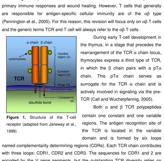
T cells bear on their surface thousands of antigen receptor molecules that recognize specific antigen determinants presented by MHC molecules (Figure 1). TCRs are made up of two membrane anchored polypeptide chains, either α and β in αβ T cells or γ and δ in γδ T cells (Garcia and Adams, 2005). Although they do not follow exactly the same differentiation processes, both αβ T cells and γδ T cells develop mostly in the thymus. In mice and humans, γδ T cells represent 1-5% of circulating T lymphocytes, but in epithelial tissues most cells express γδ receptors. The receptors of some of these epithelial γδ T cells show very restricted variability. In contrast, γδ T cells found in the blood, peripheral lymphoid organs or certain epithelial tissues as the gut, display highly diverse TCRs. Unlike αβ T cells, lymphocytes bearing a γδ TCR are not MHC restricted. They are implicated in
immunoregulation processes and tumor surveillance, as well as in some particular primary immune responses and wound healing. However, T cells that generally are responsible for antigen-specific cellular immunity are of the αβ type (Pennington et al., 2005). For this reason, this revision will focus only on αβ T cells and the generic terms TCR and T cell will always refer to the αβ T cells.
During early T-cell development in the thymus, in a stage that precedes the rearrangement of the TCR α chain locus, thymocytes express a third type of TCR, in which the β chain pairs with a pTα chain. This pTα chain serves as surrogate for the TCR α chain and is actively involved in signaling via the pre-TCR (Call and Wucherpfennig, 2005).
Both α and β TCR polypeptides contain one constant and one variable regions. The antigen recognition site of the TCR is located in the variable domain and is formed by six loops named complementarity determining regions (CDRs). Each TCR chain contributes with three loops: CDR1, CDR2 and CDR3. The sequences for CDR1 and 2 are encoded by the V gene segments, but the outstanding TCR diversity relies on CDR3, which is created by the combinatorial and junctional processes between the V, D and J gene segments. In fact, the TCRα chain has a large number of J gene segments that, together with the diversity conferred by the D segments of the TCR β chain, accounts for the almost unlimited variability of the TCRs (Arden, 1998).
3.2. The CD3 complex
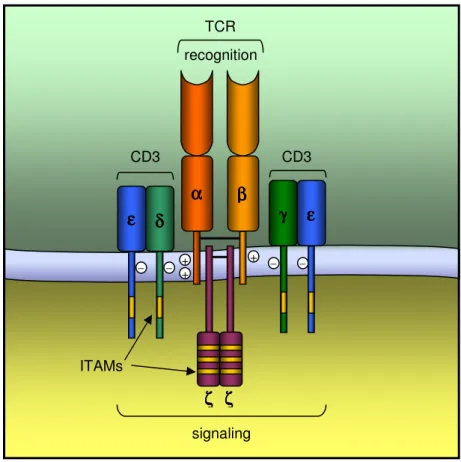
The functionally complete TCR comprises also the CD3 invariant chains, which associate non-covalently with the variable α and β chains of the TCR (Call and Wucherpfennig, 2005). The CD3 complex subunits include the γ, δ, ε and ζ chains, with all but CD3ζ being structurally related (Figure 2). Indeed, CD3γ, CD3δ
β chain α chain carbohydrate Variable region (V) disulfide bond
TCR
Constant region (C) Hinge (H) Transmembrane region Cytoplasmic tailFigure 1. Structure of the T-cell receptor (adapted from Janeway et al., 1999)
and CD3ε are encoded in adjacent genes, while ζ chains are encoded elsewhere in the genome. The CD3γ, CD3δ and CD3ε proteins contain an Ig-like extracellular domain, a transmembrane region and a cytoplasmic tail that contains one single immunoreceptor tyrosine-based activation motif (ITAM). In turn, CD3ζ is largely intracytoplasmic, having only a short extracellular portion, and own 3 ITAMs in its cytoplasmic domain.
Although the precise stoichiometry of the components that constitute a minimal TCR complex remains controversial, it is generally accepted that each αβ heterodimer associates with one CD3εγ heterodimer, one CD3εδ heterodimer and one CD3ζζ homodimer. In this way, each TCR would contain 10 ITAMs. Apparently the assemblage of TCR and CD3 chains depends largely on the interaction of the transmembrane domains, where the positively charged residues of the TCR interact with the negatively residues of the CD3 subunits (Arnett et al., 2004; Call and Wucherpfennig, 2005; Rudolph et al., 2006).
When the αβ heterodimer recognizes a specific peptide presented by the MHC complex, a cascade of signaling events are initiated via the ITAMs. The TCR αβ chains, however, have no functional relevant cytoplasmic domains and thus are not able to signal to the cell that antigen has bound. Therefore, while the αβ heterodimer recognizes and binds antigen, the CD3 complex is fundamental to transmit information from the external environment into the intracellular compartment, in form of signal transduction (Samelson, 2002).
The CD3 subunits are also required for normal surface expression of the TCR αβ heterodimer in mature T lymphocytes. The expression of TCR-CD3 complex is regulated in such a way that the absence of CD3 subunits results in defective or null TCR expression (Ashwell and Klusner, 1990). Moreover, deficiencies in the genes coding for the different CD3 subunits, which also associated with the pre-TCR, induce a blockade on thymocyte development before the CD4+CD8+ stage and impair the TCRα gene rearrangement. These evidences indicate that CD3 components have a role in the T cell differentiation (de Saint Basile et al., 2004; Fischer et al., 2005).
In summary, the TCR consists of two functionally distinct types of components: two genetically variable chains, which account for the recognition and binding to the antigen/MHC complex, and the invariant chains of the CD3 complex that interact with the intracellular proteins involved in signal transduction.
3.3. The T cell co-receptors CD4 and CD8
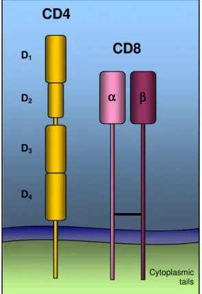
T-cell co-receptors, CD4 and CD8, are cell surface glycoproteins expressed by distinct subsets of mature T lymphocytes (Figure 3). Hence, lymphocytes are referred to as CD4+ or CD8+, depending on the type of co-receptor they express.
During antigen recognition, the co-receptors associate with the TCR and bind the lateral face of MHC molecules. While CD4 associates with a constant domain of the MHC class II molecule, CD8 binds to invariant parts of the MHC class I molecule. Both co-receptors interact with MHC molecules in a domain that is distant from the TCR binding site, enabling the simultaneous association of CD4 and TCR, or CD8 and TCR, to the same peptide-MHC complex. Binding of CD4 or CD8 to the same peptide-MHC complex than the TCR is believed to be required for T cell activation and is on the basis of the classification of CD4 and CD8 as co-receptors (Janeway, 1992).
Figure 2. Simplified structure of the TCR-CD3 complex (adapted from Janeway et al., 1999).
– + + + – – – β ββ β α αα α εεεε δδδ δ γγγγ εεεε ζ ζζ ζ ζζζζ ITAMs signaling CD3 TCR CD3 recognition
Figure 3. Structure of CD4 and CD8 molecules.
CD4 is a single polypeptide chain molecule, made of four external immunoglobulin-related domains. Domains 1 and 2 (D1 and D2) have a unique strand topology, similarly to domains 3 and 4 (D3 and D4), which are joined to the first domains by a flexible hinge. The CD8 molecule is a disulfide-linked heterodimer of two polypeptides, α and β. Both chains contain an immunoglobulin-like amino-terminal domain linked to the membrane by an extended polypeptide region containing several O-linked sugars, which helps to maintain this stretch of polypeptide in an extended conformation and to protect it from cleavage by proteases. CD8α chains can also form homodimers, although these are not seen when the β chains are available (Zamoyska, 1998).
One of the major functions of CD4 and CD8 is to increase the avidity of the interaction between TCR and MHC complex (Hampl et al., 1997; Luescher et al., 1995). Furthermore, the cytoplasmic domains of CD4 and CD8 molecules interact strongly with components involved in the signaling cascade initiated upon antigen recognition, in particular the protein tyrosine kinase Lck (Veillette et al., 1988). Hence, the simultaneous binding of the co-receptors and the TCR to the same MHC:peptide complex brings Lck into close proximity to its targets, which associate with the cytoplasmic domains of the TCR complex. This enhances the tyrosine phosphorylation and further recruitment and activation of downstream signaling effector molecules (Germain, 2001).
In summary, mature T cells that recognize antigen in the context of MHC class I molecules express CD8, whereas T cells restricted by MHC class II express CD4. Aggregation of the appropriate co-receptor with the TCR enhances enormously the sensitivity of a T cell to the antigen presented by MHC molecules. In addition, optimal signaling trough the TCR occurs only when it clusters with the co-receptors CD4 or CD8.
CD4
Cytoplasmic tailsCD8
D1 D3 D2 D4 α αα α ββββ3.3. T-cell receptor signaling
3.3.1. Signaling events initiated upon antigen recognition
The ITAMs are tyrosine-containing motifs that serve as sites of association with protein tyrosine kinases (PTKs) and other phosphotyrosine-binding moieties involved in receptor signaling. Upon antigen binding, the co-receptors CD4 or CD8 are clustered with the TCR complex. Consequently, the receptor-associated PTKs are brought together and act on each other and on the receptor cytoplasmic tails to initiate the signaling process.
The phosphorylation of the tyrosine residues of the ITAMs is the first intracellular signal indicating that specific antigen has been encountered. Two members of the Src family of PTKs mediate ITAM phosphorylation: Lck and Fyn. Lck interacts constitutively with the cytoplasmic domain of the co-receptor molecules CD4 and CD8 and is the predominant enzyme involved in ITAM phosphorylation (Rudd et al., 1989; Veillette et al., 1988). Fyn binds to the cytoplasmic domain of ζ and CD3ε chains upon receptor clustering. Both Fyn and Lck phosphorylate specific ITAMs on the accessory chains of the TCR complex (Samelson, 2002).
The activity of Src-family kinases (SFKs) is regulated by the phosphorylation status of two regulatory tyrosine residues, one activating and the other inhibitory, on the enzyme active site. In thymocytes and T cells both tyrosine residues are usually not phosphorylated. In this state, the SFKs are ready to be activated and are said to be in a “primed” state. During the receptor clustering that follows antigen recognition, the SFKs are brought in close proximity and transphosphorylation events can occur, leading to the activation of SFKs via their activating tyrosine. In contrast, phosphorylation of the inhibitory tyrosine induces SFK to adopt a closed conformation that renders the enzyme inactive (Palacios and Weiss, 2004). Lck and Fyn can, thus, be simultaneously present in the cell in three different states: in an open and non-activated conformation (primed); in an open and activated conformation (phosphorylated in the activating tyrosine); and in a closed and inactivated conformation (phosphorylated in the inhibitory tyrosine). These forms can exist in an equilibrium that might be shifted by CD4 or CD8 co-receptor ligation, or by the action of SFK regulatory enzymes, such as Csk or CD45.
3.3.1.1. The regulation of the Src-family kinases Lck and Fyn Csk
The PTK Csk (C-terminal Src kinase) is a key element in controlling the activity of SFKs due to its ability to phosphorylate their inhibitory tyrosine. In resting cells, Csk constitutively associates to transmembrane proteins. Membrane anchoring favors Csk proximity to SFKs that, in this way, are maintained in an inactive state by phosphorylation of the inhibitory tyrosine. Upon TCR ligation, Csk is released from its membrane anchor into the cytosol, which likely contributes for the activation of Lck and Fyn (Palacios and Weiss, 2004).
CD45
The transmembrane tyrosine phosphatase CD45, also known as leukocyte common antigen, has the opposite effect of Csk. This phosphatase specifically dephosphorylates the inhibitory tyrosine residue in the C-terminus of SFKs and it is considered to be a critical positive regulator of Lck and Fyn in T cells (Hermiston et al., 2003). Hence, the balance between the action of Csk and CD45 is one of the ways in which the activity of SFKs is regulated.
All hematopoietic cells, with the exception of erythrocytes, express CD45 proteins. In lymphocytes, expression of CD45 is so abundant that can reach 10% of their surface area (Dahlke et al., 2004).
Structurally, the CD45 is a transmembrane protein with an extracellular region, a single transmembrane domain and a cytoplasmic tail. Only the cytoplasmic region, which contains two tandem tyrosine phosphatase domains, is required for enzymatic activity (Trowbridge and Thomas, 1994).
CD45 exists as distinct isoforms generated by alternative splicing of the exons 4, 5 and 6 that encode part of the N-terminal extracellular domain. At least eight isoforms can be theoretically generated that are distinguished on the basis of antibody recognition. In this way, the isoforms containing the product of exon 4, 5 or 6 are named CD45RA, CD45RB or CD45RC, respectively. The isoform generated by the complete splicing out of the three exons is called CD45R0. The amino-terminal region encoded by the exons 3 to 8 contains several O-linked
carbohydrates attachment sites. Therefore, the variable use of exons 4, 5 and 6 changes the size, shape and overall charge of the extracellular domain (Trowbridge and Thomas, 1994). Alternative splicing of CD45 occurs during T-cell development and peripheral activation. The pattern of isoform expression is highly conserved amongst vertebrates, suggesting functional importance in vivo (Okumura et al., 1996).
The natural ligand for CD45 has not yet been identified and the mechanisms involved in the regulation of CD45 activity are only beginning to be understood. The high regulation of isoform expression and the abundance of CD45 on the cell surface suggest a mechanism for regulating CD45 activity based on the spontaneous and isoform-differential homodimerization. Artificial dimerization of chimeric CD45 molecules inhibits the phosphatase function apparently by blocking the catalytic domain on the paired enzymes (Desai et al., 1993; Majeti et al., 1998). Disruption of this regulatory mechanism leads to autoimmunity and lymphoproliferation in mice (Majeti et al., 2000). The extracellular domains of the different CD45 isoforms, which exhibit different numbers of O-glycosylation modifications, have a major influence in the homodimerization and, consequently, the activation status of the protein (Xu and Weiss, 2002). For instance, smaller molecular isoforms, which are less O-glycosylated, dimerize more efficiently than larger isoforms.
In human cord blood almost all CD8+ T cells express the high molecular weight (m.w.) isoform CD45RA (Hamann et al., 1997). With increasing age, CD8+ T cells express also the CD45R0 isoform. Actually, T cell activation induces a shift from high to low CD45 m.w. isoforms (Akbar et al., 1988). Consequently, the expression of CD45RA is down-regulated while CD45R0 expression is up-regulated. In addition, expression of CD45RA appears to be re-acquired later in the immune response, with the concomitant loss of CD45R0 (Champagne et al., 2001). These changes in cell-surface phenotype are one of the ways to distinguish naïve from antigen experienced cells, effector and memory, and will be discussed in more detail later in this manuscript.
3.3.2. TCR signaling final destination: the nucleus
TCR activation following antigen recognition leads to phosphorylation of ITAMs. In the phosphorylated state, ITAMs can recruit the zeta-associated kinase (ZAP-70), which binds to the ITAMs in the ζ chain via its tandem SH2 (Src homology domain 2) domains. Recruitment of ZAP-70 allows its phosphorylation and enzymatic activation by either Lck or Fyn. Active ZAP-70 then phosphorylates the membrane adapter protein LAT (linker of activation in T cells). LAT contains multiple tyrosines that, once phosphorylated, serve as SH2-domain-binding regions. The cytoplasmic adapter SLP-76 (SH2 domain containing leukocyte phosphoprotein of 76 kDa) binds to phosphorylated LAT, and together these proteins act as sites for the recruitment of additional adapters and key enzymes involved in further downstream signaling events in T cells (Germain, 2001).
Two main types of proteins containing SH2 domains participate in the signal cascade initiated by TCR triggering. The first one is phospholipase C-γ (PLC-γ), which initiates two of the main signaling pathways leading to the nucleus. The second is Ras, a small G protein that initiates the third and major pathway, which consists in a cascade of protein kinases that leads directly to the phosphorylation and activation of transcription factors.
PLC-γγγγ pathways
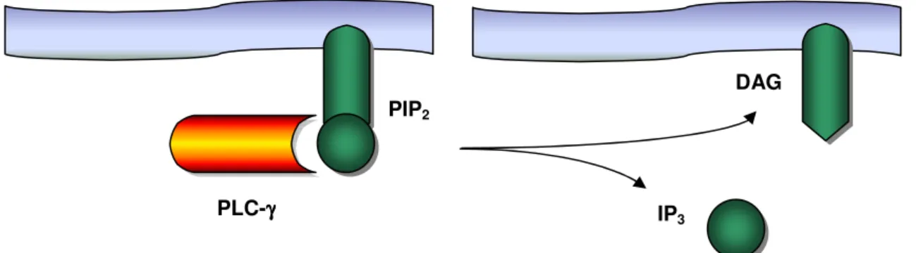
PLC-γ is recruited to the cell membrane by its SH2 domains, where it is activated by phosphorylation of a tyrosine residue. In the active state, PLC-γ cleaves a molecule of the phospholipid membrane, the phosphatidylinositol bisphosphate (PIP2), into inositol trisphosphate (IP3) and diacylglycerol (DAG,
Figure 4). One single molecule of PLC-γ is able to generate many molecules of DAG and IP3. Therefore, this and similar enzymatic steps contribute importantly to
amplify and sustain the signal initiated by TCR ligation. Noteworthy, production of DAG and IP3 by activated PLC-γ is not exclusive from the TCR-signaling cascade,
After cleavage of PIP2, diffusion of IP3 induces the release of Ca2+ from
intracellular storage sites in the endoplasmic reticulum (ER) into the cytosol. Intracellular free Ca2+ levels increase drastically, triggering the opening of calcium channels in the plasma membrane, which let in more Ca2+ into the cell, thus sustaining the signal. High levels of free Ca2+ induce activation of the Ca2+-binding protein calmodulin, which can associate to and regulate the activity of several other proteins, propagating the signal onwards along pathways that also eventually converge to the nucleus.
The other product of PIP2 cleavage, DAG, remains linked to the plasma
membrane and contributes to the activation of the protein kinase C (PKC). This serine/threonine kinase is believed to be important in initiating one of the main signaling pathways leading to the nucleus. PKC is additionally activated by the raise of Ca2+ concentration due to IP3 action. In this way, both products of the
cleavage of PIP2 reinforce each other in activating PKC (Janeway, 1999).
Ras pathway
Like the other small G proteins, Ras can be present in the cell in two different forms, depending on whether it is binding GTP or GDP. When bound to GTP, Ras is in the active state. Since this form has an intrinsic GTPase activity, Ras can turn itself off by removing a phosphate group of GTP. The GDP-bound form of Ras is therefore inactive and is the predominant form found in the cell.
Activation of small G proteins requires a guanine-nucleotide exchange factor (GEF), which exchanges GDP to GTP. In lymphocytes, Ras is recruited to
DAG
PLC-γγγγ IP3 PIP2
Figure 4. Cleavage of PIP2 into DAG and IP3, an intermediate step in the signaling cascade
the antigen receptor complex by adaptor proteins to which GEFs also bind, making therefore possible the activation of Ras.
Activated small G proteins trigger a cascade of protein kinases named mitogen-activated protein kinases (MAP kinases). Localization and activity of MAP kinases depends on their phosphorylation status: non-phosphorylated MAP kinases are inactive and stay in the cytoplasm, but once phosphorylated become active and translocate into the nucleus.
MAP kinases pathway has three major enzymatic levels. The first enzyme in the cascade is a serine/threonine kinase, generally called MAP kinase kinase kinase (MAPKKK or MAPK3), which is activated by the GTP-bound form of Ras. In the active form, MAPKKK phosphorylates the next downstream enzyme, the MAP kinase kinase (MAPKK or MAPK2). This class of proteins has the ability to phosphorylate MAP kinases, the last level enzymes, on both a tyrosine and a threonine residue, which induces MAP kinase activation.
MAP kinases directly phosphorylate and activate transcription factors, a class of proteins that bind specific sites on DNA and regulate gene transcription. Transcription factors activated through MAP kinase pathway in consequence of antigen recognition usually regulate the expression of genes involved in proliferation, cell death and survival, inflammation and DNA repair (Dong et al., 2002).
Part II. T-cell immune responses
The most remarkable feature of the acquired immune system is the capacity to generate immunological memory, i.e., a faster, stronger and more effective response against an antigen that has already been cleared in a prior immune response.
In the course of a primary immune response, antigen presented by professional antigen-presenting cells (APCs) will drive specific naïve T-cell to proliferate and further differentiate. Several different outcomes are possible. In the case of CD4+ T lymphocytes, differentiation will induce the polarization of lymphocytes toward a T helper 1 (TH1) or TH2 phenotype, originating cells highly
specialized in cytokine secretion (INFγ or IL-4, respectively). TH1 CD4+ T
lymphocytes are the main macrophage activators, potentiating inflammatory immune responses, whereas TH2 CD4+ T cells activate B lymphocytes and
promote immune responses predominantly mediated by antibodies. In the other hand, CD8+ T lymphocytes differentiation leads to the formation of cytotoxic T cells (CTL) that kill infected target cells. Upon differentiation T lymphocytes can also acquire regulatory properties, which enable them to modulate immune responses. In any case, cells generated upon differentiation are ready to respond quickly and efficiently upon encounter with the specific antigen and, thus, can be considered as “effector cells”.
In the sequence of naïve priming and differentiation, another type of cells can further be generated, which have less stringent requirements for activation than naïve cells, are able to proliferate and acquire effector functions in a reduced lag-time, and have increased survival capacity. As these cells can survive for years in the organism, they were termed “memory cells”.
1. Primary immune responses
1.1. APCs interact with naïve T cells in the secondary lymphoid organs
The effective onset of adaptive immune responses requires that naïve antigen-specific lymphocytes, being inherently rare throughout the body, rapidly encounter foreign antigens. This problem has been elegantly solved in evolution through the development of secondary lymphoid tissues as intersections in the migratory pathways of presenting dendritic cells (DCs) and antigen-specific lymphocytes. T-cell immune responses are therefore initiated in secondary lymphoid organs, where peptide-loaded APCs can encounter T cells bearing a specific TCR for the antigens they have uptaken in the periphery. Pathogens infecting peripheral tissues are captured by DCs that will be trapped in the lymph nodes directly downstream the site of infection. Nonetheless, antigen presentation can take place in other sites, such as the spleen, if infection takes place in the blood, or Peyer’s patches and tonsils, when pathogens invade mucosal surfaces.
The secondary lymphoid organs are highly organized structures where B, T and antigen presenting cells can interact and initiate immune responses. Naïve T lymphocytes are continuously circulating from one lymphoid organ to another, via blood and lymph, until they encounter antigen. While migrating and prior to contact with specific antigen, naïve T cells are metabolically quiescent and have a prolonged lifespan, which depends on the successive contact with self-peptide/MHC complexes and a cytokine, interleukin-7. Recognition of these ligands presumably delivers low-level signals, which keep T cells sufficiently metabolically active to avoid passive death (Sprent and Surh, 2002; Tanchot et al., 1997).
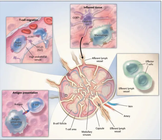
The migratory properties of naïve T cells do not enable them to enter the peripheral tissues. Therefore, naïve T lymphocytes can only recognize specific antigen and initiate immune responses in the secondary lymphoid organs (Sprent and Surh, 2002) (Figure 5).
To get in lymph nodes and Peyer’s patches, but not in the spleen, T cells must cross the walls of the high endothelial venules (HEV). This event requires the interaction of molecules present in the plasma membrane of lymphocytes, such as selectins and integrins, and their corresponding ligands expressed by the cells of the vascular endothelium. Of particular interest are the L-selectin (also known as
CD62L) and the chemokine receptor CCR7 expressed on the surface of T cells. Interaction of L-selectin and the vascular addressins mediates T-cell rolling on the endothelium, which enables the interaction between CCR7 and its ligands, SLC and ELC (standing for, respectively, secondary lymphoid-organ chemokine, also known as CCL21, or Epstein–Barr virus-induced molecule 1 ligand chemokine, also known as CCL19). SLC and ELC are also expressed by stromal cells within the T cell areas in the lymphoid tissue and thus target T cells into these sites (Cyster, 1999; Zlotnik et al., 1999). Mice lacking CCR7 (through targeted gene deletion) or that have insufficient CCL19 or CCL21 (through naturally occurring mutation) have structurally disorganized lymph-node T-cell zones and are deficient in T-cell dependent immunity (Muller et al., 2003a). Interaction with ELC or SLC differs fundamentally in the ability to induce the internalization of CCR7, which occurs when this receptor interacts with ELC, but not SLC. Based on this finding, it was proposed that T lymphocytes enter lymphoid tissues mainly in response to
Figure 5. Trafficking of cells of the immune system through the lymph node.
SLC produced by HEVs. In this way, lymphocytes would retain full chemotatic responsiveness to ELC and SLC produced at similar levels in T-cell zones, therefore maintaining the ability to migrate into those areas (Bardi et al., 2001).
When T lymphocytes reach the T cell-zones on the secondary lymphoid tissues they establish contact with APCs, in particular DCs, and scrutinize the peptides presented by MHC class I and II molecules on its surface.
Typically, DCs are dispersed in nonlymphoid tissues as resident cells in a resting, immature state. Immature DCs are very efficient in capturing antigen, but are not capable of efficiently present antigens and activate T cells. This capacity only emerges after a complex developmental process called “DC maturation”. The antigen uptake associated to other signals, like the cytokine and chemokine microenvironment generated by local inflammation, drives immature DCs to undergo phenotypic and functional changes that culminate in the complete transition from an “antigen-capturing” cell to an “antigen-presenting” cell specialization. These modifications include changes in morphology, such as formation of dendritic projections, upregulation of co-stimulatory receptors (like B7-1, B7-2, CD40 and CD58) and an overall increase in the levels of MHC class I and class II molecules on the surface. Another important property that accompanies maturation is the acquisition of high cellular motility and up-regulation of chemokine receptors, such as CCR7 (Mantovani, 1999). After antigen uptake DCs leave the inflamed tissues, enter the lymph stream and migrate to the secondary lymphoid organs. Here they are driven by gradients of CCL19 and CCL21 to the T-cell zones where contact with T T-cells takes place and the DC maturation process is completed (Trombetta and Mellman, 2005).
Interaction with T cells should start soon after DCs reach the T-cell zone because DCs are relatively short-lived cells, especially after activation, with a half-life as low as 1–2 days upon arrival in the lymph node (Ingulli et al., 1997; Kamath et al., 2002). Yet recent in situ imaging studies suggest that individual T–DC couplings may last 37 h or longer (Stoll et al., 2002). Longevity and abundance of DCs in the site of T-cell priming influence the capacity to prime and the strength of the activating signals delivered to T cells (Josien et al., 2000; Wong et al., 1997). Evidence that enhanced DC survival potentiates T-cell activation has been demonstrated by studies of TRANCE, a TNF family member expressed on T cells, which can trigger its receptor, also known as RANK, on DCs enhancing their
viability (Anderson et al., 1997; Wong et al., 1997). A role for CD40L in regulation of DC survival has also been demonstrated (Quezada et al., 2004).
The naïve T lymphocytes that do not encounter specific antigen leave the secondary lymphoid tissue by the efferent lymphatics and proceed circulation in the bloodstream and through the other secondary lymphoid organs. In contrast, if a specific peptide has been presented by the APC, T cells interrupt circulation to initiate a sequence of events that leads to the generation of effector cells. This can take several days and encompass two distinct processes: proliferation and differentiation. In the end, T cells can leave the lymphoid tissue also by the efferent lymphatics and re-enter the circulation to migrate to the sites of infection or, instead, stay within the lymph node to help B lymphocytes.
1.2. Requirements for T-cell activation
T-cell activation is a complex, multistep process involving a multitude of molecules. When T lymphocytes recognize the specific peptide on the surface of an APC, several protein-protein interactions are elicited in addition to the specific interaction between the TCR and the MHC-peptide complexes. As mentioned above, the co-receptors CD4 or CD8 are recruited to their binding sites on MHC class II or MHC class I, respectively, thereby lowering the threshold for T-cell activation. Moreover, interaction between co-stimulatory receptors, the most important of which is CD28, and their cognate ligands on the APC also occur. These co-stimulatory interactions are fundamental for effective lymphocyte activation and also enhance the immune response (Lenschow et al., 1996). Finally, it should be stressed out that for all these interactions to be possible, a stable adhesive junction must be formed between the T cell and the APC. Adhesion molecules like CD2, ICAM-3 or LFA-1, present on lymphocytes plasma membrane, play an important role in the efficient binding to APCs by interaction with molecules on their surface.
After antigen recognition, the avidity of LFA-1 for ICAM-1 (standing for intercellular adhesion molecule-1), an integrin expressed on the surface of the APC, increases rapidly (Dustin and Springer, 1989). This leads to the formation of an adhesion complex between the T cell and the APC characterized by a specific pattern of receptor segregation with a central cluster of TCRs and one of its
downstream signaling effectors, protein kinase C-θ (PKC-θ), surrounded by a ring of integrin family adhesion molecules. This complex is known as immunological synapse (Grakoui et al., 1999) or supramolecular activation cluster (SMAC) (Monks et al., 1998), and facilitates and sustains TCR engagement and signaling (Bachmann et al., 1997). Large molecules such as CD45, also interact with the APC but are excluded from the SMAC (Huppa and Davis, 2003).
In addition to the signal generated by TCR engagement, a second signal is often required to achieve full activation of T lymphocytes. This second signal is provided by ligation of molecules displayed on the lymphocyte surface and is complementary to TCR activation. For this reason, it is called co-stimulation and the molecules involved are known as co-stimulatory receptors.
The immune system has evolved in such a way that only APCs display the molecules capable of delivering co-stimulatory signals to lymphocytes. This is particularly important to avoid activation of self-reactive T cells that have escaped thymic negative selection and might recognize specific peptides displayed by MHC class I molecules of tissue cells. Hence, the need for co-stimulation constitutes also a strategy of discrimination between self and non-self. Indeed, lymphocytes receiving a TCR-mediated signal 1 in the absence of an additional, accessory molecule-mediated signal 2 become functionally tolerant (for a review, see Baxter and Hodgkin, 2002). This is generally known as the “two-signal theory” of lymphocyte activation and supports that signal 2 is important not only for complete T cell activation but also to prevent induction of unresponsiveness.
The major co-stimulatory pathway involves the binding of CD28 to its ligands, CD80 or CD86 (also known as B7-1 or B7-2, respectively). Co-stimulation by anti-CD28 antibody enhances T cell activation by decreasing the time of commitment, amplifying TCR signaling and protecting T cells from death (Iezzi et al., 1998; Lenschow et al., 1996; Viola et al., 1999). In addition to CD28, other molecules expressed by lymphocytes can mediate co-stimulation, such as several members of the tumor necrosis factor receptor (TNFR) family, like CD27 (Watts, 2005). The biology and function of CD27 and CD28, as well as their role in lymphocyte co-stimulation, will be discussed in further detail in the following section.