Cristiana Ribeiro Silva Teixeira
outubro de 2014
The Prefrontal Cortex and chronic pain:
the role of metabotropic glutamatergic
receptors in the descending modulation
of pain
UMinho|20 14 Cristiana Ribeir o Silva T eix eira The Prefront al Cor tex and chronic pain: t
he role of met abotropic g lut amatergic recep tor s in t
he descending modulation of pain
Universidade do Minho
Escola de Ciências da Saúde
Trabalho efetuado sob a orientação da
Professora Doutora Filipa Pinto-Ribeiro
Cristiana Ribeiro Silva Teixeira
outubro de 2014
Dissertação de Mestrado
Mestrado em Ciências da Saúde
The Prefrontal Cortex and chronic pain:
the role of metabotropic glutamatergic
receptors in the descending modulation
of pain
Universidade do Minho
Escola de Ciências da Saúde
iii
Agradecimentos
Este trabalho representa o final de mais uma etapa da minha formação e é muito importante para mim. Adorei integrar um trabalho de investigação no âmbito das neurociências, foi um trabalho muito enriquecedor a nível de experiência laboratorial e competências adquiridas.
Agradeço a todas as pessoas que me apoiaram, me compreenderam e acreditaram em mim neste ano de trabalho.
E agradeço inteiramente a todas as pessoas, que de alguma forma contribuíram para a realização e enriquecimento deste trabalho.
À minha mãe agradeço carinhosamente por todo o apoio, pelas palavras de incentivo, coragem, confiança,e por todo o amor e um muito obrigado pela oportunidade de dar continuidade aos meus estudos, um sonho muito desejado.
À minha irmã pela força, apoio, carinho e paciência.
À minha sobrinha querida agradeço a sua amabilidade, boa disposição e carinho.
Quero agradecer generosamente à minha orientadora Professora Doutora Filipa Pinto-Ribeiro por toda a paciência, pela exigência do trabalho, pela compreensão, pela oportunidade e pela motivação na área das neurociências.
Agradeço à Ana Pereira pela enorme ajuda neste trabalho, apoio, simpatia, paciência, e pelas sugestões das correcções deste trabalho que enriqueceram o mesmo e pela dedicação.
Agradeço a todos os colegas do grupo da Dor, à Diana Amorim pela sua simpatia, apoio, e sempre receptiva e disponível para ajudar; à Vera Cardoso pela sua simpatia e grande dedicação no apoio ao microscópio; à Sónia Puga pelo seu apoio e simpatia.
Agradeço a todos os docentes, que me transmitiram sábios conhecimentos ao longo do mestrado, e que enriqueceram o meu conhecimento científico.
Agradeço também à Patrícia pelo companheirismo sincero, apoio,por todos os momentos, pela sua autêntica amizade, carinho, pelas extensas e divertidas noites de microscópio, pelos seus úteis conselhos; à Maria pelo seu apoio a longa distância, amizade, carinho, boa disposição e pelas videochamadas sempre muito divertidas.
v
The Prefrontal Cortex and chronic pain: the role of metabotropic glutamatergic receptors in the descending modulation of pain
Abstract
In osteoarthritis (OA) the progressive degeneration of articular structures persistently activates nociceptors leading to chonic pain and subsequent cognitive and emotional impairments. Amongst the several supraspinal nuclei involved in the modulation of nociception, the medial prefrontal cortex (mPFC), a brain area part of the limbic system, is known to mediate nociception as well as emotional and cognitive functions. In addition, metabotropic glutamate receptors (mGLUR), greatly expressed in the PFC, play an important role in the neuroplasticity of brain circuits in persistent pain disorders. Amongst the three groups of mGLURs, group I (mGLUR1 and mGLUR5) increase neuronal excitability in chronic pain.
In order to evaluate the role of infralimbic cortex (IL) mGLUR5 in the facilitation of nociception, we activated and inhibited the IL with specific and unspecific agonists and antagonists of mGLUR5 while (i) behaviourally assessing nociception, (ii) analysing c-Fos expression in brainstem areas involved in the modulation of nociception with and without noxious peripheral stimulation and (iii) recording neuronal activity in the rostral ventromedial medulla (RVM) of control (SHAM) and osteoarthritic (ARTH) animals.
Our data showed the activation of IL mGluR5 receptors induced behavioural hyperalgesia in both experimental groups while its inhibition an antinociceptive effect in ARTH animals. Regarding c-Fos expression, the ipsilateral ventrolateral periaqueductal gray matter (VLPAG) and the dorsal part of the dorsal raphe nucleus (DRD) displayed a greater number of c-Fos expressing cells in ARTH animals when compared to SHAM animals with noxious stimulation enhancing c-Fos expression in the RVM of SHAM animals. Activating IL mGLUR5 increased c-Fos expression in the DRD, RVM and dorsal reticular nucleus (DRt) of SHAM animals and in the VLPAG of ARTH animals. The combination of pharmacological and mechanical stimuli increased c-Fos expression in the ipsilateral LC and RVM of SHAM animals and the contralateral DRt of ARTH animals. Data from the electrophysiological recordings showed antinociceptive ON- and pronociceptive OFF-like cells of the RVM are not involved in descending facilitation after the activation of IL mGLUR5.
In the present work a facilitatory role of mGLUR5 upon behavioural nociception has been clearly demonstrated, further studies are however needed to understand the potential role of the RVM and DRt in mediating its effect.
vii
O Córtex Pré-frontal e a dor crónica: o papel dos receptors metabotrópicos glutamatérgicos na modulação descendente da dor
Resumo
Na osteoartríte (OA) a degeneração progressiva das estruturas articulares activa continuamente nociceptores levando ao desenvolvimento de dor crónica e a déficits emocionais e cognitivos. De entre as áreas supraspinhais envolvidas na modulação da nocicepção, o córtex pré-frontal medial (mPFC), parte do sistema límbico, é uma área que medeia não só a nocicepção como também as funções emocionais e cognitivas. Mais ainda, os receptores metabotrópicos do glutamato (mGLUR), cuja expressão é intensa no PFC, desempenham um importante papel na neuroplasticidade de circuitos neuronais em doenças crónicas. Mais ainda, sabe-se que de entre os três grupos de mGLURS, o grupo I (mGLUR1 e mGLUR5) é responsável por aumentar a excitabilidade neuronal na dor crónica.
De forma a avaliar o papel dos mGLURs do córtex infralímbico (IL) na facilitação da nocicepção, o IL foi activado/inibido através da administração de agonistas/antagonistas específicos dos mGLURs durante (a) a avaliação comportamental da nocicepção, (b) a avaliação da expressão de c-Fos em áreas do tronco cerebral envolvidas na modulação da nocicepção na presença/ausência de estimulação nóxica periférica e (c) o registo da actividade neuronal no núcleo rostral ventromedial da medula oblongata em animais controlo (SHAM) e com osteoartrite (ARTH). Os nossos resultados mostraram que a activação dos mGLUR5 no IL induziram hiperalgesia comportamental em ambos os grupos experimentais enquanto a sua inibição teve um efeito antinociceptivo apenas nos animais ARTH. Em relação à expressão de c-Fos, observou-se um aumento no lado ipsilateral da parte ventrolateral da substância periaqueductal cinzenta (VLPAG) e na parte dorsal do núcleo raphe (DRD) dos animais ARTH em comparação com os animais SHAM embora a estimulação nóxica periférica apenas tivesse aumentado a expressão de c-Fos nos animais SHAM. A activação dos mGLUR5 aumentou a expressão de c-Fos no DRD, RVM e DRt de animais SHAM e na VLPAG de animais ARTH. A combinação de protocolos de estimulação aumentou a expressão de c-Fos no LC e RVM ipsilateral nos animais SHAM e no DRt contralateral nos animais ARTH. Os dados da análise electrofisiológica mostraram que as células pronociceptivas tipo-ON e antinociceptivas tipo-OFF não estão envolvidas na facilitação descendente após a activação dos mGLUR5 do IL. Com este trabalho demonstrou-se o papel facilitador dos mGLUR5 sobre a nocicepção, no entanto, são necessários mais estudos de forma a destrinçar o papel do RVM e DRt neste circuito facilitador.
ix
Index
Chapter 1: Introduction ... 1
1.1.Nociception, nociceptors ... 3
1.2. Supraspinal pain modulation ... 5
1.2.1. The Prefrontal Cortex and pain ... 7
1.2.2. Descending modulation of pain ... 10
1.3. Chronic inflammatory pain ... 13
1.3.1. Osteoarthritis ... 13
1.4. Chronic pain and Neuroplasticity ... 14
1.5. Neurotransmitters in OA ... 15
1.6. Glutamate metabotropic receptors and chronic pain ... 16
1.7. Animals models of experimental osteoarthritis ... 17
Chapter 2: Objectives ... 19
Chapter 3: Materials and Methods ... 23
3.1. Animals and ethics issues ... 25
3.2. Anaesthesia and euthanasia ... 26
3.3. Induction of experimental osteoarthritis ... 26
3.4. Behavioural analysis of nociception ... 26
3.4.1. Nociceptive behaviour ... 26
3.4.2 Pressure application measurement (PAM) ... 27
3.4.3. Paw withdrawal Test (Hargreaves model) ... 27
3.5. Procedures for intracerebral injections ... 28
3.6. Drugs ... 29
3.7. c-Fos stimulation ... 29
3.7.1.Brain processing ... 31
3.7.2 Immunohistochemistry for c-Fos ... 31
3.7.3 Stereological Quantification of c-Fos expression - Stereology ... 32
3.8.Course of the electrophysiological study ... 32
3.8.1.Electrophysiological evaluation of the modulatory action of the mPFC upon the RVM………..33
3.9.Statistics /DATA analysis ... 34
Chapter 4: Results... 35
4.1. Histological confirmation of the injection and recording sites... 36
x
4.2.1 Pressure application measurement ... 38
4.2.2. Paw withdrawal test (PWL) ... 39
4.3. c-Fos quantification ... 41
4.3.1. Locus coeruleus (LC) ... 41
4.3.2. Ventrolateral periaqueductal gray (VLPAG) ... 42
4.3.3. Dorsal raphe nucleus, dorsal part (DRD) ... 43
4.3.4. Rostral ventromedial medulla (RVM) ... 44
4.3.5. Dorsal reticular nucleus (DRt) ... 45
4.4. Electrophysiological evaluation of the modulatory action of the mPFC upon the RVM ... 47
4.4.1. Evaluation of changes in the spontaneous activity of RVM pain modulatory cells ... 47
Chapter 5: Discussion ... 51 5.1. Technical considerations ... 53 5.1.1 Animal model ... 53 5.1.2. Anaesthesia ... 53 5.1.3. Behavioural tests ... 54 5.1.4. c-Fos expression ... 56 5.1.5. Electrophysiology ... 57
5.2. Pharmacological manipulation and assessing nociception in IL ... 58
5.3. Supraspinal pain modulatory areas activated by mGLUR5 in IL ... 60
5.3.1 Locus Coeruleus (LC) ... 61
5.3.2. Periaqueductal gray (PAG) ... 61
5.3.3. Dorsal raphe nucleus, dorsal part (DRD) ... 62
5.3.4. Rostral ventromedial medulla (RVM) ... 62
5.3.5. Dorsal reticular nucleus (DRt) ... 62
5.4. Electrophysiological evaluation of the modulatory action of the mPFC upon the RVM ... 63
5.5. Brain areas mediating descending modulation pathway of the IL ... 64
Chapter 6: Conclusions and future perspectives ... 67
Chapter 7: References ... 71
xi
Abbreviations
ACC – anterior cingulate cortex
AMPA - α-amino-3-hydroxy-5-methylisoxazole-4-Proprionate AMY – amygdala
ANOVA – analysis of variance
CHPG - 2-chloro-5-hydroxyphenylglycine CNS - central nervous system
CVLM – caudal ventromedial medulla DR - dorsal raphe nucleus
DRD - dorsal raphe nucleus, dorsal part DRG – dorsal root ganglia
DRt – dorsal reticular nucleus GABA - gamma-aminobutyric acid GLU – glutamate
i.p. – intraperitoneal injection IL – infralimbic cortex K/C - kaolin/carrageenan LWT - limb withdrawal threshold
mGluR(s) – metabotropic glutamate receptor(s) MPEP - 2-methyl-6[phenylethynyl]-pyridine mPFC – medial prefrontal cortex
NMDA - N-methyl-D-aspartate NON-N – non-nociceptive neurones NS – nociceptive specific neurones OA - osteoarthritis
PAG – periaquedutal grey matter
PAM - pressure application measurement PB – parabrachial nucleus
xii PFC – prefrontal cortex
PrL – prelimbic cortex PW – paw withdrawal
PWL – paw-withdrawal latency RVM – rostral ventromedial medulla S1 - (primary somatosensory cortex) S2 - (secondary somatosensory cortex) SEM – standard error of the mean TF – tail-flick
VEH – vehicle
VLPAG - ventrolateral periaqueductal gray WDR – wide-dynamic range neurones
1
3
According to the International Association for the Study of Pain (IASP), pain is defined as “an unpleasant sensory and emotional experience associated with actual or potential tissue damage, or described in terms of such damage” (Merskey and Bogduk, 1994).
Pain is a subjective experience, varying substantially from person to person (Merskey and Bogduk, 1994), since it is influenced by past experiences, own expectations in relation to pain and cultural background (Koyama et al., 2005). In addition pain is not always associated to an identifiable pathology since in many cases the pain sensation prevails even in the absence of tissue damage or any other identifiable cause (Savage et al., 2008).
Pain can be divided in acute and chronic pain (Loeser and Melzack, 1999). Acute pain is elicited by injury to the body which activates local nociceptive receptors (Loeser and Melzack, 1999). The first response to pain comprises a motor protective reaction intended to immediately discontinue contact with the source of pain (Millan, 1999). However, chronic pain is persistent or recurrent pain that lasts for more than 3 to 6 months without an identifiable cause or long after the original cause has been treated (Merskey and Bogduk, 1994).
Chronic pain can be further divided in (i) nociceptive pain, if it results from the activation of nociceptors secondary to the actual area damaged, as in arthritis (Nicholson, 2006), (ii) neurogenic pain, when a cause cannot be identified but the pain sensation persists due to abnormal activity of the central nervous system, and (iii) neuropathic pain when the lesion or dysfunction of the peripheral or central nervous system occurs as in sciatic pain (Nicholson, 2006).
1.1. Nociception, nociceptors
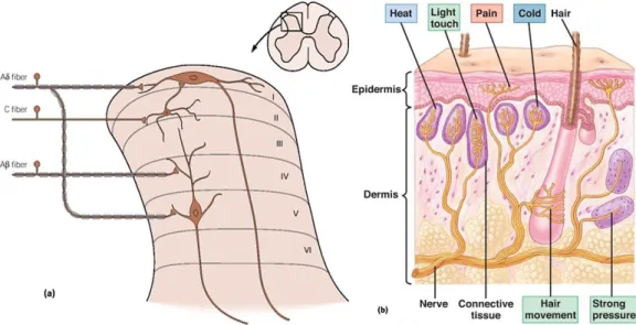
Nociception is the neuronal process that comprises the transduction and transmission of noxious (painful) stimuli from the periphery to dorsal horn of the spinal cord (D’Mello and Dickenson, 2008). Nociceptors or primary afferents, (figure 1) are specialized peripheral sensory neurones that respond to potentially damaging stimuli, such as thermal, either high (over 40°C–45°C) or low temperatures (below 15°C), mechanical or chemical stimuli (Dubin and Patapoutian, 2010).
4
Figure 1 – Schematic representation of nociceptor projections to the spinal cord (Basbaum and Jessell, 2000) (a); and its distal adaptations (Martini and Nath, 2009) (b). While the nociceptive afferent fibers terminate on projection neurons in the dorsal horn of the spinal cord, the proximal arm of the nociceptor synapses in the dorsal horn so that the information can be transmitted upwards to the brain (Basbaum and Jessell, 2000) (a); while the distal arm innervates peripheral structures and is activated by thermal, mechanical and chemical stimuli (Dubin and Patapotian, 2010) (b).
Nociceptors are pseudounipolar neurones as a unique process arises from the cell body (Dubin and Patapoutian, 2010). Soon after leaving the dorsal root ganglia (DRG) the process divides in two arms, a distal one that innervates the periphery and a proximal one that synapse mainly on second-order neurones in the dorsal horn of the spinal cord (Basbaum et al., 2009). Primary afferents can be classified according to their degree of myelinization (unmyelinated and myelinated), conduction velocity (slow or fast transmission) and response threshold to thermal, mechanical and chemical stimuli (figure 1 b)),(unimodal or polymodal) (Dubin and Patapoutian, 2010).
The transmission speed is directly proportional to the degree of myelination transmitting it (Dubin and Patapoutian, 2010). In summary, primary afferents can be divided (Woolf and Ma, 2007) into (i) C-fibers, slow non-myelinated fibers (0.4 -1.4 m/s; Djouhri and Lawson, 2004), that are responsible for the “second” or slow pain (Basbaum et al., 2009), and (ii) Aδ-fibers, fast but small myelinated neurones (about 5-30 m/s; Djouhri and Lawson, 2004), that transmit the “first” or fast pain (Basbaum et al., 2009). Severe authors have also suggested that, a third type of fibers, the Aβ-fibers, fast myelinated fibers, can also be included in this group. Although, in acute conditions, these fibers respond only to innocuous mechanical stimulation (light touch; Basbaum et al., 2009),
5
they have been shown to participate in nociceptive transmission in chronic pain disorders (Basbaum et al., 2009). Since Aβ-fibers are considerably larger than Aδ-fibers, its conduction velocity is also considerably higher (Dubin and Patapoutian, 2010). As shown in figure 1 a) nociceptors project to distinct laminae in the dorsal horn of the spinal cord, with most projection targeting laminae I and II of the superficial dorsal horn (Basbaum and Jessell, 2000). Interestingly, Aδ fibers project not only to lamina I, but also to deep lamina V that target lamina II neurons. That also target neurones not identically, Aβ nociceptors project to deep laminae IV. Finally C nociceptors project more superficial to lamina II, neurones whose projections also are target lamina I neurones (Basbaum and Jessell, 2000).
There are several neuronal types of cell in the spinal cord receive projections from primary afferents that can be classified as (i) non-nociceptive (NON-N) when they only respond to touch and receive inputs mainly from Aβ-fibers (D’Mello and Dickenson, 2008), (ii) nociceptive-specific (NS) neurones that responde exclusively to noxious stimulation (D’Mello and Dickenson, 2008) mainly located in the superficial in lamina I, and receiving projections from Aδ- and C-fibers (Basbaum and Jessell, 2000), and (iii) wide-dynamic range (WDR), neurones that receive input of all fiber types and respond to various modalities of stimuli such as innocuous or noxious mechanical, heat and chemicals stimuli (D’Mello and Dickenson, 2008).
1.2. Supraspinal pain modulation
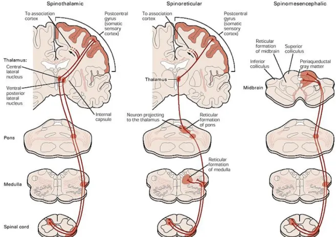
After the primary afferent nociceptors convey nociceptive information to spinal projection neurones forward the nociceptive signal to the brain through several ascending tracts: spinothalamic, spinoreticular, spinomesencephalic, cervicothalamic and spinohypothalamic tracts (Basbaum et al., 2009). However, the most relevant tracts are represented in figure 2.
6
Figure 2 – Schematic representation of three of the main ascending tracts in the spinal cord: spinothalamic, spinoreticular and spinomesencephalic tracts involved in the ascending transmission of pain (Basbaum and Jessell, 2000).
The cervicothalamic tract arises from neurones in the lateral cervical nucleus, and receives inputs from nociceptive neurones from laminae III and IV (Basbaum and Jessell, 2000). The spinohypothalamic tract comprises neurones from laminae I, V and VIII and projects to supraspinal autonomic control centers (Basbaum and Jessell, 2000).The spinothalamic tract, the most prominent ascending nociceptive pathway (Willis, 1995), comprises, neurones from laminae I and V-VII (Basbaum and Jessell, 2000) and targets the somatosensory cortex, via the lateral thalamus, providing information about the location, duration and intensity of the painful stimulus and initiating the behavioural response (Basbaum et al., 2009).
In parallel, the spinoreticular tract which comprises axons from neurones in laminae VII and VIII activates circuits in the brainstem (Basbaum and Jessell, 2000), mediating the sensory-discriminative components of the pain experience (Basbaum et al., 2009). Additionally, the spinomesencephalic tract comprises axons from neurones in laminae I and V (Basbaum and Jessell, 2000) and projects mainly to pain modulatory nuclei mediating the affective component of the pain experience (Basbaum and Jessell, 2000), located in the midbrain and the brainstem, such
7
as the locus coeruleus (LC) (Willis and Westlund, 1997), the periaqueductal gray area (PAG), the amygdala (Amy) (Basbaum et al., 2009), a major component of the limbic system and the neural system involved in emotion, and the dorsal reticular nucleus (DRt) (Willis and Westlund, 1997). Pain results from activations of brain areas associated with the sensory-discriminative properties such as somatosensory cortex and emotional component such as the anterior cingulate gyrus and insular cortex (Basbaum et al., 2009). With sensory-discriminative aspects the primary somatosensory cortex (S1) (Craig, 2003) is generally associated. Relatively to affective/cognitive aspects, the secondary somatosensory cortex (S2) is involved, the insula and anterior cingulate cortex is related for affective-motivational and cognitive aspects of pain as anticipation, attention and evaluation (Seminowicz et al., 2004; Apkarian et al., 2005; Ohara et al., 2005; Zhuo, 2008, Neugebauer et al., 2009). At emotional level, amygdala has an important role in the emotional-affective dimension of pain and it is connected to the cortical areas (Neugebauer et al., 2009).
1.2.1. The Prefrontal Cortex and pain
The prefrontal cortex (PFC) is a brain area associated to emotional and cognitive functions that include attention, decision making, goal-directed behavior and working memory (Gusnard et al., 2001; Phelps et al., 2004). In terms of nociceptive processing, the PFC is involved in the detection and response to noxious electrical (Tanaka et al., 2008), chemical (Porro et al., 2002), mechanical (Zhang et al., 2004) and thermal (Matre et al., 2010; Tran et al., 2010) stimuli.
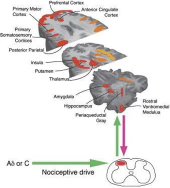
Amongst the areas involved in pain processing human imaging studies, leave shown that during an acute pain experience, the principal frontal areas activated are the primary and secondary somatosensory, the insular, the anterior cingulate and the prefrontal cortices, the thalamus and the amygdala (Apkarian et al., 2005) (figure 3).The intensity and affective quality of perceived pain leads of the interaction between ascending nociceptive inputs and antinociceptive controls, and dysregulations in these controls may contribute to susceptibility factors for the development of chronic pain and comorbid conditions (Apkarian et al., 2005).
8
Figure 3 – Brain areas activated during an acute painful experience. Orange areas represented bilaterally activation in the brain; red areas means areas activated contralaterally; and yellow areas are activated ipsilaterally (Tracey and Mantyh, 2007).
In humans, the PFC divided into:
(i) the medial prefrontal cortex (mPFC) –involved in the unpleasantness of pain sensation (Lorenz et al., 2002) and in anticipation (Porro et al., 2002)
(ii) the anterior cingulate cortex (ACC), that in involved in the mediation the affective component of pain responses (Rainville et al., 1997), the autonomic responses to pain (Craig, 2003) and the placebo effect (Wager et al., 2004);
(iii) the dorsolateral cortex that mediates the detection of conflicting information (Medalla and Barbas, 2009);
(iv) the insular cortex involved in initiation of autonomic response (Augustine, 1996; Verbene and Owens, 1998) and in mediating the encoding of nociceptive stimulus intensity (Craig et al., 2000).
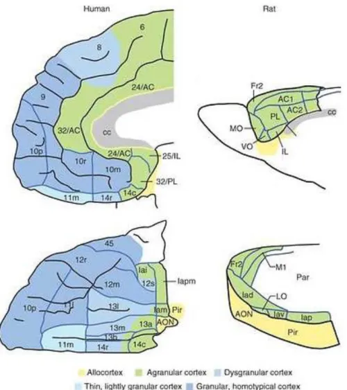
In rodents, however, only three different regions can distinguished (i) the mPFC in the cortical zone, (ii) the orbital prefrontal cortex in the ventral area and (iii) the agranular area more laterally (Heidbreder and Groenewegen, 2003) (figure 4). Another difference in rodents, in relation to humans, it is the fact that mPFC comprises by four areas, the medial precentral, the anterior cingulate cortex (ACC), the prelimbic (PrL) and the infralimbic (IL) areas (Vertes, 2004).
.
In rodents, amongst the four areas, the ACC is the more frequently studied as it has been shown that in acute conditions, the electrical or chemical stimulation of this area facilitates nociception
9
(Calejesan et al., 2000). In inflammatory conditions an increase expression of excitatory neurotransmitters (glutamate) has been observed (Wu et al., 2005) while in neuropathic pain, the ACC undergoes profound morphological and functional changes (Metz et al., 2009). Some evidences suggest that plasticity in the ACC is involved in the development of persistent pain (Zhuo, 2008). Other studies suggest that in neuropathic pain there is a structural modification of the microcircuitry (layer 5) in the ACC (Blom et al., 2014). Other study in arthritis showed that increased neuronal activity in the amygdala was accompanied by decreased mPFC activation and impaired decision-making (Gaungchen et al., 2010).
Figure 4 – Schematic representation of corresponding areas of the prefrontal cortex in humans and rodents (AC, anterior cingulate area; AON, anterior olfactory nucleus; c, caudal; cc, corpus callosum; Fr2, second frontal area; I, insula; i, inferior; Ia, agranular infralimbic cortex; IL, infralimbic cortex; l, lateral; LO, lateral orbital area; m, medial; M1, primary motor area; MO, medial orbital area; o, orbital; p, posterior;Par, parietal cortex; Pir, Piriform cortex; PL, prelimbic cortex; r, rostral; s, sulcal; v, ventral; VO, ventral orbital area. Numbers indicate cortical fields, except that after certain areas, such as Fr2 and AC1, they indicate subdivisions of cortical fields) (adapted from Wallis, 2011).
10
By comparison, studies on the PrL or the IL are almost non-existent even though the patterns of afferent and efferent projections to these areas, in rodents suggest some homology between the PrL and the orbitofrontal PFC in humans and the IL and the dorsolateral cortex in primates (Vertes, 2004). Regarding the PrL it is known that this area projects to the ACC, the AMY, the hypothalamus, the PAG and the RVM (Sesack et al., 1989). In terms of nociception, neurones on IL respond to visceral stimuli in both rodents and humans but only in women (Wang et al., 2009).
In what concerns the IL, this nucleus projects mainly to the PrL, the ACC, the hypothalamus, the AMY, the PAG, the PB, and to the dorsal horn of the spinal cord (Hurley et al., 1991; Floyd et al., 2001). In behavioral terms, IL is associated with anxious behavior (Jinks and McGregor, 1997) and behavioral flexibility (Delatour and Gisquet-Verrier, 2000). In males, both in rodents and in humans, the IL is responsive to noxious visceral stimulation (Wang et al., 2009).
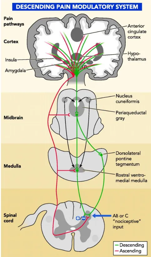
1.2.2. Descending modulation of pain
The existence of a descending pain modulatory system was first hinted by observations in the Second World War, the soldiers with severe wounds reported no or moderate pain, meaning where pain sensation was not proportional to the extent of the injury (Beecher HK, 1946). More importantly, it was also observed that pain sensation could be heightened or dampened depending of the emotional state of the subject (Beecher HK, 1946; Ossipov, 2012).
The pioneering work of Sherrington showed that transmission in the spinal cord is modulated by descending pathways originating in the brain, based on the fact that nociceptive reflexes were enhanced after transection of the spinal cord (Sherrington, 1906; Bingel and Tracey, 2008). The first studies on descending pain modulation using animal models were performed in the 60s and showed that electrical stimulation of the PAG induced profound analgesia (loss of pain sensitivity) (Fields, 2000; Milan, 2002; Bingel and Tracey, 2008). Later, several electrophysiological and pharmacological studies showed that the antinociceptive action of the PAG depended on the RVM, because almost none of PAG’s neurones projected to the spinal cord (Fields et al., 2000; Millan, 2002) (figure 5). Later in the 80s, Fields and colleagues (1983) showed that the RVM could either facilitate or inhibit spinal nociceptive transmission (Gebhart, 2004; Bingel and Tracey, 2008) and more importantly that this bidirectional control of the RVM was associated with the activity of RVM different cell types, the ON-, OFF- and NEUTRAL-cells (Fields, 1992; Fields et al., 1983; Fields and Heinricher, 1985).
11
12
These studies showed that RVM ON-cells were pronociceptive, facilitated nociception, since their activity increased immediately before a behavioral avoidance response occurred after the application of a noxious stimulus (Fields et al., 1995). By contrast, RVM OFF-cells were considered antinociceptive, to inhibit pain, as their activity decreased immediately before a motor avoidance response occurred after the application of noxious stimulus (Fields et al., 1995). Interestingly, although at that time RVM NEUTRAL-cells were shown not alter their activity during the acute application of noxious stimuli (Khasabov et al., 2012) more recently, Miki and collaborators (2002) showed that, these cells would shift their phenotype towards mimicking ON- and OFF-cells in chronic pain disorders.
Later other caudal areas, such as the caudal ventromedial medulla (CVLM) (Pinto-Ribeiro et al., 2011) and the dorsal reticular nucleus (DRt) (Almeida et al., 1999), have also been demonstrated to enhance the nociceptive transmission. CVLM like the RVM, the as part of the descending pain modulation, is able either to enhance or inhibit nociception (Pinto-Ribeiro et al., 2011) targeting neurones in laminae I, IV to V (Tavares and Lima, 2002).
By contrast, the DRt is an area that exclusively enhances nociception (Almeida et al., 1999). Besides sharing reciprocal projections with spinal neurones (Almeida and Lima, 1997), it transmits nociceptive signals to the PAG and RVM areas, as well as to the thalamus and the amygdala (Almeida et al., 2006; Lima and Almeida, 2002; Almeida et al., 1993).
At the spinal cord level, the facilitation or inhibition of nociceptive transmission is achieved by three different mechanisms: (i) the inhibition of the release of excitatory neuropeptides by primary afferents, (ii) the inhibition of transmission at the level of projection neurones of the spinal cord and, (iii) the activation of inhibitory interneurones in the spinal cord (Fields et al., 1995).
In evolutionary terms, the ability to inhibit pain can alleviate pain in conditions where antinociception is necessary for survival (Bingel and Tracey, 2008). However, the posterior pain facilitation is also, an important protective mechanism to alert for the need to protect injuried region, invoking protective behaviours thus ensuring it recovery (Bingel and Tracey, 2008). Nonetheless, when pain is prolonged in time, far beyond the recovery time from the injury that gave rise to it, then pain loses all its evolutionary value and becomes a serious illness (Porreca et al., 2002).
13 1.3. Chronic inflammatory pain
Chronic inflammatory pain is a debilitating disease that affects 20% of the world’s adult population (McGuire and Kennedy, 2013). As previously mentioned, in chronic pain states, the nociceptive transmission is altered and due to increased excitability of primary afferents (Basbaum et al., 2009) and projection neurones that lead to neuroplasticity and the development of chronic pain syndromes, such as allodynia, hiperalgesia and spontaneous pain (Dubin and Patapoutian, 2010). While spontaneous pain is characterized by intense painful sensation of short duration without an apparent cause, allodynia is characterized by an increased sensitivity to stimuli that are usually considered innocuous, and hyperalgesia is an exacerbation of pain sensation to the application of noxious stimuli (Cheng, 2010).
Chronic inflammatory conditions include diseases that involve musculoskeletal disorders, consisting of more than 100 different conditions involving the gradual degeneration of joints, bones, muscles and cartilage, impairing physical movement and causing pain (Arthritis Foundation, 2014). The most common inflammatory disorders are rheumatoid arthritis and osteoarthritis (Merskey and Bogduk, 1994).
1.3.1. Osteoarthritis
Osteoarthritis (OA) is one of the most common forms of arthritis, affecting 27 million people in America alone (Lawrence et al., 2008) and whose cause is not fully understood (Maldonado and Nam, 2013). It is a degenerative disease that affects various tissues within and surrounding joints including the articular cartilage, the subchondral bone, the synovial membrane and surrounding ligaments.
The most common symptom reported by OA patients is painful or stiff joints (particularly the hips, knees and lower back) after inactivity or overuse physical movement and is usually worse after activity or at the end of the day (Arthritis Foundation, 2014).
According to the World Health Organization, OA is already one of the ten most disabling diseases in developed countries with estimates showing that 9.6% of men and 18.0% of women aged over 60 years have symptomatic osteoarthritis. Of these, 80% display limitations in physical movement
14
and 25% cannot perform their major daily activities (World Health Organization, 2014). Although aging contributes greatly to the degeneration of cartilages, other risk factors are also importatnt such as inappropriate mechanical stress, trauma, obesity, metabolic syndrome and genetic predisposition (Houard et al., 2013).
In the initial phases of OA acute inflammation of synovia is a subtle process that can last minutes or hours and is accompanied by heat, pain, redness and swelling. Recurrent episodes of inflammation lead to chronic inflammation that develops over a longer period of time and that may persist for days, weeks or months, resulting in biochemical changes within the cartilage components (Bonnet and Walsh, 2005).
1.4. Chronic pain and Neuroplasticity
In chronic pain, structural changes are accompanied by biochemical changes including the expression of neurotransmitter receptors. In situations of pain, after injury the neurotransmitter glutamate induced plasticity that is a key step in the increased synaptic efficacy occurring in the dorsal horn of the spinal cord between primary afferent terminals and second order neurons (Chiechio and Nicoletti, 2012).
The predominance of the neurotransmitter glutamate in CNS is well documented. The L-glutamate form activates both ionotropic and metabotropic glutamate receptors and regulates a wide variety of CNS functions and effector systems (Conn and Pin, 1997; Schoepp et al., 1999). However, the predominance of metabotropic glutamate receptors is mostly associated with periphery (Neugebauer, 2001). Some studies suggest the involvement of peripheral mGluR1 and mGluR5 in prolonged pain (Bhave et al., 2001), with the injection of mGluR1 and mGluR5 antagonists inhibiting the second phase of the formalin test. The second phase is usually associated with peripheral and central sensitization, while the first phase is associated with acute nociception (Taylor et al., 1995; Puig and Sorkin, 1996; Neugebauer, 2001).
The metabotropic glutamate receptors are also expressed on pre- and post-synaptic elements in the dorsal horn of the spinal cord (Bleakman et al., 2006). Generally the mGluRs has pronociceptive effects, however it has been observed both excitatory and inhibitory effects in terms of nociception at the level of the spinal cord (Neugebauer, 2002). At the supraspinal level of the brain, the mGluRs has extensively being studied, specifically group I of mGluRs has been studied
15
in supraspinally areas such as the ventrobasal thalamus, the amygdala, the PAG, the RVM (Palazzo et al., 2014) and the mPFC (Ji and Neugebauer, 2011).
Neuroplasticity can be defined as the ability of the nervous system to adapt and response to persistent intrinsic or extrinsic stimuli and often involves structural and functional reorganization of neuronal pathways (Cramer et al., 2011). Indeed, neuroplasticity of circuits modulating pain is considered one of the factors responsible for the development of hyperalgesia and allodynia as it promotes an, imbalance between descending facilitator and inhibitory actions towards pain exacerbation. In the early stages of chronic pain, there is an increase in the excitability of primary afferent pathways and consequently, increased signaling of areas involved in the central integration and processing of nociceptive information. It is activating descending inhibitory pathways in order to decrease nociception (Cramer et al., 2011). In this context several works, have shown that the activation of mGlu receptors can either increase or decrease cell excitability depending on the specific receptor subtype of mGluRs being expressed and the anatomical and cellular localization (Conn and Pin, 1997; Yamakura and Shimoji, 1999).
However, the persistent activation of these pathways leads to gradual neuroplasticity at the morphofunctional level (cyto-architecture, expression of receptors and neurotransmitters, ion channels and intracellular metabolic pathways) and a transition from antinociception to pronociception is observed (Von et al., 1988).
1.5. Neurotransmitters in OA
In the case of OA, in McNearney and colleagues, analyzed the synovial fluids of joints patients, and verified that the levels of glutamate were significantly elevated (McNearney et al., 2004). Glutamate is an excitatory neurotransmitter that activates ligand-gated ion channels, ionotropic glutamate receptors (AMPA, α-amino-3-hydroxy-5-methylisoxazole-4-Proprionate, and NMDA, N-methyl-D-aspartate) and G-protein coupled metabotropic receptors (Dray and Read, 2007). In OA, glutamate receptors were found not only in primary afferents surrounding joints (Dray and Read, 2007) but also, on non-neural cells, such as osteoblasts that mediate bone remodeling and osteoclasts and chondrocytes that mediate mechanic-transduction (Dray and Read, 2007).
16
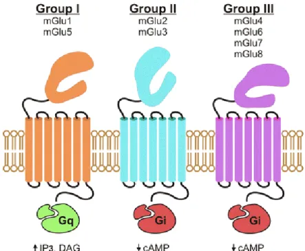
1.6. Glutamate metabotropic receptors and chronic pain
Glutamate metabotropic receptors (mGluRs) are coupled G-proteins receptors that can be divided into three subfamilies (Meldrum, 2000) (figure 6). (i) Group I receptors (mGlu1 and mGlu5) that receptors are coupled to G αq proteins and activate phospholipase C (PKC) (Chiechio and Nicoletti, 2012) stimulating phosphoinositide hydrolysis and subsequent formation of inositol 1,4,5-triphosphate and diacylglycerol (Conn, 2003; Niswender and Conn, 2010; O’Connor and Cryan, 2010). (ii) Group II (mGlu2 and mGlu3) and III (mGlu4, mGlu6, mGlu7 and mGlu8) that receptors are coupled to Gαi proteins, that inhibit adenylate cyclase (Chiechio and Nicoletti, 2012) and decrease the intracellular levels of cAMP upon activation (Conn, 2003; Niswender and Conn, 2010; O’Connor and Cryan, 2010). Aditionally, mGlu receptors can also acts at the level of ions channels and alter the ions flow through G βγ subunits (Chiechio and Nicoletti, 2012).
Figure 6 – Schematic representation of the three types of metabotropic glutamate receptors (mGlu) (Julio-Pieper et al., 2011). The mGluR are divided in three distinct groups: group I (mGluR1 and mGluR5), group II (mGluR2 and mGluR3) and group III (mGluR4, mGluR6, mGluR7 and mGluR8)(Conn, 2003; Niswender and Conn, 2010; O’Connor and Cryan, 2010).
17
The mGlu receptors in the CNS have normal brain functions, but they are also involved in persistent pain (Neugebauer, 2001).
The mGluRs, specifically group I (mGluR1 and mGluR5) have been associated to play a key role in and increase of excitability in chronic pain comparatively a minimal involvement in acute nociception (Dray and Read, 2007).
The activation of group’s mGlu receptors is associated with pain mechanisms, where these receptors can either promote pronociceptive and antinociceptive effects depending on the site of activation (Chiechio and Nicoletti, 2012). In rodents, the intraplantar injection of group I mGlu receptor agonists induces pronociceptive effects (Bhave et al., 2001; Walker et al., 2001). In arthritic animals, the inhibition of the mPFC neurones results in the activation of GABAA receptors
an action, mediated by mGluR1 receptors. In healthy animals, mGluR1/5 receptors inhibit GABAergic action (Gaungchen and Neugebauer, 2011). This observation can lead a therapeutic strategy based on manipulation of metabotropic glutamate receptors in persistent pain (Gaungchen and Neugebauer, 2011).
Numerous studies correlate the activation of mGluRs with, changes in the modulation of nociceptive transmission at the spinal cord and the brain levels in models of inflammatory and neuropathic chronic pain (Ren et al., 2011). In fact, increased expression of mGluRs has been associated with the development of central neuroplasticity and sensitization in both pathologies (Chiechio and Nicoletti, 2012).
1.7. Animals models of experimental osteoarthritis
Several animal models of osteoarthritis have been developed in an attempt to mimic all the aspects of the human disease (Bendele, 2001). The available animal models of OA included, various species such as, mice, rats, guinea pigs, Syrian hamsters, primates, dogs, rabbits (Bendele, 2001), sheep, goats and horses (Gregory et al., 2012).
In rodents, experimental OA can be induced be (i) mechanically, by inducing joint instability by partial meniscectomy combined with transection of collateral and/or cruciate ligaments (Neugebauer et al., 2007), (ii) chemical, through the intra-articular injection of compounds that cause damage of ligaments and tendons and (iii) genetic by overexpression some compounds such as, IL 1B over expression (Little and Zaki, 2012).
18
The animal model (K/C model) used in this work is a chemical model, in which OA is induced, through the intraarticular injection of a solution of kaolin and carrageenan into the synovial cavity of the knee joint (Radhakrishnan et al., 2003). As in human OA, this model causes damage to the cartilage, inflammation of the synovia and synovial fluid exudate. And arthritis develops within hours and persists for weeks (Neugebauer et al., 2007). In addition, the K/C model of experimental OA leads to the gradual degeneration of the articular structures such as medial femoral plateaux, and there is an increase in subchondral bone volume and a decreased bone marrow area and subchondral cyst formation (Amorim et al., 2014).
19
21
Taking into account that the mPFC is an area strongly involved in pain modulation in both humans and rodents, and that metabotropic glutamate receptors have been proposed to mediate the effects in pain modulation, in the work herein we propose, through the use of an experimental model of OA to:
- Assess the activation of the infralimbic area of the mPFC at rest and during acute noxious peripheral stimulation;
- Compare the activation levels of the infralimbic neurones between healthy rodents and animals with experimental OA;
- Identify the supraspinal pain modulatory areas activated by the administration of an mGLU5 receptor agonist in the infralimbic cortex;
- Evaluate the impact of the administration of an mGLU5 receptor agonist in the infralimbic upon the activity of RVM pain modulatory cells.
23
25 3.1 . Animals and ethics issues
In this work, adult male Wistar han rats obtained from Charles Rivers (Barcelona, Spain) weighting between 300-460g, were housed in pairs in standard polycarbonate cages (45.4x25.5x20 cm) in the animal house of the Life and Health Sciences Research Institute (ICVS). All regulations established by the local veterinarian committee (in accordance to the European Community Council Directive 86/609/EEC) and the 2010/63/EU decree concerning the use of animals for scientific purposes were approved by the ICVS Ethical Commission.
Animals were kept under a light cycle of 12:12 h (with lights ON at 8:00 am) at 22°C ±1°C and 30% relative humidity. Water and food were available ad libitum. One week before the induction of OA, all animals were placed in the experimental room for an hour and handled for 10 minutes by the experimenter. After habituation, the animals did not vocalize in response to handling, restraint, or movement of a healthy joint. Before the beginning of the experiment, rats were divided in two experimental groups (figure 7), a group with experimental osteoarthritis (ARTH, n=16) and a control group (SHAM, n=26).
26 3.2. Anaesthesia and euthanasia
For the induction of experimental OA and during the implantation of intracerebral cannulae, animals were anaesthetized with a solution of ketamine, a NMDA receptor agonist (0.75mg/kg, Imalgene®,
Merial Lyon, France) and medetomidine, an α2-adrenergic receptor agonist (0.5mg/kg, Dorbene®,
Esteve, Carnaxide, Portugal), injected intraperitoneally (i.p.). After the surgical procedures, the anaesthesia was reverted by administering atipamezole hydrochloride (1mg/kg, i.p.; Antisedan, Pfizer, Seixal, Portugal), a synthetic α2-adrenergic antagonist, and the animals were monitored until fully recovered (grooming and eating).
For the c-Fos stimulation protocol, the implantation of intracerebral cannulae in the IL, and to record neuronal activity in the RVM, animals were anaesthetized with pentobarbitone (50mg/kg; i.p.; Eutasil®, Ceva, Algés, Portugal). After the end of the experimental sessions, animals received a lethal dose of pentobarbitone (80mg/kg; i.p.).
3.3. Induction of experimental osteoarthritis
The induction of experimental OA followed a protocol described in detail by Ansah and Pertovaara (2007). Briefly, ARTH animals (n=16) were injected with a solution of 3% carrageenan (Sigma-Aldrich, St.Louis, MO, USA) and 3% kaolin (Sigma-(Sigma-Aldrich, St.Louis, MO, USA) dissolved in distilled saline (0.9%NaCl, Brown, Bracarena, Portugal) in the synovial cavity of the right knee joint, at a volume of 0.1 mL, followed by five extension of the right hind paw. SHAM animals were injected with the vehicle solution (distilled saline) in the synovial cavity of the right knee joint, followed by five extension of the right hind paw.
3.4 . Behavioural analysis of nociception 3.4.1. Nociceptive behaviour
The development of experimental OA was verified 1-2h prior to each experimental session in each animal individually. Only the rats that vocalized every time after five flexion-extension movements of the knee joint were considered to have experimental osteoarthritis and were included in the
27
ARTH group. SHAM animals did not vocalize to any of the five consecutive flexion-extension movements of the knee joint (Pinto-Ribeiro et al., 2008).
3.4.2 Pressure application measurement (PAM)
The pressure application measurement test was specifically designed and validated for mechanical stimulation of joints. In arthritis research it is especially suited to assess joint hypersensitivity in rodent knees or ankles (Barton et al., 2007) by measuring the force necessary to evoke a paw withdrawal response. The apparatus (38500, Ugo Basile, Comerio, Italy) consists of a force transducer mounted on a unit fitted to the operator’s thumb that is connected to a display device. During the test the animals are held by the experimenter that places the transducer unit (fitted to the experimenter’s thumb) on one side of the animal’s knee joint and the forefinger on the other, applying pressure to the joint until a behavioural response is observed (paw withdrawal, wriggling or vocalization).
The results are presented in grams of force (gf) and correspond to the peak force applied immediately prior to limb withdrawal - limb withdrawal threshold (LWT). Two measurements of both the ipsilateral and contralateral limbs with 1 min intervals were recorded during the testing sessions and the mean value was used for further statistical analysis (Barton et al., 2007). The animals were returned to their respective home cages after the testing session.
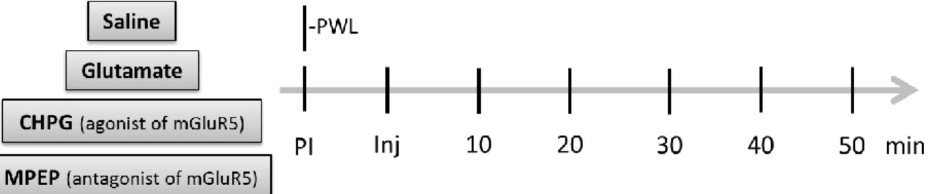
3.4.3. Paw withdrawal Test (Hargreaves model)
For assessing nociception in unanesthetized animals, thermal hyperalgesia was determined by measuring the latency to paw withdrawal (PWL) following the application of a radiant heat stimulus to the plantar surface of the right hind paw (Hargreave’s Test; Plantar Test Device Model 37370, Ugo Basile, Comerio-VA, Italy) in freely moving animals. Briefly, in each behavioural session, the rats (SHAM, n=8 and ARTH, n=8) were placed in a clear plastic chamber with a glass floor 5 minutes before testing to allow habituation to the environment. After this period, the radiant heat source was positioned under the glass floor directly beneath the hind paw, and the PWL was measured prior to drug administration and at various intervals following the intracerebral injections in the IL. At each time point (figure 8), the measurements were repeated twice at an interval of 1
28
minute. The cut-off time was 15s (Pinto-Ribeiro et al., 2008). The animals were returned to their respective home cages after the testing session.
Figure 8 - Schematic representation of timepoints of paw withdrawal testing before and after drug administration in the IL (PWL -; PI - ; Inj - ; CHPG - ; MPEP - ).
3.5. Procedures for intracerebral injections
For intracerebral drug administration, cannulae were implanted according to the procedure described by Pinto-Ribeiro and colleagues (2008). Briefly, the rats (ARTH n=8; SHAM n=8) were placed in a stereotaxic frame (KOPF instruments, Tujunga, California, USA), a longitudinal incision was made, the skull was exposed and drilled and a sterilized stainless-steel guide cannula (26 gauge; Plastics One, Roanoke, Virginia, USA) was implanted in the brain.
The tip of the guide cannula was positioned 1mm above the desired injection site in the IL (figure 9) ( RC: +2,76mm; LM: -0.6mm; DV: -4.9mm; RC – rostro-caudal to the bregma; LM – latero-medial to the sagital suture; DV – dorsoventral to the brain surface), according to the coordinates of the rat brain atlas by Paxinos and Watson (2007). The guide cannula was fixed to the skull with two screws and dental acrylic cement and the skin sutured around it. A dummy cannula (Plastics One) was inserted into the guide cannula to prevent contamination. The animals were allowed to recover from the surgery for at least one week before any handling was performed. The tip of the guide cannula was positioned 1mm above the desired injection site in the IL (RC: +2,76mm; LM: -0.6mm; DV: -4.9mm; RC – rostro-caudal to the bregma; LM – latero-medial to the sagital suture; DV – dorsoventral to the brain surface), according to the coordinates of the rat brain atlas by Paxinos and Watson (2007). The guide cannula was fixed to the skull with two screws and dental acrylic cement and the skin sutured around it. A dummy cannula (Plastics One) was inserted into
29
the guide cannula to prevent contamination. The animals were allowed to recover from the surgery for at least one week before any handling was performed.
Test drugs were administered in the IL through a 33-gauge injection cannula (Plastics One) protruding 1 mm beyond the tip of the guide cannula. The microinjection was performed using a 5.0 μL Hamilton syringe (Hamilton, Nevada, USA) connected to the injection cannula by a polyethylene catheter (PE-10; Plastics One). The injection volume was 0.5 μL and therefore, the spread of the injected drugs within the brain was expected to have a diameter of 1 mm (Myers, 1966). The efficacy of injection was monitored by observing the movement of a small air bubble through the tubing. The injection lasted at least 20 seconds and the injection cannula was left in place for an additional 30 seconds to minimize the possibility of drug solution returning to the injection cannula.
3.6. Drugs
CHPG (2-chloro-5-hydroxyphenylglycine, 100 nmol, (Ansah et al., 2009) Tocris, Bristol, United Kingdom), an agonist of mGluR5 (metabotropic receptor of group I), was prepared with sterilized saline (0.9%NaCl, Brown, Bracarena, Portugal). Glutamate solution (50 nmol (Pinto-Ribeiro et al., 2008), Merck, Darmstadt, Germany) was prepared with sterilized saline. MPEP (2-methyl-6[phenylethynyl]-pyridine, 50 nmol (Movseyan et al., 2001), Tocris, Bristol, United Kingdom), an antagonist of mGluR5, was dissolved in 10% DMSO solution (dimethyl sulfoxide Hybri-Max®,
Sigma-Aldrich, St. Louis, MO, USA). Control injections with saline and 10% DMSO were performed as a control for the potential effect of injecting the solution itself.
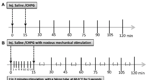
3.7. c-Fos stimulation
The evaluation of the expression of the oncogene c-Fos was studied in animals (ARTH n=8; SHAM n=8), in order to assess which downstream brain areas might be activated after the administration of an agonist of metabotropic glutamate receptor type 5 (mGluR5) in the IL.
30
(i) Injection of saline in the IL – to verify basal expression of c-Fos in the brain of SHAM (n=2) and ARTH animals (n=2);
(ii) Peripheral noxious stimulation of the right hind paw – to analyse changes in basal expression of c-Fos in the brain of SHAM (n=2) and ARTH (n=2) animals after the application of noxious peripheral stimuli;
(iii) Injection of CHPG in the IL – to verify which areas increase their c-Fos expression after activation of the IL in the SHAM (n=2) and ARTH animals (n=2) ;
(iv) Injection of CHPG in the IL with simultaneous application of noxious peripheral stimulation – to verify which areas might be involved in descending modulation of nociception by the IL in the SHAM (n=2) and ARTH animals (n=2).
Figure 9 - Schematic representation of the protocols for c-Fos stimulation. A- c-Fos protocol without stimulation, at 0 minutes and 15 minutes saline or CHPG was injected in the IL. Animal remains without stimulation for the following time. B- c-Fos Protocol with stimulation, at 0 minutes saline or CHPG was injected, and every 2 minutes after the first injection, animal was stimulated for 5 seconds. The stimulus consisted of noxious heat stimulation with a falcon tube containing water at 60°C, 15 minutes after the protocol begins a second injection of saline or CHPG was given, while the noxious heat stimulation continued every 2 minutes until the 2 hours were complete.
The protocol had a total duration of two hours. Drugs were injected at minutes 0 and 15 in SHAM (n=8) and ARTH (n=8) animals. The stimulation protocol consisted in the application of a 5 second noxious heat stimulus every 2 minutes using a falcon tube containing water at 60°C (Pertovaara
31
et al. 1998) to the right hindpaw of the rat. The stimulation was performed every 2 minutes until the end of the protocol (2 hours).
3.7.1. Brain processing
At the end of the experiment, rats are sacrificed and transcardially perfused with a 4% paraformaldehyde solution (PFA solution at 4% (m/v) was prepared with paraformaldehyde (Panreac, Barcelona, Spain) and distilled water, pH=7.4 and it is dissolved in hot and shaking. Brains were then excised and preserved in 4% PFA for at least 72h, followed by a period of 48 h in 8% sucrose solution. Brains were then sectioned in a vibratome (VT1000, Leica Biosystems, Freiburg, Germany) and 50μm thick coronal sections were stored in processing 12 wells plates with 0.1M phosphate buffer saline solution (PBS) at 4ºC until further processing.
3.7.2 Immunohistochemistry for c-Fos
c-Fos immunohistochemistry was performed in brain sections representative of the whole brain (one section evaluated and mounted every 0.41 mm). And the immunohistochemistry protocol was described according Morgado and Tavares (2007).
Brain sections were first washed twice with PBS 0.1M, that was prepared with PBS (1M) stock solution and distilled water (PBS (1M) stock solution was prepared with 160 g of NaCl, 4 g of KCl, 28.8 g of Na2HPO4 all reagents are from Panreac (Barcelona, Spain)). Sections were then incubated
for 30 minutes with 0.03% hydrogen peroxide solution (H2O2,Panreac, Barcelona, Spain) prepared in PBS 0.1M, in order to inhibit endogenous peroxidase activity. After incubation, sections were washed twice with PBS/T (0.3% (v/v) was prepared with Triton-X 100 (Sigma-Aldrich, St. Louis, MO, USA) and PBS 0.1M, pH=7.2). Brain sections were then incubated for 2 hours in fetal bovine serum (FBS 2.5% (v/v), Biochrom, Cambridge, United Kingdom), followed by overnight incubation with rabbit anti-Fos polyclonal antibody (1 µL antibody: 2.000 µL PBS/T + 2% FBS (v/v), Calbiochem, Merck Milipore, Algés, Portugal), at room temperature.
The following day, sections were washed in PBS/T (thrice), followed by incubation for 1 hour in biotinylated swine anti-rabbit secondary antibody (1 µL: 200 µL PBS/T, Dako, Lisboa, Portugal).
32
Sections were washed in PBS/T (thrice), followed by incubation with avidin-biotin complex solution (ABC, was prepared in PBS/T 1 µL:200 µL Vectastain, Vector Laboratories, Peterborough, United Kingdom) for 1 hour. Then, the sections were washed consecutively with PBS/T (twice), PBS (thrice) and Tris buffer (thrice, Tris buffer was prepared with trizma base 0.05M (Sigma-aldrich, St.Louis, MO, USA) and distilled water). Finally, the sections were stained with diaminobenzidine solution (DAB, 0.5 mg/mL, Sigma Aldrich, St. Louis, MO, USA) dissolved in Tris buffer for 2-5 minutes (20 mg DAB+40 mL Tris+8 µL H2O2). At the end of the reaction, the sections were washed with Tris (twice) and PBS (twice). After the immunohistochemistry protocol, sections were mounted in glass slides, dehydrated, diaphanized, fixed and mounted with Entellan (Entellan New, Merck, Darmstadt, Germany).
3.7.3 Stereological Quantification of c-Fos expression
The stained brain sections were analyzed using a stereology Olympus Golgi microscope coupled to a computer, using the Stereo Investigator 10 Software® (MicroBrightField Williston, VT, USA) that
allowed to outline the brain section and the areas of interest and the quantification of c-Fos stained cells. The cells were marked red in the ipsilateral side of the injection and blue in the contralateral side. The brain regions analyzed were: the locus coeruleus (LC), the periaqueductal gray (PAG), the rostral ventromedial medulla (RVM), the dorsal raphe nucleus (DRN), and the dorsal reticular nucleus (DRt). The quantification of c-Fos expression was performed blind.
3.8. Course of the electrophysiological study
In order to further assess the involvement of the IL in nociceptive processing, the evaluation of changes in the activity of RVM pain modulatory ON- and OFF-like cells (spontaneous, innocuous- and noxious-evoked activity), during the pharmacological activation of the IL cortex was evaluated in SHAM (n=10) animals.
33
3.8.1. Electrophysiological evaluation of the modulatory action of the mPFC upon the RVM
A guide cannula was placed 1 mm above the desired injection site in the IL (RC: +2,76mm; LM: -0.6mm; DV: -4.9mm) and a recording electrode was placed in the RVM (RC: -10.92mm; LM: 0mm; DV: -10.4mm, Paxinos and Watson, 2007). During the RVM recordings (figure 10), the response properties of neurones were assessed by determining its spontaneous activity and its response to (i) innocuous stimulation, (ii) noxious heat stimulation of the tail (thermal heat) and (iii) noxious mechanical stimulation of the tail.
These parameters were then reassessed after the microinjection of 0.5 μL of CHPG and saline solution into the IL. Each recording had the duration of 50 minutes. Two recordings were performed by session - the search for a second recording site started 30 min after the end of the previous recording. Spike 2 Software®2 (Cambridge Electronic Design Limited, Cambridge, England) was
used to record electrophysiologically data.
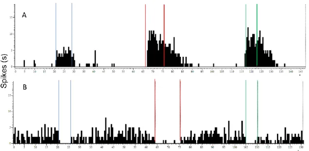
Figure 10 - Example of a recording of RVM cells from Spike 2 software. A- Example of a recording of an ON-WDR-like neurone discharge frequency. The activity of this cell increases in the innocuous and noxious stimulation. After stimulation, the cell returns to basal levels. B- Example of a recording of an OFF-WDR-like neurone discharge frequency. Before and after stimulation cell has an increased activity, and in the moment of the innocuous and stimulation the firing rate decreases substantially and. In the end of stimulation, cell returns to normal activity. (Blue lines - delimitation of the period of innocous stimulation (brushing); red lines - delimitation of the period of noxious heat stimulation by tail-flick; green lines - delimitation of the period of noxious mechanical stimulation by pinch).
34 3.9. Statistics /DATA analysis
The GraphPad Prism 6 (GraphPad software Inc., Lajolla, CA, USA) was used to perform a statistical analysis. A Two-away analyses of variance (ANOVA2w) followed by t-test with a Bonferroni correction for multiple comparisons was used to compare results from the behavioral tests and resukts from the c-Fos quantification. An ANOVA repeated-measures (ANOVARM) test followed by t-test with a
Bonferroni correction for multiple comparisons was used to compare results from the RVM neuronal alterations after drug injection in the IL. P<0.05 was considered to represent a significant difference. The results are expressed as mean±standard error (SEM).
35
37
4.1. Histological confirmation of the injection and recording sites
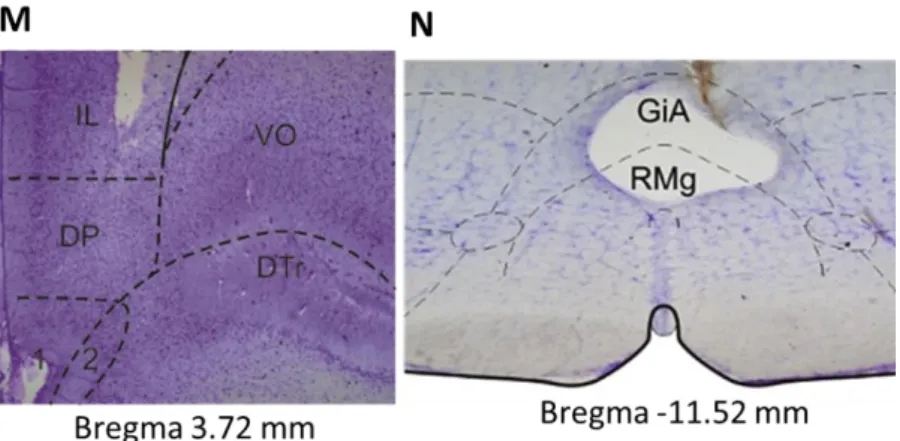
In the paw withdrawal test, the anatomical confirmation of the drug microinjection sites in the infralimbic cortex (IL) is presented in the figures 11 A-D. In the electrophysiology study, the sites of microinjection in IL are presented in figure 11 E-H while a histological example is shown in figure 11M. Finally, representations of recording sites in the RVM are presented in figures 11 I-L and a histological example of the recording site in the RVM is shown in figure 11N. The location of the injection/recording sites was confirmed on brain coronal sections counterstained with Cresyl Violet.
38
Figure 11 – Histological confirmation and schematic representation of microinjection/recording sites in the IL (Pharmacological treatment, A: 3.24mm; B: 3.00mm; C: 2.76mm; D: 2.52mm, Electrophysiological recordings, E: 3.72mm F: 3.24mm; G: 3.00mm; H: 2.76mm and Histological section, M: 3.72mm) and the RVM (I: -10.44mm; J: -11.16mm; K: -11,28mm; L: -11.52mm, Histological section, N: -11.52mm). (Gray dots correspond to SHAM animals; black dots represent ARTH animals). (IL - ; DP - ; DT - ; VO - ; GiA - ; RMg - ).
4.2. Nociceptive behavior
4.2.1 Pressure application measurement
In the PAM test, ARTH animals displayed a significantly lower limb withdrawal threshold (LWT) when compared to SHAM animals (F(1,26) =12.53, P=0.015). In addition, the LWT of the ipsilateral knee of ARTH animals was significantly lower when compared with the LWT of the contralateral knee (F(1,26)=27.13, P<0.0001).
Figure 12 - Evaluation of limb-withdrawal latency (LWT) during the pressure application measurement test in SHAM (n=8) and ARTH (n=8) animals. Data presented as mean±SEM. (***P<0.001). (SHAM – control animals injected with saline in the synovial cap of the right hind paw knee; ARTH – animals injected with kaolin and carrageenan in the in the synovial cap of the right hind paw knee; PAM - pressure application measurement).
39 4.2.2. Paw withdrawal test (PWL)
4.2.2.1. Vehicle injection
To control for a potential effect of the microinjection upon infralimbic (IL)-mediated pathways, we evaluated PWL after injecting a vehicle solution in the IL. As shown in figure 13, the administration of a volume of 0.5µL did not alter nociceptive behavior (F(1,66)=0.3612, P=0.5499).
Vehicle injection also had no effect throughout the duration of the experimental session (F(5,66)=0.1420,P=0.9817).
Figure13 - Effect of the administration of the vehicle (saline) to the IL in SHAM (n=8) and ARTH animals (n=8). No changes in PWL could be observed either between experimental groups or throughout time. Data presented as mean±SEM.
4.2.2.2. Glutamate injection
Overall, the administration of glutamate in the IL significantly decreased PWL (F(5,40)=10.91, P<0.0001) (Fig.14) although the nociceptive threshold of ARTH animals was not significantly different from SHAM animals (F(1, 40) =0.0284, P=0.8671).
Post hoc tests showed that glutamate decreased PWL in the SHAM group 10 and 30 minutes after drug administration while in the ARTH group this effect was observed 30 minutes after GLU microinjection in the IL (Fig. 14).
40
Figure 14 - Effect of the administration of GLU in the IL upon PWL in SHAM (n=8) and ARTH (n=8) rats. Data presented as mean±SEM. (*P<0.05). (IL – infralimbic cortex; PWL – paw-withdrawal latency; GLU - ; SHAM – control animals injected with saline in the synovial cap of the right hind paw knee; ARTH – animals injected with kaolin and carrageenan in the in the synovial cap of the right hind paw knee).
4.2.2.3. CHPG injection
The administration of CHPG (2-chloro-5-hydroxyphenylglycine), a metabotropic glutamate receptor 5 (mGluR5) agonist, in the IL significantly altered PWL in rats (F(5,120)=16.38, P<0.0001) (Fig.15) although no differences were found between SHAM and ARTH animals (F(1,120)=0.2725, P=0.6026).
Interestingly, CHPG microinjection enhanced nociception in both experimental groups from 10 to 40 minutes in SHAM animals, and from 20 to 40 minutes in ARTH animals. Animals fully recovered from CHPG injection 50 minutes after its administration.
Figure 15 - Effect of the administration of CHPG in the IL upon PWL in SHAM (n=8) and ARTH (n=8) animals. Data presented as mean±SEM. (*P<0.05; **P<0.01; ***P<0.001). (IL – infralimbic cortex; PWL – paw-withdrawal latency; CHPG - ; SHAM – control animals injected with saline in the synovial cap of the right hind paw knee; ARTH – animals injected with kaolin and carrageenan in the in the synovial cap of the right hind paw knee).
41 4.2.2.4. MPEP injection
Overall, the administration of MPEP, a mGluR5 antagonist, into the IL significantly altered the PWL of rats but this effect was dependent on the development of arthritis (F(5,82)=7.864, P<0.0001). Post-hoc tests showed PWL of ARTH animals were significantly increased when compared to SHAM animals but only 30 minutes after MPEP injection in the IL.
Figure 16 - Effect of the administration of MPEP in the IL upon PWL in SHAM (n=8) and ARTH (n=8) animals. Data presented as mean± SEM.(***P<0.001). (IL – infralimbic cortex; PWL – paw-withdrawal latency; MPEP - ; SHAM – control animals injected with saline in the synovial cap of the right hind paw knee; ARTH – animals injected with kaolin and carrageenan in the in the synovial cap of the right hind paw knee).
4.3. c-Fos quantification
4.3.1. Locus coeruleus (LC)
Overall, in SHAM animals, the number of c-Fos expressing cells was significantly increased in the ipsilateral side (F(1,24)=54.92, P<0.0001) an effect that depended on the protocol (F(3,24)=17.99, P<0.0001). In ARTH animals, c-Fos expression was also increased in the ipsilateral side (F(1,22)=9.761, P=0.0049) but this effect was independent of the protocol performed (F(2,22)=1.525, P=0.2361).
In the ipsilateral side, the number of c-Fos expression was significantly altered between protocols (F(3,23)=5.940, P=0.0037) with no differences observed between SHAM and ARTH animals (F(1,23)=0.8171, P=0.3754) (Fig.17A). Post-hoc tests showed that c-Fos expression was only significantly increased in SHAM animals after the simultaneous administration of CHPG in the IL and peripheral application of a noxious stimulus (Fig.17A).