Olívia Alexandra Elias Pontes
New Chromene-Based Drug Candidates for
Cancer Treatment
outubro de 2016Olívia Ale
xandr
a Elias P
ont
es
Ne
w Chromene-Based Drug Candidates for Cancer T
reatment
Minho | 20
1
6
i
O
LÍVIAA
LEXANDRAE
LIASP
ONTESN
EW CHROMENE
-
BASED DRUG CANDIDATES FOR
CANCER TREATMENT
Master Thesis
Applied Biochemistry
Under the supervision of:
Professor Maria Fernanda de Jesus Rego Paiva Proença, PhD Marta Sílvia Freitas da Costa, PhD
October 2016 University of Minho School of Sciences
iii
Acknowledgments/Agradecimentos
Muitas foram as pessoas que, das mais variadas formas, contribuíram para a elaboração desta tese de mestrado, às quais quero manifestar a minha gratidão.
Em primeiro lugar, agradeço à Professora Doutora Maria Fernanda Proença, orientadora desta tese, pela oportunidade e voto de confiança que me deu para a elaboração deste trabalho. Obrigada pela paciência, sabedoria, compreensão e disponibilidade. Obrigada pela ajuda em todas as horas.
À Doutora Marta Costa por ser co-orientadora deste trabalho, pela atenção, delicadeza e incentivo à minha progressão, obrigada por toda a ajuda na química e, sobretudo, pelo seu tempo.
À Professora Doutora Fátima Baltazar, obrigada pelo conhecimento transmitido e rigor. Agradeço, ainda, a sua disponibilidade e o incentivo ao meu crescimento científico.
Obrigada a todas as técnicas de laboratório. Em especial à Elisa pelo apoio técnico e pela prontidão com que sempre me disponibilizou os espectros de NMR.
A todos aqueles que sempre me ajudaram no laboratório, tanto no Departamento de Química como no ICVS, onde as dúvidas e o receio persistiam.
A TODOS os meus amigos, dos mais recentes aos mais antigos. A todos eles que, de alguma forma, foram pilares muito importantes durante este ciclo. Obrigada por se manterem por perto, pelos sorrisos, pela força, pelas palavras de incentivo e por acreditarem sempre que eu conseguiria.
À Célia, uma irmã de coração, obrigada pela amizade inabalável, pelo carinho de todos estes anos e por me ouvir nas horas mais difíceis.
E, sobretudo, obrigada aos meus pais. Pelo esforço que sempre fizeram por mim, pelo apoio que me deram quando mais precisei, pela educação e valores que me transmitiram, por me terem mostrado que o melhor caminho nem sempre é o mais fácil, pelo exemplo de força e, principalmente, pelo amor incondicional. Por tudo isto e muito mais, um enorme Obrigada.
iv
v
Abstract
Cancer is a devastating disease worldwide, with millions of diagnoses per year and many people living with this pathology. Breast cancer is one of the major causes of death in women while male breast cancer represents less than 1% of the total male diagnosed population. Due to this increasing cancer incidence, research in this area has been growing at the same rate. Thus, there is an urgency in discovering new drugs for cancer treatment.
The chromene scaffold has been identified in several compounds with anticancer activity. The substitution pattern highly influences the activity and the mode of action and the synthesis of new derivatives is an important element in the search for improved drug candidates. The major aim of this Master thesis was the synthesis of new chromene derivatives with enhanced anticancer activity.
Chalcone and chromene derivatives were isolated in good yield through clean reactions using innocuous solvents such as water and ethanol and highly effective aldol condensations. Generally, the reactions were performed at room temperature, leading to the isolation of highly pure compounds.
Newly synthetized compounds were tested on cancer cells and a non-tumoral cell line. For the first screening, a range of 51 compounds was assessed for cell survival by SRB assay on MCF-7 cells. Then, only the 22 most active compounds were tested against another breast cancer cell line (HS578t) and cytotoxicity was evaluated for MCF-10 normal cells. Then, a selection of the eight most promising chalcones and chromenes had their IC50 determined for all three cell lines. Finally, some more specific assays were performed and it was found that a selected chromene acted as a cell migration inhibitory agent. Some preliminary results, with protein expression, also showed that this chromene might be causing its anticancer activity through induction of cell-apoptosis. For chalcones, the results suggest an anti-proliferative ability and reduction of membrane integrity.
Generally, compounds with halogenated substituents presented enhanced activity comparing to methoxy or methyl groups. More specifically, the bromine atom was often present in the bioactive molecules that proceeded to the final assays and showed to be promising candidates for further studies.
vi
vii
Resumo
O cancro é uma doença devastadora que em todo o mundo apresenta milhões de diagnósticos por ano e muitas pessoas vivem com esta patologia. O cancro da mama é uma das maiores causas de morte entre a população feminina enquanto que, o cancro da mama masculino afeta menos de 1% de todos os diagnósticos oncológicos nos homens. Devido à crescente incidência de cancro, a investigação farmacológica nesta área tem vindo a crescer na mesma proporção. Assim, a descoberta de novas drogas para tratamento oncológico representa uma urgência.
O núcleo de cromeno foi identificado em diversos compostos com atividade anticancerígena. O padrão de substituição demonstra afetar a atividade e o modo de ação e a síntese de novos derivados é um elemento importante em investigação para melhorar a ação do candidato. O principal objetivo desta tese de Mestrado era sintetizar novos derivados de cromeno com atividade anticancerígena melhorada.
Vários derivados de calconas e cromenos foram isolados com bons rendimentos, através de reações limpas usando solventes inócuos tais como água e etanol, em condensações aldólicas altamente efetivas. Na generalidade, as reações foram realizadas à temperatura ambiente, levando ao isolamento de compostos altamente puros.
Os recém-sintetizados compostos foram testados em linhas celulares de cancro e numa linha celular não-tumoral. Num primeiro screening, um lote de 51 compostos foi testado para a viabilidade celular por ensaio com sulforodamina em células MCF-7. Então, apenas os 22 compostos mais ativos foram testados para outra linha celular (HS578t) e a citotoxicidade foi avaliada em células normais MCF-10. Uma série dos oito compostos mais promissores para as três linhas celulares, foi selecionada e teve o IC50 determinado. Finalmente, alguns ensaios mais específicos foram elaborados e registou-se que o único cromeno selecionado apresentou poder para inibição da migração celular. Alguns resultados preliminares, para expressão proteica, clarificaram que este cromeno poderá estar a agir como agente anticancerígeno através da indução da apoptose. Para as calconas testadas, os resultados sugerem uma capacidade antiproliferativa e a redução da integridade membranar.
Em geral, os compostos com substituintes halogenados exibiram melhor atividade em comparação com grupos metilo ou metoxilo. Mais especificamente, o átomo de bromo esteve por diversas vezes presente nas moléculas bioativas que procederam para os ensaios finais e demostraram ser candidatos promissores para estudos futuros.
viii
ix
I
NDEX
Acknowledgments/Agradecimentos ... iii Abstract ... v Resumo ... vii List of Figures ... xiList of Tables ... xii
List of Schemes ... xiii
Abbreviations ... xv
CHAPTER I
–
INTRODUCTION ... 11. Cancer ... 2
1.1. Breast cancer ... 3
1.1.1. Breast cancer treatments ... 6
2. Chalcones ... 10
2.1. Anticancer properties of chalcones ... 10
3. Chromenes and coumarines ... 12
3.1. Anticancer activity of chromenes ... 13
3.1.1. Natural chromenes as anticancer agents ... 13
3.1.2. Synthetic chromenes as anticancer agents ... 16
Aims ... 22
CHAPTER II
–
CHEMICAL SYNTHESIS: RESULTS AND DISCUSSION... 231. Chalcone derivatives ... 24
1.1. Synthesis and mechanistic discussion ... 25
1.2. Spectroscopic characterization ... 29
2. 4H-chromene derivatives ... 35
2.1. Synthesis and mechanistic discussion ... 36
2.2. Spectroscopic characterization ... 38
CHAPTER III
–
BIOLOGICAL ACTIVITY: RESULTS AND DISCUSSION ... 471. Initial compound viability screening ... 48
2. Survival curves and IC50 determination ... 51
3. Effect of selected compounds on cell migration ... 54
4. Effect of chalcones and chromenes on cell proliferation ... 57
5. Effect of selected compounds on membrane integrity ... 58
x
CHAPTER IV
–
CONCLUSIONS AND FUTURE WORK ... 63Conclusions ... 64
Future work ... 65
CHAPTER V
–
EXPERIMENTAL SECTION ... 671. Experimental procedures ... 68
1.1. General procedure for the synthesis of chalcone derivatives ... 68
1.2. Synthesis of 4H-chromene derivatives ... 76
2. Biological assays ... 82
2.1. Cell lines and culture conditions ... 82
2.2. Cell viability assays ... 82
2.2.1. Sulforhodamine B assay ... 82
2.2.2. Trypan blue assay ... 83
2.3. Proliferation assay ... 84
2.4. Migration assay ... 84
2.5. Protein extraction and Western Blot ... 85
2.6. Statistical analysis ... 86
xi
List of Figures
Figure 1. The Hallmarks of Cancer ... 2
Figure 2. Estimated incidence for all types of cancer worldwide . ... 4
Figure 3. Hormone therapy agent: tamoxifen. ... 5
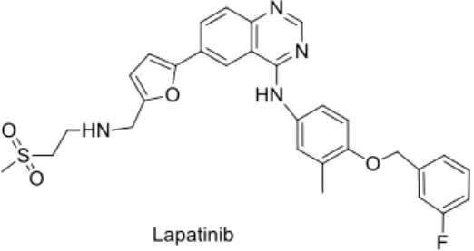
Figure 4. Chemical structure of drugs used in targeted therapy: lapatinib. ... 6
Figure 5. Chemical structures of chlorambucil and busulfan ... 7
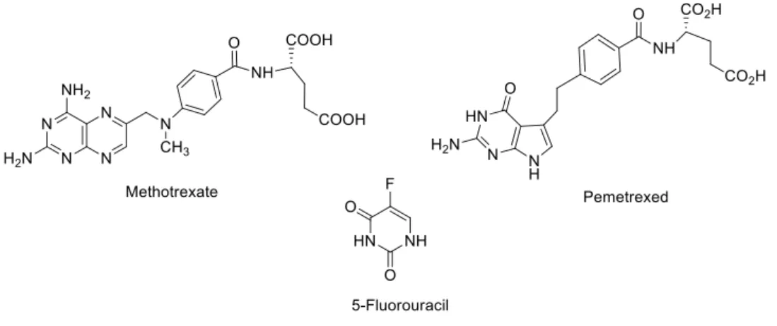
Figure 6. Antimetabolites chemical structures: the examples of methotrexate, pemetrexed and 5-fluorouracil. ... 8
Figure 7. Chemical structures of doxorubicin and daunorubicin. ... 8
Figure 8. Two examples of topoisomerase inhibitors: chemical structures of etoposide and teniposide. ... 8
Figure 9. Chemical structures of paclitaxel, docetaxel and eribulin, some mitotic inhibitors. ... 9
Figure 10. Chalcone. ... 10
Figure 11. Natural and synthetic chalcones with anticancer properties. ... 11
Figure 12. 1-benzopyrans and 2-benzopyrans. ... 12
Figure 13. Chroman, 2H-chromene, 4H-chromen, 2H-chromen-2-one and 4H-chromen-4-one ... 13
Figure 14. Series of natural products with observed anticancer activity. ... 14
Figure 15. Numerous synthetic chromene derivatives with reported anticancer properties. ... 17
Figure 16. Interaction by bidimensional correlation of Hα and Hβ with the vicinity, for compounds 2.3... 35
Figure 17. Interaction by bidimensional correlation of CH2 and C4H with the vicinity, for compounds 2.10 and 2.11. ... 40
Figure 18. Series of previously synthetized chromenes for first screening with MCF-7 cell line. ... 48
Figure 19. Selected compounds for IC50 determination in MCF-7, HS578t and MCF-10 cell lines. ... 52
Figure 20. Effect of chalcones and chromenes on MCF-7, HS578t and MCF-10 cells for total cell biomass ... 53
Figure 21. Selected compounds for cell migration assay in MCF-7 cells ... 55
Figure 22. Representative images of control and effect of chalcones 2.3e, 2.3g and 2.3i and chromene 2.11e on MCF-7 cell migration by wound healing assay ... 55
Figure 23. Effect of chalcones 2.3e, 2.3g and 2.3i and chromene 2.11e on MCF-7 cell migration by wound healing assay.. ... 56
Figure 24. Effect of chalcones 2.3e, 2.3g and 2.3i and chromene 2.11e on MCF-7 cell proliferation ... 57
xii
Figure 25. Membrane integrity of MCF-7 cell line treated with the respective ¼IC50, ½IC50 and
IC50 of chalcones 2.3e, 2.3g and 2.3i and chromene 2.11e... 59
Figure 26. Membrane integrity of MCF-10 cell line treated with the respective ¼IC50, ½IC50 and IC50 of chalcones 2.3e, 2.3g and 2.3i and chromene 2.11e ... 60
Figure 27. Representative blot of effect of compounds 2.3e, 2.3g, 2.3i and 2.11e on MCF-7 cells - caspase-9, PARP and CAIX ... 62
Figure 28. Most promising chromene structure. ... 64
List of Tables
Table 1. Molecular subtypes of breast cancer ... 5Table 2. General substitution pattern of chalcones 2.3a-z ... 26
Table 3. Physical and analytical data for chalcones 2.3a-z ... 27
Table 4. Attempted reactions, experimental conditions and obtained products. ... 28
Table 5. Infrared spectroscopic data for chalcones 2.3a-p and 2.3y ... 30
Table 6. 1H NMR spectroscopy data for chalcones 2.3 ... 31
Table 7. 13C NMR spectroscopy data for chalcones 2.3. ... 33
Table 8. Physical and analytical data for 4H-chromenes 2.10 ... 37
Table 9. Physical and analytical data for 4H-chromenes 2.11 ... 37
Table 10. Infrared spectroscopic data for 4H-chromenes 2.10 and 2.11 ... 38
Table 11. Spectroscopic data of 1H NMR for 4H-chromenes 2.10 ... 41
Table 12. Spectroscopic data of 13C NMR for 4H-chromenes 2.10 ... 42
Table 13. Spectroscopic data of 1H NMR for 4H-chromenes 2.11 ... 43
Table 14. Spectroscopic data of 13C NMR for 4H-chromenes 2.11 ... 44
Table 15. Cell viability for MCF-7 cell line after 72 hours of treatment with two compound concentrations ... 49
Table 16. Cell viability for HS578t and MCF-10 cell lines after 72 hours of treatment at 10 and 30 µM. ... 51
xiii
List of Schemes
Scheme 1. 2-Hydroxychalcone synthesis by a LiHMDS-mediated aldol condensation ... 24
Scheme 2. “ thesis of ’-hydroxychalcones under basic catalysis. ... 24
Scheme 3. Chalcone synthesis under borontrifluoride-etherate catalysis. ... 25
Scheme 4. Acidic catalysis of acetophenone with 3,4-dimethoxybenzaldehyde reaction. ... 25
Scheme 5. Synthesis of chalcones 2.3 from substituted acetophenones and benzaldehydes .. 27
Scheme 6. Preparation of 8-methoxy-2-(4-fluorophenyl)-3-nitro-2H-chromene and analogues. ... 35
Scheme 7. Proposed mechanism for the synthesis of 4H-chromenes from chalcones 2.3 and the active methylene compounds. ... 36
Scheme 8. Simplified reaction of chalcones 2.3 with methyl cyanoactetate ... 36
xiv
xv
Abbreviations
1H NMR Proton Nuclear Magnetic Resonance Spectroscopy 13C NMR Carbon-13 Nuclear Magnetic Resonance spectroscopy
ºC Celsius degree Ar Aromatic comp compound
CSC Cancer stem cell
d doublet (relative to NMR peaks)
dd doublet of doublets (relative to NMR peaks) dt doublet of triplets (relative to NMR peaks) DMSO Dimethyl sulfoxide
DMSO-d6 Deuterated dimethyl sulfoxide DNA Deoxyribonucleic acid
EC50 half maximal effective concentration ED50 median effective dose
ER Estrogen receptor
EWG electron withdrawing group GEP gene expression profiling
GI50 half maximal growth inhibition concentration HER2 Human epidermal growth factor receptor 2
HIF Hypoxia-inducible factor
IC50 half maximal inhibitory concentration IHC Immunohistochemistry
IR Infrared Spectroscopy
LiHMDS Lithium bis(trimethylsilyl)amide
m Medium (relative to IR spectra absorption bands) m multiplet (relative to NMR peaks)
M molar
MBC Male breast cancer MeOH methanol
m.p. melting point ppm parts per million
PR Progesterone receptor
q quartet (relative to NMR peaks ROS Reactive oxygen species
rt room temperature
s singlet (relative to NMR peaks)
s Strong (relative to IR spectra absorption bands) SRB Sulforhodamine B
xvi t triplet (relative to NMR peaks)
td triplet of doublets (relative to NMR peaks) THF tetrahydrofuran
TLC Thin Layer Chromatograph
tt triplet of triplets (relative to NMR peaks) w Weak (relative to IR spectra absorption bands)
η yield
1
C
HAPTER
I
–
I
NTRODUCTION
2
1.
Ca er
Cancer is the name given to a group of related diseases where part of the body cells start to grow uncontrollably and spread into contiguous tissues [1,2]. Cancer is the second leading cause of death in contemporary society, preceded by cardiovascular diseases. In 2012, approximately 14 million people around the world were diagnosed with cancer, about 8.2 million deaths were registered due to this disease and 32.6 million people live with cancer (within 5 years of diagnosis) [1,3].
For the first time, in 2000, Hanahan and Weinberg defined the succession of acquired abilities which allow cancer cells to survive, proliferate and disseminate, as hallmarks of cancer [2,4]. Self-sufficiency in growth signals, insensitivity to growth-inhibitory signals, evasion of programmed cell death, unlimited replicative potential, sustained angiogenesis and tissue invasion and metastasis were the six hallmarks proposed [2].
Recently, these hallmarks were reviewed and modified by the same authors, including the ability to alter or reprogram cellular metabolism and the capability of evasion to immunological destruction [5]. Consequently, two more consequential features were defined: the inflammation by innate immune cells and the genomic instability and mutation. Additionally, in 2012, Floor and co-workers mentioned another essential characteristic – loss of differentiation [6].
Figure 1. The Hallmarks of Cancer. Schematic illustration of the acquired capabilities of tumor cells essential
3
These hallmark abilities are acquired through deregulation of signaling pathways which, in combination with the interconnections and crosstalk between the individual sub-circuits, can lead to alteration of multiple capabilities through a certain oncogenic event [5].
Over the years, the knowledge about carcinogenesis has increased and two models were suggested to describe the development of cancer cells. The stochastic model defends that any type of cell is able of initiating and promoting tumor development and the cancer stem cell (CSC) model which suggests that tumors are hierarchically structured and cancer-promoting potential is only present in CSCs [7,8].
According to this information, cancer is a rather heterogeneous pathology related to defects in terms of the regulatory circuits that control cellular homeostasis, including death, proliferation, differentiation and cell migration and, due to this dynamic system, there is an explicit difficulty to reach an effective treatment to this disease [9,10].
1.1.
Breast cancer
Breast cancer is the most frequent cancer in women worldwide and the fifth cause of death from cancer, with 522.000 deaths in 2012. Furthermore, with an estimated 1.67 million new cases, it is considered the second most common cancer in the world (Figure 2) [3]. Moreover, successive demographic and epidemiologic transitions, predominantly in less developed countries, have a huge contribution in the increased incidence of cancer and, until 2025, it is expected an occurrence of 20 million cancer cases every year [11]. On the other hand, male breast cancer (MBC), with less than 1% of the total cancer diagnoses in male population, is very uncommon [12,13]. Over the years, MBC mortality is increasing due to lack of male population concern about this pathology. Consequently, male patients are diagnosed in a more advanced stage of the disease [13,14].
In Portugal, for both sexes, breast cancer had an incidence and mortality of 12,4% and 6.5%, respectively, in 2012 [3]. Due to its high incidence, breast cancer is one of the diseases that shows more concern by the scientific community. This fact led to an increasing search for new anticancer drugs with increasing potential for prevention and treatment.
As prevention for breast cancer, a leading medical screening procedure was implemented: mammography. However, not all the cases of breast cancer have successful
4
endings so it is important to understand this cancer heterogeneity and improve prevention, diagnosis and survival for the specific subtypes [15–18].
Over the last five decades, the inter and intravariability of breast cancer tumors have been studied by histopathology methods [19]. Recently, new techniques like microarrays and next-generation sequencing, by advances in genetic sequencing and molecular analysis, were combined and employed to describe the multiplicity of breast tumor subtypes [20,21]. Hence, luminal A, luminal B, HER2-overexpressing and triple negative were the breast cancer tumor subtypes defined through genetic and molecular methods (Table 1) [22–24]. This classification was possible according to the evaluation of progesterone (PR) or estrogen receptors (ER) by immunohistochemistry (IHC) and fluorescent in situ hybridization. In recent years, as a form to differentiate aggressiveness or tumor cell proliferation in the several types of breast cancer, new tumor procedures for classification, such as Ki-67 status or histologic grade, were labored [22]. This novel established approach aims at more effective treatments in terms of surgery, chemotherapy, radiotherapy or new targeted therapies [23,25,26].
According to the different subtypes of breast cancer, the treatment used will also be changed. From all the four subtypes of breast tumors, luminal A tends to have a best prognosis, with high rates of survival and equally low recurrence rates. Due to the presence of ER-positivity, treatment for these tumors generally includes a diversified hormone therapy as tamoxifen (Figure 3) and/or aromatase inhibitors [27,28].
5
Figure 3. Hormone therapy agent: tamoxifen.
Before discovering that HER2 subtype tumors could have a target for treatment, women with these tumors had a very poor prognosis. However, nowadays, for tumors HER2-positive, it is common to treat this cancer subtype with anti-HER2 drugs such as trastuzumab [29].
Table 1. Molecular subtypes of breast cancer (adapted from [30]).
Intrinsic subtypes (GEP) IHC classification (St Gallen) Agreement IHC/GEP
Luminal A Lu i al A
ER and/or PR positive HER2 negative Ki-67<14%
73%-100%
Luminal B Lu i al B HER egati e
ER and/or PR positive HER2 negative Ki- ≥ % Lu i al B HER positi e ER and/or PR positive Any Ki-67
HER2 over-expressed or amplified
73%-100%
HER2-enriched HER positive (non-lu i al HER2 over-expressed or amplified ER and PR absent
41%-69%
Basal-like T iple egati e
ER and PR absent HER2 negative
80%
(ER, estrogen receptor; PR, progesterone receptor; GEP, gene expression profiling; HER2, human epidermal growth factor receptor 2)
Basal-like tumors are the most aggressive subtype of breast cancer. Because of their negativity for ER and HER2, hormone therapy and trastuzumab cannot be used as treatments. So, generally, a combination of surgery, radiotherapy and chemotherapy is used, making the treatment with larger scope but also more aggressive to the patients [31]. Targeted therapies for basal-like tumors do not exist however treatment options are currently being studied. As
6
possible targets for coming therapies, epidermal growth factor receptor and androgen receptor were mentioned [32].
1.1.1.
Breast cancer treatments
Treatment for breast cancer depends on the general health of the patient as well as the stage and grade of cancer and may include a combination of surgery and radiation, chemical, immunological and targeted therapies. The typical treatment for the patients with stage I tumors involves surgery to remove the cancer and a small amount of tissue around the tumor (lumpectomy) [33]. Stage II patients are considered for adjuvant chemotherapy as a preventive strategy against cancer recurrence [33]. Patients with stage III disease, having a high rate of recurrence and cancer related death, are usually considered for adjuvant chemotherapy [28,29]. However, as referred previously, breast cancer is divided into four different subgroups according to the expression of PR, ER or both. This differential expression on the surface of cancer cells will condition adjuvant chemotherapy. Regarding targeted therapies, tumors with over-expression of HER2 are treated with an anti-HER2 antibody, such as trastuzumab, or a tyrosine kinase inhibitor, such as lapatinib (Figure 4) [30,31]. Triple negative tumors, with absent expression of PR, ER and HER2 and increased rates of metastasis, relapse and death, compared to other breast cancer types, are treated with cytotoxic chemotherapy due to the lack of targeted therapies [31].
Radiotherapy treatment uses X-ray or other types of radiation to kill and inhibit the continuous proliferation of cancer cells [37]. Two types of radiation therapy may be performed: external radiation which sends radiation to the cancer from a machine outside the body and internal radiation, that injects radioactive substances into or near the tumor [38]. This procedure will vary depending on the type and stage of treating cancer [35–37].
7
The cancer treatment that uses drugs to stop tumor cellular growth is defined as chemotherapy. This procedure acts essentially in the hallmark of cancer characterized by the evasion from growth suppressors and its action leads to cellular division arrest (mitosis) or cell death (apoptosis) [42]. This type of therapy has two different ways of administration: systemic chemotherapy, in which the drug enters into the bloodstream and regional chemotherapy, that places the drug directly in the local of action [38,39]. However, as tumor cells share mechanisms of action with normal cells, there are many side effects that limit the patient life during the treatment [44]. Chemotherapy, because of its cytotoxic and non-selective properties, has limited clinical applications [45]. So, hematological malignancies, metastatic solid cancers and primary cancers as an aide to surgery, by their high proliferative index and as an attempt of localized treatment, are the main applications of this type of treatment [42].
The drugs administrated through chemotherapy are divided into classes according to the drug target and its mechanism of action [43]. Hence, some of the highlighted classes with current and common use are:
i. Alkylating agents which damage the DNA by formation of covalent bonds, inducing single-strand or double-strand breaks that lead to DNA cross-linking, preventing cellular proliferation. Chlorambucil [46] and busulfan [47] are two examples of the many alkylating agents currently available (Figure 5).
ii. Antimetabolites affect the RNA and DNA growth by the introduction of similar pyrimidine and purine analogues into the strand. S phase is generally affected by these drugs. As examples, methotrexate [48], pemetrexed [49] and 5-fluorouracil (5-FU) are represented in Figure 6 [48].
8
iii. Anthracyclines are anti-tumor antibiotics that alter the cancer cells DNA, keeping them from proliferate and replicate uncontrollably by DNA intercalation or formation of free radicals. Two of these antibiotics are doxorubicin [50] and daunorubicin [51] (Figure 7).
iv. Topoisomerase inhibitors influence the DNA strand separation, done by topoisomerase enzymes, that arises during S phase and would result on DNA copies at the end of cell cycle. Etoposide [52] and teniposide [53] are both inhibitors of this enzyme (Figure 8).
Figure 6. Antimetabolites chemical structures: the examples of methotrexate, pemetrexed and 5-fluorouracil.
Figure 7. Chemical structures of doxorubicin and daunorubicin. Two anti-tumor antibiotics.
9
v. Mitotic inhibitors, also known as tubulin-binding drugs, interfere with numerous functions of the cell such as mitosis, meiosis, intracellular transport and others. Paclitaxel [54], docetaxel [55] and eriblin [56] illustrate this class of drugs (Figure 9).
Due to the heterogeneity of most cancers, chemotherapy is usually administrated as a combination of the above referred classes of drugs. This procedure intends to overcome cell resistance and prevents the generation of new resistant subclones [57]. As a benefit to patients, it reduces the side effects of single agents that would have to be used in higher doses in monotherapy [38,53].
In the last few years, research for cancer cures and treatments have made progresses. Targeted agents were recently discovered as potential drug candidates [54,55]. These agents widen the therapeutic window, reducing many side effects of traditional procedures as chemotherapy and radiotherapy [58]. In 2013, der Hollander and research group described that targeted drugs have potential uses as preventive agents [59]. It was revealed that ER-targeting drugs, such as tamoxifen, have shown preventive efficacy for ER-positive breast cancer leading to and effective reduction in incidence. However, the major problem still remains for triple negative breast cancer in which it is necessary to identify possible novel targets, develop
10
toxic drugs that successfully interrupt the activity of these targets and find possible delivery systems for these novel preventive therapies [59].
2.
Chal o es
Chemically described as 1,3-diphenyl-2-propen-1-ones, chalcones 1.1 result from the connection of two aromatic rings by a three-carbon α,β-unsaturated carbonyl bond [60] (Figure 10). An aldol condensation between benzaldehyde and acetophenone in the presence of sodium hydroxide as a catalyst is the most efficient method that leads to the preparation of chalcones.
Figure 10. Chalcone.
Chalcones are naturally occurring compounds in edible plants that exhibit a wide-range spectrum of biological activities as anti-inflammatory [57,58], antioxidant [63], anti-tubercular [64], anti-HIV [65], antimalarial [66], anti-allergic [67], anti-diabetic [68], cardioprotective [69] and also anticancer agents through multiple mechanisms [70].
2.1.
Anticancer properties of chalcones
A general overview of recent works reveals a constant search for naturally occurring chalcones with anticancer activity and novel mechanisms of action. Thus, many natural chalcones with promising anticancer effectiveness against a diversity of cancer cell lines have been identified.
11
Figure 11. Natural and synthetic chalcones with anticancer properties.
Found in Angelica keiskei roots, 4-hydroxyderricin 1.2 (Figure 11) was described as an inducer of apoptotic cell death via death receptor-mediated and mitochondrial pathways by topoisomerase II inhibition in leukemia cells (HL60) [71]. Furthermore, this chalcone also exhibited great cytotoxic properties against another three cancer cell lines: A459 (lung), AZ521 (stomach) and CRL1579 (melanoma).
Ye et al. found a novel pyranochalcone 1.3 (Figure 11) with strong cytotoxic effect isolated from the seeds of a Chinese medicinal plant, Milletia pachycarpa, showing great minimal effective concentration in several cancer cell lines [72]. Moreover, this pyranochalcone showed strong apoptosis inducing effects at a concentration of 2 μM ithi h.
HTMC 1.4 ’-hydroxy- , , ’, ’-tetramethoxychalcone) (Figure 11) was extracted from a medicinal plant Caesalpinia pulcherrima and demonstrated great in vitro selectivity for many human lung cancer cell lines [73]. In A549 (lung adenocarcinoma cells), HTMC presented a cytotoxic activity by causing G1 phase cell-cycle arrest.
Naturally occurring chalcones revealed promising anticancer properties that led to many synthetically inspired chalcones with enhanced anticancer activity. The manipulation of the aryl rings or replacement by scaffolds with heteroaryl/steroidal/alicyclic and conjugation of pharmacologically interesting scaffolds through molecular hybridization are the three main used strategies to manipulate and enhance the characteristics of chalcones as anticancer agents [74].
Zhang et al. described that chalcone 1.5 (Figure 11) presented a remarkable antiproliferative potential with very low IC50 values of 0.03 and 0.95 µg/mL for MCF-7 (breast) and A549 (lung) cell lines, respectively [75]. At 1.42 µg/mL, this compound also demonstrated to be a promising antitubulin polymerization inhibitor.
12
A series of 20 brominated chalcones were designed, prepared and tested in four different gastric cancer cell lines [76]. From all the compounds, H72 1.6 (Figure 11) was the one that presented more potential with lower IC50 and less cytotoxicity to the normal gastric cell line GES-1. The treatment with H72 in MGC803 and HGC27 seems to induce the generation of ROS which causes mitochondria mediated apoptosis and activates the caspase 9/3 cascade.
A synthetic flavonoid derivative, 2'-hydroxy-2,3,5'-trimethoxychalcone 1.7 (DK143) (Figure 11) showed evidences to be involved in apoptosis induction by unfolded protein response in the breast cancer cell line MDA-MB-231 [77]. The production of ROS (Reactive Oxygen Species) and upregulation of ER stress sensors expression are the mechanisms involved in the anticancer activity of this synthetic chalcone. DK143 does not affect viability for the non-transformed cell line MCF10A. In vivo, DK143 suppressed mouse tumor growth.
3.
Chro e es a d ou ari es
The fusion of a benzene ring with a heterocyclic pyran ring results in a polycyclic compound named benzopyran. Chromene was the nomenclature adopted by IUPAC to define these compounds. 1-benzopyran (1.8 and 1.9 in Figure 12) and 2-benzopyran (1.10 and 1.11 in Figure 11) were the two classes of benzopyrans named by the position of the oxygen atom on the pyran ring [78]. Depending on the level of oxidation and saturation, 1-benzopyran may be named chroman 1.12, 2H-chromene (2H-1-benzopyran) 1.13, 4H-chromen (4H-1-benzopyran)
1.14, 2H-chromen-2-one 1.15 and 4H-chromen-4-one 1.16 (Figure 12) [79].
Coumarin, also known as 2H-1-benzopyran-2-one or 1,2-benzopyrone (Figure 13), and its derivatives are largely disseminated in nature [80]. Present as secondary metabolites in the roots, leaves and seeds of several plant species, coumarines have various and advantageous biological activities [81]. Regulation of plant growth, bacteriostasis, fungistasis and waste products exemplify the not yet completely clear functions of coumarines [81].
13
Owing to their natural occurrence, chromenes and coumarin derivatives were studied and many therapeutic applications were found including antitumor [60,61], anti-HIV therapy [84], antibacterial [85], antifungal [86], anti-inflammatory [87], anti-coagulant [88], antioxidant [65,67] and antidepressant [68,69]. It is also reported that coumarines present lipid-lowering properties with moderate triglyceride-lowering potential [70,71]. Previously used as fixative and flavoring agents, Food and Drug Administration regulated coumarines as food adulterants because of their side effects including hepatotoxicity. Several European countries use coumarin type drugs for therapeutic purposes and the compounds are administrated to patients with lymphedema [72–74]. Additionally, perfumes, cosmetics, dyes and herbicides were also some applications of coumarine derivatives in industry [97].
3.1.
Anticancer activity of chromenes
As described above, several studies were conducted to determine the potential therapeutic applications of chromenes. Since cancer is a disease that affects the population worldwide and chromenes have already proved some anticancer activity, this became an interesting research area. Several natural chromenes and synthetic derivatives were tested for their anticancer properties both in vivo and in vitro. Their cytotoxic effects and mechanism of action were also studied.
3.1.1.
Natural chromenes as anticancer agents
Isolated from Calophyllum dispar fruits and stem barks, coumarins 1.17 and 1.18 (Figure 14) inhibited 50% of cellular growth in nasopharyngeal carcinoma KB cell lines. The cytotoxic effect was found to be more effective in a 4 to 8 µg/mL concentration range [98].
Figure 13. Chroman 1.12, 2H-chromene (2H-1-benzopyran) 1.13, 4H-chromen (4H-1-benzopyran) 1.14,
14
Scio et al. extracted coumarins 1.19a and 1.19b with dichloromethane from the stem bark of a Brazilian plant, Kielmeyera albopunctata (Figure 14). These compounds showed active anticancer activity against numerous and varied human cancer cell lines such as Lu1 (lung), Col2 (chondrosarcoma), KB (nasopharyngeal) and LNCaP (prostate), with ED50 from 11.1 to 19.8 µg/mL [99].
Figure 14. Series of natural products with observed anticancer activity.
The ethanol extract from the root of Salvia miltiorrhiza led to the isolation of a minor compound, neo-tanshinlactone 1.20 (Figure 14). The anticancer properties of this chromene were assessed for many breast cancer cell lines and a molecular route was proposed for this chromene. In terms of ED50, the molecule showed low values with
15
concentrations ranging from 0.2 to 0.6 µg/mL for MCF-7 and ZR-75-1, both ER+ overexpressing cell lines, and for one ER-/HER2 (SK-BR-3) [78,79].
17 Chromene-based compounds were extracted from the leaves of a tree native from Panama, Costa Rica and Colombia, Marila pluricostata. From this set of compounds, some mammeisin derivatives were recognized (chromenes 1.21, 1.22 and 9 examples of chromenes
1.23) (Figure 14) and some molecules were newly discovered (compounds 1.21, 24a and 1.24b
with R1 = H and R2 = CH2CHC(CH3)2). This range of compounds exhibited great cytotoxic effect (0.1 µg/mL GI50 value) in human cancer cell lines MCF-7, H-480 and SF-268 leading to a promising anticancer activity [102].
Naturally occurring in the Egyptian plant Polygonum senegalense, 5 chromenes 1.25 (Figure 14) were isolated from the fungal endophyte Alternaria sp. The cytotoxic activity of these molecules was studied against lymphoma cells L5178Y and relevant results were found for compounds 1.25a-c, with EC50 values of 1.7 to 7.8 µg/mL. The inhibitory potential of these chromenes was also assessed in 24 different protein kinases leading to an effective inhibitory power for several enzymes. An interesting inhibition of cellular growth was also found for concentrations lower than 10 µg/mL [81,82].
Du et al. isolated 15 coumarins 1.26 (Figure 14) from Mammea Americana, a tropical/subtropical plant. These compounds showed anticancer activity in human breast and prostate cancer cell lines through the inhibition of the activation of HIF-1 (hypoxia-inducible factor-1). These molecules demonstrated to have EC50 values lower than 10 µM for human breast tumor (T47D) and prostate tumor (PC-3) cell lines. The inhibition of HIF-1 seemed to be enhanced by coumarins bearing a 6-prenyl-8-(3-methyloxobutyl) substituent [105].
Extracted from the roots of Mallotus resinosus, coumarins 1.27a-c (Figure 14) demonstrated a potent antitumor effect by their activity in cleaving DNA. The effectiveness of these molecules for DNA cleavage was not extraordinary, however Ma et al. considered that these compounds were valuable leads for future synthetic approaches, because of the simplicity of their structure and also due to their importance as DNA strand scission agents [106].
In 1996, a potent anticancer chromene was isolated from Psoralea corylifolia, psoralidin
1.28 (Figure 14). This molecule exhibited cytotoxic effects on colon and breast cancer cell lines
however its activity was more effective against gastric tumor cell lines (SNU-1 and SNU-16) [107]. This chromene was also found to have antioxidant [108], antibacterial[109] and antidepressant activity [90].
16
Aiello et al. isolated two new prenylated benzoquinones, thiaplidiaquinones A (1.29) and B (1.30) (Figure 14), from Aplidium conicum an ascidian from the Sardinia coast (Italy) [110]. These compounds were studied by propidium iodide (PI) staining and flow cytometry assessing their cytotoxicity against a human T lymphoma cell line. Thiaplidiaquinones 1.29 and 1.30 were demonstrated to have a pro-apoptotic character. The treated cells showed that both compounds induced a strong production of intracellular ROS that led to the appearance of apoptotic cells.
Recently, flemingins were isolated from leaves of a tropical native plant of Africa, Flemingia grahamiana [111]. Flemingins 1.31a and 1.31c (Figure 14) seemed to present cytotoxic activity when tested against MCF-7 breast human cancer cell line with IC50 of 8.9 and 7.6 µg/mL, respectively. For this reason, authors describe that these compounds could be interesting for the development of novel anticancer agents.
Millepachine 1.32 (Figure 14) is a compound isolated from a leguminosae, Millettia pachycarpa, for the first time in 2013 [112]. This compound showed a great anti-proliferative activity in HepG2 (human hepatocellular carcinoma) cell line and flow cytometry showed that millepachine presents the ability of arresting cells in G2/M phase. When this arrest was investigated, the authors found a relation with the inhibition of cyclin-dependent kinase 1 activity and suggested millepachine induced apoptosis through a mitochondrial apoptotic pathway. Consequently, this compound also induced the formation of ROS. Hence, compound
1.32 may be a potential lead compound for antitumor drugs.
3.1.2.
Synthetic chromenes as anticancer agents
A general overview of some recent articles shows that a large number of chromene derivatives have been prepared with enhanced anticancer activity comparing to the natural analogues. The main advantage of synthetic chromenes is the reduction of concentrations needed to induce an improved cytotoxic effect.
4-Arylcoumarins 1.33 (Figure 15) were synthetized and tested for their anticancer potential [113]. The best results were found for coumarins bearing the substituents R2 = H, R3 = OMe, R4 = OH and R1 = H or R1 = OMe that inhibited cellular growth of human T cell leukemia cell line (CEM) at a reduced concentration (0.083 and 0.52 µM, respectively). These two compounds showed inhibition of microtubule formation through a pro-apoptotic mechanism, however a nonapoptotic pathway also demonstrated to be involved in the induction of cell death.
17
Figure 15. Numerous synthetic chromene derivatives with reported anticancer properties.
Di Braccio et al. prepared chromene derivatives 1.34 (Figure 15) and assessed their ability to inhibit proliferation and cellular growth in two cell lines (Ehrlich and HeLa cells) [114]. In general, the compounds were only active in one of these tests. However, several molecules were recognized for their antiproliferative and cytotoxic activities, with low IC50 values in the 1.74 to 12.9 µM concentration range. The amine moiety and the substituent in the aromatic ring affected the activity of the chromenes. The phenylamino and cycloalkylamino groups proved to
18
be the most active amino substituents and the tricyclic nucleus, formed by an extra aromatic ring in the chromene moiety, also favored the results.
A series of DNA-dependent protein kinase (DNA-PK) inhibitors was prepared as an attempt to enhance cytotoxicity of DNA-damaging therapies used in cancer therapy [115]. Chromenes 1.35 (Figure 15) carrying a heterocyclic amine in position 4 were studied and the IC50 varied between 1.75 and 11.31 µM. The lower IC50 value (1.75 µM) was found for the chromene bearing a morpholine group in C4 and a methoxyl group in position 6 of the aromatic ring.
Schmidt and co-workers prepared nonesteroidal chromene derivatives 1.36a and 1.36b (Figure 15) and described, after assessment against primary human vein endothelial cells, that these compounds presented specific inhibitory activity for endothelial cell proliferation [116]. These molecules have such relevance because inhibitors of endothelial cell proliferation are used as treatment of angiogenic pathologies, including solid tumors.
Blagg et al. [95–97]and Bras and co-workers[120] prepared and evaluated novobiocin derivatives 1.37 and 1.38 (Figure 15) for their antiproliferative ability and potential heat shock protein 90 (Hsp90) inhibitory properties, both as potential targets for new anticancer agents. Blagg et al. proved that derivative 1.37, bearing a methyl group in R2 and in C8 of R1, were potent inhibitors of the protein-folding mechanism for Hsp90 [117]. These authors studied the antiproliferative action of chromenes 1.37 carrying a substituted biaryl or 2-indolyl group using numerous cell lines of breast (SkBr3 and MCF-7), prostate (LNCaP and PC-3), pancreatic (PL45) and colon (HCT116) cancer and several promising compounds with low IC50 values were identified [96,97].On the other hand,Bras and co-workers tested derivatives 1.38 without the noviose moiety. Varying the substitution pattern in positions 4 and 7 was critical and a tosyl moiety in both positions led to the best results. These compounds not only led to degradation of Hsp90 client proteins, but also to cell death in MCF-7 cell line [120]. Coumermycin A1 derivatives 1.39 (Figure 15) were also tested and showed inhibitory power against Hsp90 protein folding process [121].
Compounds 1.40 (Figure 15) showed antiproliferative effect and inhibitory activity for Hsp90, by suppressing HIF-1α h po ia i du i le fa to alpha lie t p otei . These h o e es exhibited excellent antiproliferative activity in the nano-molar concentration range for H1299 cell line (lung carcinoma). The antiangiogenic properties were also studied using zebrafish embryos and a dose dependent manner was observed (0.25- . μM [122].
A variety of neo-tanshinlactone analogues 1.41 (Figure 15) were synthetized and tested for their anticancer activity in several breast cancer cell lines (MCF-7, ZR-75-1, MDA-MB-231,
19
HS587T and SK-BR-3), non-small cell lung cancer (A549), skin cancer (A431), prostate cancer (LN-CaP and PC-3), colon cancer (SW620), nasopharyngeal carcinoma (KB) and vincristine-resistant KB subline (KB-VIN) [123]. Chromene 1.41 bearing a methyl group in the R1 position and a methyl or ethyl group in the R3 position led to the best inhibition of cell growth and also to the best selectivity values, showing that the substitution pattern in the furan and the neighboring aromatic ring strongly influenced the activity. This compound showed ED50 of 0.45 µg/mL for MCF-7 and 0.18 µg/mL for ZR-75-1. In a different work, the same research group investigated a new set of neo-tanshinlactone analogues 1.41, varying the substitution pattern in different ring positions [124]. Once again several chromenes were identified as promising anticancer agents with low ED50 values (lower than 1 µg/mL). The methyl group in the R1 position and alkyl groups in R3 led once again to the best results.
Dong et al. prepared analogous chromenes 1.42 (Figure 15), bearing an amine moiety in C4 instead of a furan ring. The cytotoxicity of these compounds was assessed for several tumor cell lines [125]. In terms of substitution pattern, the most advantageous in the tested lines showed to be the aromatic, due to the low ED50 values (0.01-5.8 µM). Later, the same authors synthetized similar chromenes 1.43 (Figure 15) [126]. These molecules involved a five or six-membered ring appended to the aromatic chromene unit. On the other hand, chromenes 1.42 had a second aromatic ring. Compounds combining a six-membered ring and a 4-methoxyphenyl group in the amine unit, with R3 = H or Me, presented great cytotoxic effect against different human tumor cell lines from diverse tissues, with ED50 values from 0.008 to 0.32 µM.
Compound 1.44 (Figure 15) described by Chandra and co-workers was studied as an inhibitory of hyperplasia formation agent using estradiol-induced rat uterine hyperplasia [127]. Atypical hyperplasia and consequent cancer formation in the endometrium was induced by estrogen hormones which primarily change the structure of the tissue in the uterus. The molecule was injected in the ovariectomized rats at 100 and 200 µg/kg concentrations. The results suggest that this chromene induces apoptosis via the intrinsic pathway and inhibits the formation estradiol-induced hyperplasia in rat uterus.
Xing et al. evaluated the cytotoxic effect of 4H-chromene 1.45 and derivatives in various tumor cell lines (Figure 15). These authors were mostly interested in compounds that selectively target drug-resistant cancer cells to overcome this problem. Cancer cells overexpressing the antiapoptotic Bcl-2 family proteins were treated with compound 1.45, with R1 = R2 = Et and R3 = H, and they perform an antagonist role for malignant cells indicating selective cytotoxicity and overcome drug resistance [106,107]. This compound induced cell death through the endoplasmatic reticulum (ER) pathway, causing stress and ER Ca2+ release. Compound 1.45 with
20
R1 = R2 = Et and R3 = 3,5-OMe led to more promising results and demonstrated lower micromolar cytotoxicity against a series of hematologic and solid tumor cells (11 different cancer cell lines) [108,109]. The performance of this chromene was also assessed in two drug-resistant cancer cells for camptothecin (CCRF-CEM/C2) and mitoxantrone (HL-60/MX2) and selectively killed these resilient cells over parent cancer cells.A structure-activity relationship study resulted in the synthesis of other analogues of compound 1.45, bearing similar substitution pattern in R3 as the former compound, but with R1 = R2 = CH2C≡CH [131]. This compound presented improved activity in the drug resistant HL60/MX2 human leukemia cell line with an IC50 value of 640 nM.
The activation of HIF pathway aids tumor cells to adapt to low levels of oxygen (hypoxia), leads to therapeutic resistance and has become a vital target for several cancer therapies. 2H-Chromenes 1.46 (Figure 15) demonstrated to inhibit HIF-1, when assessed against LN229-HRE-Lux (human glioma cell line) using hypoxia conditions [110,111]. The substitution pattern was studied and when R2 = phenyl group and the R1 substituent was varied between a 3,4-dimethoxyphenyl, 4-methoxyphenyl or 3,5-dimethylphenyl moiety, IC50 values were lower (nM range). It was demonstrated that when compound 1.46 bare a sulfone group a 3,4-dimethoxyphenyl and an aromatic ring in the amine moiety (IC50 value of 650 nM) the levels of HIF-1 were not modified, though the ability of HIF-1α/HIF-1β to interact with cofactors p300/CBP was affected interfering with the creation of an active transcriptional complex.
2H-Chromene 1.47 (Figure 15) were also evaluated as possible anticancer agents, using four human cell lines: chronic myelogenous leukemia (K562), alveolar basal epithelial adenocarcinoma (A549), acute lymphoblastic leukemia (MOLT-4) and breast adenocarcinoma (MCF-7) [134]. The chromene with R1 = 6-Br and a hydantoin moiety in the 3-position (X = O, Y = NH and R2 = H) led to the best results, with similar IC50 values to a common anticancer agent, cisplatin.
A series of homoisoflavonoid derivatives 1.48 (Figure 15) were investigated against three human breast cancer cell lines including MCF-7, T47D and MDA-MB-231 by a MTT assay and acridine orange/ethidium bromide double staining technique [135]. From the results of the MTT assay results, researchers concluded that these derivatives had high activity against MCF-7, T47D and MDA-MB-231 cell lines with IC50 alues of . ± . , . ± . , a d . ± . μg/ l, respectively. However, the cytotoxic power of the compound seems to be influenced by the presence of benzo[d][1,3]dioxole in the benzylidene moiety, decreasing it. According to the results of the double staining technique with acridine orange and ethidium bromide, these compounds may induce apoptosis in cancer cell lines by nuclear DNA fragmentation.
21
Hematoxylin is a natural compound that demonstrated to have an inhibitory power against tyrosine kinases and in vitro antitumor activity. Hematoxylin analogue DW534 (compound 1.49 in Figure 15) was synthetized and tested in different cell lines such as HT-29 (human colorectal adenocarcinoma), K562 (human erythromyeloblastoid leukemia), T47D (human breast cancer), MCF-7 (human breast adenocarcionoma), PC-3 (prostate cancer), HCT-116 (human colon carcinoma), A549 (human lung carcinoma) and MDAMB-231 (human breast cancer), by ELISA assay and Western-Blot. According to the results obtained, DW532 inhibits cell growth, arrests cell cycle and leads to apoptosis selectively in cancer cells. It was also demonstrated that compound 1.49 suppressed angiogenesis in vitro and in vivo.
Some derivatives of compound 1.32 (Figure 14) were recently synthetized and these included compound 1.50 (Figure 15) [136]. As it happened with 1.32, this novel compound presented activity through cellular arrest in G2/M phase and also a pro-apoptotic effect. The major advantage achieved with this synthesis was the reduction of the IC50 value from 2.86 µM, in the initial compound 1.32, to 18 nM in 1.50.
Due to the increasing cancer incidence, research for novel and effective anticancer drugs has been growing at the same rate. Thus, chromenes have proven their anticancer properties as natural product and the enhanced abilities when synthetized in laboratory.
22
Ai s
The skills developed over the years by the organic chemistry group at the Chemistry Research Center of the University of Minho in the area of chromene chemistry resulted in the development of a variety of novel and potentially bioactive molecules, including chromene derivatives [82,133–136]. This project was conducted with the general aim to develop synthetic methodologies to generate new chromene derivatives with anticancer properties.
Inspired by the known anticancer potential of chromenes as well as chalcones and also on the results of an in silico screening of the library of chromene-based compounds prepared by the research group, a sequential synthetic method will be thought and optimized to allow the isolation of these molecules and test them in breast cancer cell lines. The chalcones and chromene scaffolds were identified as promising core units by preliminary results and a number of derivatives will be prepared by varying the substitution pattern.
The synthesized chalcones and chromenes will be first-screened for their anticancer potential inhibiting cell survival using luminal and basal-like breast cancer models, MCF-7 and HS578t cell lines, respectively [141]. The effects of the most interesting and promising compounds will be further characterized by assessing cell viability, cell proliferation, cell migration and expression levels of several proteins. Evaluation of the toxicity for the most active compounds to normal cells will be also performed using a non-neoplasctic breast MCF-10 cell line.
23
C
HAPTER
II
–
C
HEMICAL SYNTHESIS
:
RESULTS AND DISCUSSION
24
1.
Chal o e derivatives
Our synthetic approach to prepare chromenes requires the synthesis of substituted α,β-unsaturated carbonyl compounds and this section emphasizes the different procedures that have been used to generate these molecules from aromatic aldehydes and acetophenones. Several synthetic approaches have been reported, based on the aldol condensation catalyzed by base or acid. The aldol condensation catalyzed by base is still the most commonly used method for the formation of these structures. This procedure is simple and environmentally benign (water and ethanol are the most commonly used solvents) and the product is usually isolated in good yield. The basic catalysis is often, due to NaOH, KOH or Ba(OH)2. However, for a wider perspective of the extent and importance of the synthesis of these derivatives, some relevant studies mediated by catalysts of a different nature will be mentioned.
In 1999, Daskiewicz et al. synthetized 2-hydroxychalcones by a LiHMDS-mediated (LiHMD) aldol condensation combining appropriate acetophenones and benzaldehydes [142]. The reaction with the aldehyde occurred after metalation of the ketone, in dry THF. As an example, Scheme 1 shows one of these synthesis, carried out during 12 hours at -78ºC.
Scheme 1. 2-Hydroxychalcone synthesis by a LiHMDS-mediated aldol condensation
In 2013, Dias et al. obtained a wide range of ’-hydroxychalcones from the combination of 2-hydroxyacetophenone and appropriate aromatic aldehydes [138]. The reaction proceeded under the typical conditions for the aldol condensation with aqueous base (NaOH), at room temperature and an example is presented in Scheme 2. (E)-3-(3-Fluorophenyl)-1-(2-hydroxyphenyl)prop-2-en-1-one (2.3II) was isolated with a 99.2% yield. This synthetic method was very effective and eco-friendly and, as consequence, this will be the main method optimized in this thesis.
25
One the other hand, acid catalysis for chalcone synthesis is not as common in the literature as reactions catalyzed by base. Nevertheless, two reported examples will be presented, using Lewis acids (Scheme 3) or protic acids (Scheme 4).
In 2007, Narender et al. reported the synthesis of numerous chalcones through the reaction of different acetophenones and substituted benzaldehyde in dioxane using 0.5 eq of BF3-Et2O. The final products (2.3III) were isolated in 75 to 96% yield [143]. Scheme 3 shows the synthetic procedure for one of the analogs, synthetized at room temperature.
Scheme 3. Chalcone synthesis under borontrifluoride-etherate catalysis.
In the same year, Konieczny et al. described the synthesis of α,β-unsaturated carbonyl compounds with an oxathiolone ring, under acetic acid and sulfuric acid catalysis [144]. As the oxathiolone ring was alkali-sensitive in both the starting material and final product, this approach was preferable instead of the traditional base-catalyzed Claisen-Schmidt condensation. A substituted acetophenone was combined with a substituted benzaldehyde and led to the isolation of (E)-6-(3-(3,4-dimethoxyphenyl)acryloyl)-7-methoxybenzo[d][1,3]oxathiol-2-one (2.3IV) in 50% yield (Scheme 4).
Scheme 4. Acidic catalysis of acetophenone with 3,4-dimethoxybenzaldehyde reaction.
1.1.
Synthesis and mechanistic discussion
In this work, the synthesis of α,β-unsaturated carbonyl compounds was performed under base-catalyzed aldol condensation conditions. Thus, the reaction of aromatic benzaldehydes with acetophenones occurred in EtOH and aqueous NaOH (3M) with magnetic stirring, generally, at room temperature (2.3c, 2.3d, 2.3e, 2.3f, 2.3g, 2.3h, 2.3i, 2.3j, 2.3k, 2.3l,
26
2.3m, 2.3n, 2.3o, 2.3p, 2.3r, 2.3s, 2.3t, 2.3u, 2.3v, 2.3w, 2.3x and 2.3z). However, some reactions
required heating at 40ºC (2.3a, 2.3b and 2.3q) or 60ºC (2.3y) (Table 2).
Table 2. General substitution pattern of chalcones 2.3a-z.
Comp R1 R2 R3 R4 R5 R6 2.3a OH OCH3 H Br H H 2.3b OH OCH3 H Br Cl H 2.3c OH OCH3 H Br H OCH3 2.3d OH H H Cl H OCH3 2.3e OH H H Br H OCH3 2.3f OH F H F H H 2.3g OH H H Cl CH3 H 2.3h OH OCH3 H Br CH3 H 2.3i OH H H Br Br H 2.3j OH OCH3 H H Cl H 2.3k OH H H Br CH3 H 2.3l OH F H F CH3 H 2.3m OH OCH3 H Br Br H 2.3n H H H Br CH3 H 2.3o OH H H H H H 2.3p OH OCH3 H H H H 2.3q OH H H Br H H 2.3r OH H H Cl Cl H 2.3s OH F H F Cl H 2.3t OH H H Cl H H 2.3u OH H H CH3 Cl H 2.3v OH H H Br Cl H 2.3w OH F H F H OCH3 2.3x OH F H F H Br 2.3y OH H OCH3 H Cl H 2.3z OH H H Br F H
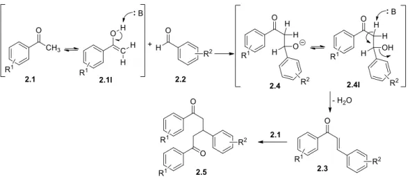
The synthetic mechanism proposed to obtain these compounds was already extensively studied and reported in the literature. It involves nucleophilic attack of the enol form from the ketone 2.1I to an electrodeficient carbon from the carbonyl of the aldehyde 2.2. This attack leads to an intermediate β-hydroxycarbonilic compound 2.4. Finally, dehydration produces the respective conjugated enone 2.3 (Scheme 5). As will be described later, some reactions led to
27
the isolation of structure 2.5, where the α,β-unsaturated carbonyl compound further incorporated another unit of acetophenone 2.1 by Michael additio to the β-carbon atom.
Scheme 5. Synthesis of chalcones 2.3 from substituted acetophenones and benzaldehydes Table 3. Physical and analytical data for chalcones 2.3a-z.
Comp R1 R2 η % m.p. (ºC) Molecular
Formula/Weight 2.3a H 2-OH;3-OCH3;5-Br 99.7 175-177 C16H15BrO3/335.2gmol-1
2.3b ’-Cl 2-OH;3-OCH3;5-Br 65.6 163-165 C16H15BrClO3/369.6gmol-1
2.3c ’-OCH3 2-OH;3-OCH3;5-Br 99.7 148-150 C17H17BrO4/365.2gmol-1
2.3d ’-OCH3 2-OH;5-Cl 86.0 158-159.5 C16H15ClO3/290.1gmol-1
2.3e ’-OCH3 2-OH;5-Br 81.8 162-164 C16H15BrO3/335.2gmol-1
2.3f H 2-OH;3,5-F 75.0 180-181.5 C15H14F2O2/262.1gmol-1
2.3g ’-CH3 2-OH;5-Cl 87.3 179-181 C16H15ClO2/274.1gmol-1
2.3h ’-CH3 2-OH;3-OCH3;5-Br 76.6 192.6-195 C17H17BrO3/349.2gmol-1
2.3i ’-Br 2-OH;5-Br 70.0 171-173 C15H12Br2O2/384.1gmol-1
2.3j ’-Cl 2-OH,3-OCH3 83.8 141.5-143 C16H15ClO3/290.7gmol-1
2.3k ’-CH3 2-OH;5-Br 77.8 191-193 C16H15BrO2/319.2gmol-1
2.3l ’-CH3 2-OH;3,5-F 78.8 202-204 C16H14F2O2/276.3gmol-1
2.3m ’-Br 2-OH;3-OCH3;5-Br 80.3 189-191 C16H14Br2O3/414.1gmol-1
2.3n ’-CH3 5-Br 96.8 127-128.5 C16H15BrO/302.2gmol-1
2.3o H 2-OH 48.4 169-171 C15H14O2/226.3gmol-1
2.3p H 2-OH,3-OCH3 94.0 262-264 C16H16O3/256.3gmol-1
2.3q H 2-OH;5-Br 85.2* - C15H13BrO2/304.2gmol-1
2.3r ’-Cl 2-OH;5-Cl 165-167 C15H12Cl2O2/295.2gmol-1
2.3s ’-Cl 2-OH;3,5-F 63.4* - C15H14ClF2O2/296.7gmol-1
2.3t H 2-OH;5-Cl 87.3* - C15H13ClO2/260.7gmol-1
2.3u ’-Cl 2-OH;5-CH3 43.0* - C16H15ClO2/304.2gmol-1
2.3v ’-Cl 2-OH;5-Br 75.2* - C15H12BrClO2/339.6gmol-1
2.3w ’-OCH3 2-OH;3,5-F 39.2* - C16H14F2O3/292.3gmol-1
2.3x ’-Br 2-OH;3,5-F 38.6* - C15H11BrF2O2/340.1gmol-1
2.3y ’-Cl 2-OH;3-OCH3 10.4 156-158 C16H15ClO3/290.1gmol-1
2.3z ’-F 2-OH;5-Br 96.7* - C15H12BrFO2/323.2gmol-1
28
One of the main aims was to vary the substitution pattern in both aromatic rings. However, many limitations in the optimization of the reaction conditions were observed. Particularly, the reaction of 5-chloro-2-hydroxybenzaldehyde with substituted and non-substituted acetophenone (Table 4) led usually to complex product mixtures in aqueous base and ethanol. Table 4 summarizes the reaction conditions that were used and the corresponding product mixtures identified by 1H NMR.
Table 4. Attempted reactions, experimental conditions and obtained products.
Experiment Reagent 2.2 Reagent 2.1 Reactional conditions Product by 1H NMR
1 3.24mmol R1=Cl (2.95mmol, 0.9eq)
EtOH (10mL) + 3M NaOH (10mL) 1: 40ºC, 4h
2: 60ºC, 12h30
Dark yellow solid
2.3t : 2.5 : 2.2 (1:0.34:0.33) 2 3.52mmol R1=Cl (3.52mmol,
1.0eq)
EtOH (10mL) + 3M NaOH (10mL)
IKA, 300rpm, 50ºC, 4 days Dark oil – complex mixture 3 3.33mmol R1=Cl (3.67mmol, 1.1eq) EtOH (12mL) + 3M NaOH (10mL) rt, 19h Yellow solid 2.3t : 2.5 : 2.2 (1:0.41:0.06) 4 (as 2.3t in Section V) 1.88mmol R1=Cl (2.07mmol, 1.1eq) EtOH (6mL) + 3M NaOH (6mL) rt, 6h Yellow solid η= . % 2.3t and traces of 2.5 5 1.68mmol R1=Cl (1.84mmol, 1.1eq) EtOH (10mL) + 3M NaOH (10mL) rt, 19h
Dark yellow solid
2.3t : 2.5 : 2.2 (1:0.70:0.06) 6 1.37mmol R1=H (1.50mmol, 1.1eq) EtOH (5mL) + 3M NaOH (5mL) rt, 19h Yellow solid 2.3r : 2.5 : 2.2 (1:0.15:0.06) 7 2.05mmol R1=H (2.25mmol, 1.1eq) EtOH (12mL) + 3M NaOH (12mL) rt, 19h
Dark yellow solid 2.3r : 2.5 (1:0.43) 8 2.05mmol R1=H (2.05mmol,
1.0eq)
EtOH (10mL) + 3M NaOH (10mL) rt, 19h
Dark yellow solid 2.3r : 2.5 (1:0.39) 9 2.05mmol R1=H (2.05mmol,
1.0eq)
EtOH (12mL) + 0.5M NaOH (12mL) rt, 16h
Light yellow solid 2.3r : 2.2 (1:0.02) 10 (as 2.3r in Section V) 3.17mmol R1=H (3.49mmol, 1.1eq) EtOH (10mL) + 3M NaOH (10mL) rt, 24h Yellow solid η= . % 2.3r and traces of 2.5 11 2.05mmol R1=H (2.25mmol, 1.1eq) EtOH (14mL) + 3M NaOH (14mL) rt, 5h Yellow solid 2.3r : 2.5 : 2.1 (1:0.06:0.10)
Comparing experiments 3 and 5, in which the experimental conditions were identical in terms of time (19h) and temperature (rt) however, the concentration of the reagents in experiment 5 was approximately half of the concentration used in experiment 3. Regarding the final products, the molar ratio shows that in experiment 5 the proportion of 2.5 is almost
![Figure 1. The Hallmarks of Cancer. Schematic illustration of the acquired capabilities of tumor cells essential for tumor growth and progression (adapted from [5])](https://thumb-eu.123doks.com/thumbv2/123dok_br/17575080.818327/19.892.289.604.708.1007/figure-hallmarks-schematic-illustration-acquired-capabilities-essential-progression.webp)
![Figure 2. Estimated incidence for all types of cancer worldwide (adapted from [3]).](https://thumb-eu.123doks.com/thumbv2/123dok_br/17575080.818327/21.892.152.588.193.411/figure-estimated-incidence-types-cancer-worldwide-adapted.webp)