Dissertação de Mestrado
Mestrado Integrado em Engenharia Biológica
Ramo de Tecnologia Química e Alimentar
Trabalho efetuado sob a orientação da
Professora Doutora Lígia Raquel Marona Rodrigues
Trabalho na empresa efetuado sob a orientação do
Doutor Leonardus Kluskens
janeiro de 2015
Ana Paula Pacheco Marques da Silva
Selection of specific colon cancer tumor
peptides from combinatorial phage display
libraries
Curso: Mestrado Integrado em Engenharia Biológica Ano de conclusão da dissertação: 2015 Área de Especialização: Ramo de Tecnologia Química e Alimentar
Escola de Engenharia, Departamento/Centro: Engenharia Biológica
TÍTULO DA DISSERTAÇÃO/TRABALHO DE PROJETO
Título em PT: Seleção de péptidos específicos para células do cancro do colon através do uso de
bibliotecas de péptidos expressas na superfície de fagos
Título em EN: Selection of specific colon cancer tumor peptides from combinatorial phage display
libraries
Orientador: Doutora Lígia Raquel Marona Rodrigues Coorientador: Doutor Leonardus Kluskens
DE ACORDO COM A LEGISLAÇÃO EM VIGOR, NÃO É PERMITIDA A REPRODUÇÃO DE QUALQUER PARTE DESTA TESE
Universidade do Minho, ____/____/_______
“Learn from yesterday, live for today, hope for tomorrow.
The important thing is to not stop questioning.”
vii
AGRADECIMENTOS
Aos meus orientadores, Professora Doutora Lígia Rodrigues e Doutor Leon Kluskens agradeço primeiramente por me terem dado a incrível oportunidade de trabalhar com este tema, um desafio que se mostrou imensamente gratificante e que me abriu novos horizontes e perspetivas para o futuro. Quero também agradecer toda a disponibilidade e amabilidade que sempre demonstraram ao longo destes meses de trabalho.
Ao Franklin Nóbrega e à Ivone Martins, que me acompanharam durante todo o trabalho, e que se mostraram incansáveis em atender a todas as minhas dúvidas. Sem eles não teria conseguido chegar até aqui, e por isso lhes estou extremamente grata.
Um muito obrigada a todos aqueles que tive o prazer de conhecer e que durante estes meses se tornaram verdadeiros amigos! Débora, obrigada por seres a pessoa que és, por me teres ajudado em todos os momentos e por teres esse coração enorme! João, obrigada por te disponibilizares sempre para me ajudar em tudo o que podias, por teres sempre uma coisa parva para dizer que me fazia rir mesmo nos momentos de maior stress, e por seres uma das melhores (e mais parvas) pessoas que conheci . Mariana, obrigada por seres essa pessoa bondosa, sempre preocupada e sempre com uma palavra amiga para dar. Adelaide, fizeste de mim uma “filha” no laboratório :p. Obrigada por estares sempre lá comigo, por me ajudares a ultrapassar momentos difíceis, e por me apoiares em tudo, mesmo tudo o que precisei. És uma pessoa incrível, a quem devo muito. Ritinha, obrigada por teres sempre um sorriso na cara, por teres sempre um otimismo fantástico e um coração do tamanho do mundo. Obrigada, ao Rui pela amizade, ao Zé, por ter animado os meus dias com os factos mais (des)interessantes ;), e pelas conversas que ajudavam a passar as longas horas de trabalho. Ao Sílvio, por ter sempre alegria para dar (ou cantar :p). Ao Franklin, por tudo.
Aos meus amigos destes 5 anos, que se tornaram a minha família e que vou levar comigo para sempre na minha vida. Vocês, que desde o início me acompanharam, que me ajudaram a crescer e que são o melhor que levo desta Universidade.
Aos meus amigos de sempre, um grande obrigada por serem quem são, com todas as particularidades que nos fazem ser nós. Um obrigada em especial à Dulce Silva, és como uma irmã, que cresceu comigo, viveu comigo e que sempre me ajudou, e que tenho a certeza me vai acompanhar pela vida fora.
viii
Ao Carlos Martins, obrigada por seres quem és e por fazeres parte de quem sou. Por toda a paciência que tiveste comigo durante estes meses e porque és o único que me sabe fazer sorrir, sempre.
Obrigada à minha mãe, por fazeres por mim tudo o que podes, por me fazeres sempre ambicionar ser mais e melhor, por me fazeres acreditar que sou capaz, por me ensinares o valor do esforço. Ao meu pai, que tem o maior coração do mundo, por todos os valores que sempre me transmitiste. Ao meu irmão, por me aturares sempre, nas minhas maluqueiras e nos meus stresses. Um grande obrigada a toda a minha família, que esteve sempre presente e unida durante o meu crescimento e que sempre foi um pilar para mim. Quero agradecer particularmente à minha madrinha e ao meu padrinho, que sempre me apoiaram em tudo, tanto no percurso académico como pessoal!
Por último, o meu maior obrigada ao meu avô Zé, que partiu mas que viverá sempre nas nossas memórias. Que estará sempre presente enquanto a minha família se mantiver unida, enquanto vivermos com base nos seus princípios que atravessaram e, com certeza, continuarão a atravessar gerações. Um grande obrigada, por me teres dado a minha família.
ix
ABSTRACT
Colorectal cancer (CRC), localized to the large intestine and rectum, is the third most commonly diagnosed cancer and the second leading cause of cancer death. At the molecular level, CRC is a heterogeneous disease. This heterogeneity, currently not covered by available screening tests, translates into differences in the disease progression and also in the response to chemotherapeutic agents which proves that there is an undeniable need for improved diagnostic and screening tools capable of distinguish cancer at a molecular level.
The main goal of this thesis was the development of a multifunctional phage-based nanoparticle to detect and report the presence of cancer cells on a given sample through the use of a biosensor. Particularly, M13KE phage particle was genetically modified to accomplish a recombinant product capable of recognizing poorly differentiated colon cancer cells (RKO cell line) and report their presence using bioluminescence. The element of this particle that allowed targeting RKO cells was obtained using phage-displayed random peptide libraries to screen the surface of these cells towards the identification of biomarkers (i.e. membrane proteins) and specific peptide ligands. For that purpose, two types of random peptide libraries (presenting seven linear peptides - Ph.D. 7 and twelve linear peptides - Ph.D. 12) were cloned into the genome of the M13KE filamentous phage. Constructed libraries and a commercial one (Ph.D. C7CTM from New England Biolabs®) were used to perform two methodologies of phage display and biopanning in order to find specific peptide ligands with affinity to RKO cells. The affinity of the phage pools obtained through biopanning was assessed using ELISA methodology. As a result of these experiments, a new specific peptide, CIGNSNTLC, with high affinity towards RKO cells was discovered. In order to confer to the phage the ability to produce a bioluminescent signal, the insertion of NanoLucTM gene into the M13KE genome was also attempted, at the 5’ end of gene VIII in order to obtain 2700 copies of NanoLucTM luciferase coupled to pVIII M13KE major coat protein. The presence of this enzyme on the phage would function as a reporter of the presence of a specific biomarker on a given sample and the amount of signal could be related to the amount of that biomarker.
In summary, the results herein gathered highlight that phage display is a powerful tool to find new peptide ligands capable of targeting cancer cells. In addition, the construction of a phage particle capable of recognizing and report the presence of biomarkers on a given sample will be a major advance on the use of biosensors for cancer diagnosis.
xi
SUMÁRIO
O cancro colorectal, localizado no intestino grosso e no recto, é o terceiro cancro mais diagnosticado e a segunda causa de morte relacionada com doenças oncológicas. O cancro colorectal é uma doença muito heterogénea a nível molecular. Esta heterogeneidade que se traduz em diferenças na progressão da doença e na resposta dos pacientes aos agentes quimioterápicos, não é, atualmente, abrangida pelos testes diagnósticos usados o que prova, inegavelmente, que há uma necessidade de encontrar métodos de diagnóstico mais eficazes e capazes de distinguir esta doença ao nível molecular. O objetivo principal desta tese foi o desenvolvimento de uma nanopartícula fágica multifuncional para detetar e reportar a presença de células cancerígenas numa dada amostra, através do uso de um biossensor. Em particular, foi planeada a modificação do fago M13KE para obter um produto recombinante capaz de reconhecer células pouco diferenciadas do cancro do colon (de uma linhagem celular denominada RKO), e reportar a sua presença através de bioluminescência. O elemento desta partícula fágica que possibilita o reconhecimento das células RKO foi obtido usando bibliotecas de fagos acopladas à cápside da partícula fágica para rastrear a superfície celular destas células e identificar biomarcadores (proteínas membranares) e ligandos específicos. Com esse objetivo, dois tipos de bibliotecas fágicas (com uma sequência peptídica linear de 7 peptidos – Ph.D. 7, e de 12 péptidos – Ph.D. 12) foram geneticamente construídas usando o fago filamentoso M13KE. As bibliotecas construídas, em conjunto com uma terceira biblioteca comercial (Ph.D. C7CTM da New England Biolabs®) foram usadas para proceder a duas metodologias de phage display e biopanning para encontrar ligandos peptídicos específicos com afinidade para as células RKO. A afinidade dos complexos fágicos obtidos através dos ensaios de biopanning foi avaliada através de ensaios de ELISA. Como resultado das atividades experimentais apresentadas descobriu-se uma nova sequência peptídica, CIGNSNTLC, com grande afinidade para as células RKO. Com o objetivo de conferir a capacidade de produzir um sinal luminescente ao fago, tentou-se inserir o gene NanoLucTM no vetor M13KE por forma a conseguir o acoplamento da NanoLuc® luciferase às 2700 cópias da proteína VIII da cápside do fago. Em suma, os resultados desta tese evidenciaram que o phage display é uma ferramenta poderosa para encontrar novos ligandos capazes de reconhecer células cancerígenas. Adicionalmente, a construção de uma partícula fágica capaz de reconhecer e reportar a presença de biomarcadores numa dada amostra será um grande avanço no uso de biosensores para o diagnóstico de cancro.
xiii
TABLE OF CONTENTS
AGRADECIMENTOS ... vii
ABSTRACT ... ix
SUMÁRIO ... xi
LIST OF FIGURES ... xvii
CHAPTER 2:STATE OF THE ART ... xvii
CHAPTER 3:MATERIALS AND METHODS ... xvii
CHAPTER 4:RESULTS AND DISCUSSION ... xviii
LIST OF TABLES ... xxi
CHAPTER 2:STATE OF THE ART ... xxi
CHAPTER 3:MATERIALS AND METHODS ... xxi
CHAPTER 4:RESULTS AND DISCUSSION ... xxii
LIST OF ABREVIATIONS ... xxiii
CHAPTER 1: MOTIVATION AND OBJECTIVES………..1
1. Motivation ... 1
2. Objectives ... 2
CHAPTER 2: STATE OF THE ART………..3
1. Colon cancer ... 3
2. Cancer Diagnosis –The Biosensors Era ... 5
2.1. Biomarkers ... 6
2.1.1. Strategies for the discovery of novel cancer biomarkers ... 7
xiv
2.1.3. Proteins as cancer biomarkers - oncoproteomics ... 9
2.2. Non-invasive biomarkers for the Early Detection of CRC... 10
2.3. Molecular recognizing elements ... 13
3. Specific cancer cells ligands identification by Phage display technology ... 15
3.1. Filamentous phages for phage display: phage structure and propagation ... 16
3.2. Phage display random peptide libraries ... 19
3.3. Selection of ligand-receptors in complex biological systems - Biopanning ... 20
4. Reporter Phages ... 24
CHAPTER 3: MATERIALS AND METHODS………..………27
1. Phage Display and in vitro biopanning ... 27
1.1. Colon Cancer Cells ... 27
1.1.1. cell culture initiation and subculture methodologies ... 27
1.1.2. Cell counting and cell viability assessment ... 29
1.2. M13KE phage ... 30
1.2.1. Bacterial cell culture ... 31
1.2.2. Phage production and isolation ... 32
1.2.3. Drop and Titer assays ... 33
1.3. Random peptide libraries construction ... 34
1.3.1. M13KE double-stranded dna extraction ... 35
1.3.2. M13KE size confirmation ... 35
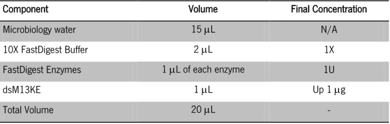
1.3.3. M13KE double digestion ... 36
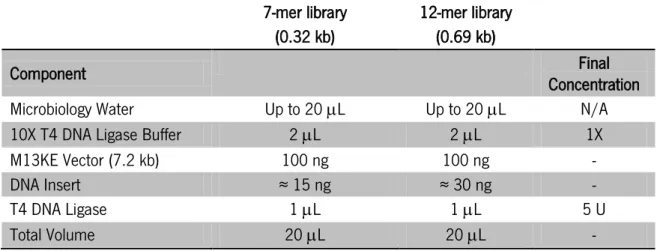
1.3.4. Inserts construction ... 38
1.3.5. Ethanol precipitation of nucleic acids ... 39
xv
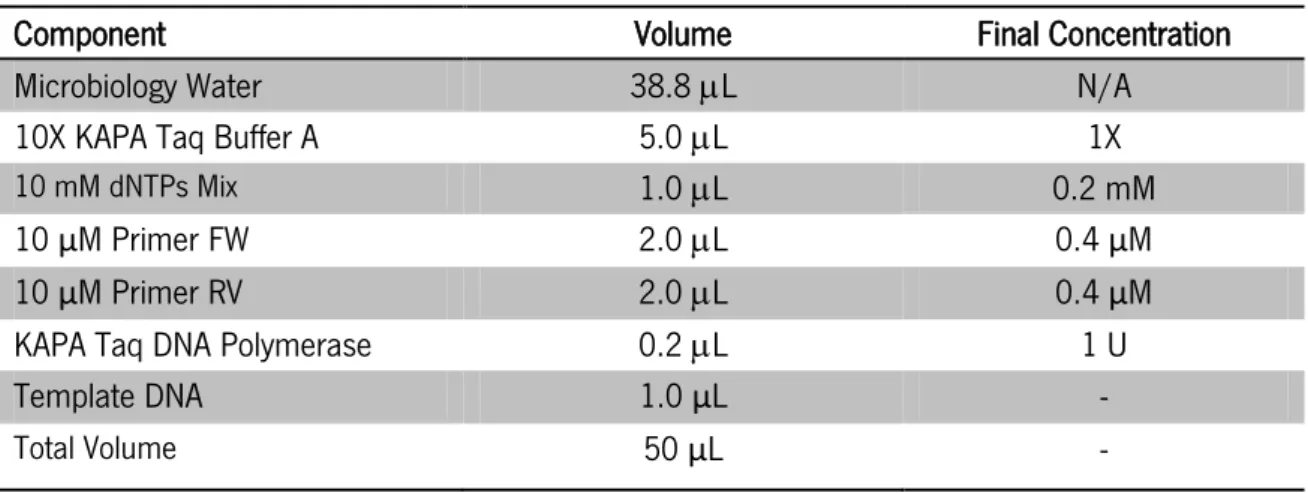
1.3.7. Cloning confirmation by PCR amplification ... 41
1.3.8. Agarose gel electrophoresis... 42
1.3.9. Chemical competent cells preparation and transformation ... 43
1.4. In vitro biopanning ... 44
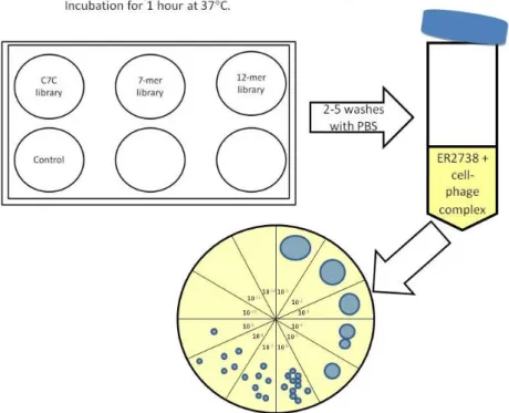
1.4.1. Conventional Panning ... 44
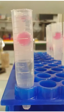
1.4.2. BRASIL biopanning ... 46
1.4.3. ELISA ... 47
1.4.4. Ligand sequencing: samples preparation ... 50
2. NanoLucTM gene insertion on M13KE ... 53
2.1. NanoLucTM gene amplification ... 54
2.2. NanoLucTM gene clean-up ... 55
2.3. M13KE and NanoLucTM double digestion ... 56
2.4. DNA Dephosphorylation ... 57
2.5. M13KE DNA::NanoLucTM Ligation ... 57
2.6. Bacteria chemical competent cells transformation ... 58
2.7. DNA extraction and cloning confirmation ... 58
CHAPTER 4: RESULTS AND DISCUSSION………..61
1. Random peptide libraries construction ... 61
1.1. M13KE vector and inserts construction ... 62
1.2. M13KE::insert(s) ligation – PCR confirmation ... 64
1.3. Bacterial chemically competent cells transformation ... 66
1.4. Libraries Amplification ... 67
2. In vitro biopanning experiments ... 68
xvi
2.2. BRASIL biopanning ... 70
2.3. ELISA ... 72
2.4. Clones sequencing ... 79
3. NanoLucTM insertion on M13KE ... 82
CHAPTER 5: CONCLUSIONS AND FUTURE PERSPECTIVES………..…...87
1. Conclusions ... 87
2. Future Perspectives... 91
REFERENCES………..…93
APPENDIXES…………..………..103
xvii
LIST
OF
FIGURES
C
HAPTER2:
S
TATE OF THE ART
Fig. 1 – The biology of cancer: tumorigenesis process. ... 4 Fig. 2 – Schematic representation of a single element biosensor containing a molecular recognizing element, a transducer and the physical output whose magnitude is related with the biomarker concentration. ... 6 Fig. 3 – Outline of strategies for biomarker discovery through utilization of emerging technologies. Abbreviation: MS, mass spectrometry [taken from: (Kulasingam & Diamandis 2008)]. ... 8 Fig. 4 – Phage Display expression system (M13 phage): the ligand-encoding oligonucleotide inserted next to gene III that encodes a coat protein of the phage generates a recombinant phage, presented on the right, with the ligand displayed coupled to the respective coat protein [taken from: (Bakhshinejad et al. 2014)]. ... 16 Fig. 5 – The general structure of M13 phage particle. ... 17 Fig. 6 – M13 particles life cycle [taken from: (Wilson & Walker 2010)]. ... 18 Fig. 7 – Schematic diagram of how foreign peptide domains are fused to coat proteins pIII in phage-display vectors [adapted from: (Smith & Petrenko 1997)]. ... 20 Fig. 8 – Schematic representation of the screening of a phage peptide library through biopanning and cloning analysis [taken from: (Huang et al. 2011)]. ... 21 Fig. 9 – Schematic representation of a BRASIL selection round [adapted from: (Giordano et al. 2001)]. ... 23
C
HAPTER3:
M
ATERIALS ANDM
ETHODSFig. 10 – RKO cell line, passage 13 at ≈ 60% confluence (Scale bar 200 m). ... 27 Fig. 11 – Trypan blue exclusion test – the cells that present dark coloration are excluded i.e. not counted. ... 30 Fig. 12 – Schematic representation of the titer assay. ... 34
xviii
Fig. 13 – M13KE DNA restriction map of Acc65I and EagI restriction enzymes. Schematic representations of M13KE double digestion with Acc65I and EagI; the excluded fragment has a length of 20 bp. ... 37 Fig. 14 – Excision of M13KE genome map. In the boxes it is presented the Ph.D. Primer FW and RV used for PCR cloning confirmation. The zone in color corresponds to the cloning site for the construction of Ph.D. libraries 7 and 12. ... 41 Fig. 15 – Schematic representation of a conventional biopanning round. ... 45 Fig. 16 – BRASIL tube containing the organic phase of dibutyl phthalate:cyclohexane 9:1 (v:v) and the non-organic phase of 500 μL of DMEM + 1% BSA. ... 47 Fig. 17 – Collection of phage particles by polyethylene glycol (PEG) precipitation [taken from: (Brown 2010)]. ... 48 Fig. 18 – Schematic representation of phage ELISA. RKO cells incubated with DMEM + 5mg/mL of BSA and 0.01% of PBS + 0.1% EDTA - RKO cells ( ), BSA ( ) [1]; RKO cells incubated with phage pool [2]; RKO-phage complexes incubated with HRP/Anti-M13 Monoclonal Conjugate – RKO cell specific peptide [3]; RKO-phage complexes incubated with HRP/Anti-M13 Monoclonal Conjugate and OPD substrate [4] and quantitative analysis performed through absorbance measurements [5]. ... 50 Fig. 19 – pNL1.1 [Nluc] Vector [Promega, N1001]. ... 53 Fig. 20 – Detection of 50 attomoles of purified enzyme; Reagents: NanoLuc™/Nano-Glo™, Firefly/ONE-Glo™ Renilla/Renilla-Glo™ [taken from: (Binkowski 2012)]. ... 53 Fig. 21 – M13KE DNA restriction map of SnaBI and BspHI restriction enzymes. Schematic representations of M13KE double digestion with SnaBI and BspHI; the excluded fragment has a length of 29 bp. ... 56 Fig. 22 – Excision of M13KE genome map. In the boxes it is presented the Ph.D. Primer FW and RV used for PCR cloning confirmation. The zone in color corresponds to the cloning site for the construction of insertion of NanoLucTM gene. ... 59
C
HAPTER4:
R
ESULTS ANDD
ISCUSSIONFig. 23 – M13KE vector and inserts construction. M13KE digestion with HindIII (a), M13KE digestion with HindIII and double digestion with Acc65I and EagI (b) and 7-mer and 12-mer
xix encoding fragments before and after double digestion with Acc65I and EagI (c), on 1% agarose gels. a) NZYTech ladder III (1) and M13KE linear vector digested with HindIII (2); b) NZYTech ladder III (1), M13KE linear vector digested with HindIII (2) and M13KE double digested vector with Acc65I and EagI (3); c) NZYTech ladder VI (1), 7-mer encoding sequence non-digested insert (2), 7-mer encoding sequence insert digested with Acc65I and EagI (3), 12-mer encoding sequence non-digested insert (4), 12-mer encoding sequence insert digested with restriction enzymes Acc65I and EagI (5). ... 62 Fig. 24 – Gel electrophoresis results of 7-mer and 12-mer encoding sequences insertion into M13KE vector amplified using PCR, on 1% agarose and SGTB gel. NZYTech ladder VI (1); amplified DNA fragment of M13KE:: 7-mer insert, ligation conditions: 2 hours at 22 C (2); amplified DNA fragment of M13KE:: 12-mer insert, ligation conditions: 2 hours at 22 C (3); amplified DNA fragment of M13KE:: 7-mer insert, ligation conditions: overnight at 16 C (4); amplified DNA fragment of M13KE:: 12-mer insert, ligation conditions: overnight at 16 C (5); amplified DNA fragment of M13KE without insert, ligation conditions: 2 hours at 22 C (6); amplified DNA fragment of M13KE without insert, ligation conditions: overnight at 16 C (7). ... 65 Fig. 25 – Outgrowth SOC plate containing X-Gal/IPTG with ER2738 chemically competent cells transformed with M13KE::7-mer library. ... 66 Fig. 26 – Libraries amplification and titer. a) Ph.D. 7 and b) Ph.D. 12 phage PFUs plate with 8 drops of successive dilutions of the amplified phage libraries. ... 67 Fig. 27 – Initial phage input and final phage recover concentration, from conventional biopanning rounds, assessed by plaque-counting for a) Ph.D. C7CTM, b) Ph.D. 7 and c) Ph.D. 12 libraries. Round 1 ( ); Round 2 ( ); Round 3 ( ); Round 4( ). ... 69 Fig. 28 – Binding efficiency of the conventional biopanning phage pools for the four rounds of affinity selection with Ph.D. C7CTM ( ); Ph.D. 7 ( ) and Ph.D. 12 ( ) libraries. ... 70 Fig. 29 – Initial phage input and final phage recover concentration, from BRASIL biopanning rounds, assessed by plaque-counting for a) Ph.D. C7CTM, b) Ph.D. 7 and c) Ph.D. 12 (c) libraries. ... 71 Fig. 30 – Binding efficiency of the BRASIL biopanning phage pools for the four rounds of affinity selection with Ph.D. C7CTM ( ); Ph.D. 7 ( ) and Ph.D. 12 ( ) libraries. ... 72 Fig. 31 – ELISA results for amplified phage pools obtained from four rounds of conventional biopanning with a) Ph.D. C7CTM, b) Ph.D. 7 and c) Ph.D. 12 libraries. Mean abs raw values with standard deviation ( ); background signal ( ); specific signal of the phage pool ( ). ... 74
xx
Fig. 32 – ELISA results for amplified phage pools obtained from four rounds of adapted BRASIL biopanning with a) Ph.D. C7CTM, b) Ph.D. 7 and c) Ph.D. 12 libraries. Mean abs raw values with standard deviation ( ); background signal ( ); specific signal of the phage pool ( ). ... 75 Fig. 33 – Normalized affinity rate of amplified phage libraries for the four rounds of selection with a) conventional biopanning and b) adapted BRASIL biopanning for Ph.D. C7CTM ( ), Ph.D. 7 ( ) and Ph.D. 12( ) libraries. ... 76 Fig. 34 – Comparison of binding affinity of the three Ph.D. libraries for the fourth round of conventional biopanning ( ) and adapted BRASIL biopanning ( ) using normalized values of affinity rates from the amplified phage pools retrieved from the fourth selection round. ... 77 Fig. 35 – Comparison of binding affinity for convention biopanning and adapted BRASIL biopanning for Ph.D. C7CTM ( ), Ph.D. 7 ( ) and Ph.D. 12 ( ) libraries using normalized values of affinity rates from the amplified phage pools retrieved from the fourth selection round. ... 78 Fig. 36 – Gel electrophoresis results of NanoLucTM gene amplification through PCR on a 1% agarose gel: NZYTech ladder III (1); amplified NanoLucTM gene (2-5). ... 83 Fig. 37 – Gel electrophoresis results of M13KE digestion with HindIII and double digestion with SnabI and BspHI and amplified NanoLucTM on a 1% agarose gel: NZYTech ladder III (1); amplified NanoLucTM gene (2-5). NZYTech ladder III (1); linear M13KE digested with HindIII (2); linear M13KE double digested with SnabI and BspHI (3); amplified NanoLucTM gene samples (4,5). ... 84 Fig. 38 – Gel electrophoresis results cloning confirmation using PCR amplification on a 1% agarose and SGTB gel: NZYTech ladder III (1); amplified fragments from the picked clones (2-5); amplified fragments from M13KE control (6). ... 85
xxi
LIST
OF
TABLES
C
HAPTER2:
S
TATE OF THE ARTTable 1 – Non-invasive molecular markers for the detection of CRC [adapted from: (Kim et al. 2008; Tanaka et al. 2010)] ... 12
C
HAPTER3:
M
ATERIALS ANDM
ETHODSTable 2 – Volumes of complete growth medium and Trypsin/EDTA solution; number of cells at 80% - 90% confluency for the correspondent culture vessels used to RKO cells culture ... 28 Table 3 - Standard mixture for digestion of dsM13KE samples ... 36 Table 4 – Standard mixture for double digestion of dsM13KE samples with Acc65I and EagI from Thermo Scientific ... 37 Table 5 – Primer’s sequence and melting temperature used for libraries construction. The melting temperatures above were calculated using Modified Breslauer's thermodynamics, dH and dS parameters. The underlined zones on the reverse primers are the restriction sites for EagI and Acc65I, respectively. Bold zones are the overlapping sequences ... 38 Table 6 – Extension reaction mixture for the random inserts construction ... 39 Table 7 – Ligation reaction mixture for M13KE::7-mer and M13KE::12-mer libraries construction ... 40 Table 8 – Primers used for insertion confirmation on pIII gene. PhD.FW – Primer Forward; PhD.RV – Primer Reverse. The melting temperatures were calculated using Modified Breslauer's thermodynamics, dH and dS parameters ... 41 Table 9 – PCR standard reaction mixture with KAPA Taq DNA Polymerase ... 42 Table 10 – PCR cycling protocol to amplify M13KE pIII cloning site ... 42 Table 11 – Primers used to amplify the cloning region. Ph.D.sequencing.FW – Primer Forward; Ph.D.RV – Primer Reverse. The melting temperatures were calculated using Modified Breslauer's thermodynamics, dH and dS parameters ... 51 Table 12 – PCR mixture used to amplify sequencing templates ... 52
xxii
Table 13 – Primers used to amplify NanoLucTM gene from pNL1.1. NlucM13.FW – Primer Forward with the restriction site for SnaBI (underlined); NlucM13.RV – Primer Reverse with the restriction site for BspHI (underlined). The melting temperatures were calculated using Modified Breslauer's thermodynamics, dH and dS parameters ... 54 Table 14 – PCR mixture used to amplify NanoLucTM gene ... 55 Table 15 – PCR cycling protocol for NanoLucTM amplification with DNA polymerase KAPA HiFi.. 55 Table 16 – Standard mix for M13KE vector and NanoLucTM gene double digestion with SnaBI and BspHI from New England Biolabs®... 57 Table 17 – Ligation reaction mixture for insertion of NanoLucTM gene on M13KE vector ... 58 Table 18 – Primers used to confirm the insertion on pVIII gene. M13KE_pVIII.FW – Primer Forward; NlucM13.RV – Primer Reverse. The melting temperatures were calculated using Modified Breslauer's thermodynamics, dH and dS parameters ... 59
C
HAPTER4:
R
ESULTS ANDD
ISCUSSIONTable 19 – Sequencing results of 12 random selected clones after 4 rounds of selection with RKO colon cancer cell line (Acc65I restriction site (GGTACC); EagI restriction site (CGGCCG); bold zone corresponds to the peptide codifying sequence) ... 80
xxiii
LIST
OF
ABREVIATIONS
2D-PAGE Two Dimension Polyacrylamide Gel Electrophoresis
abs Absorbance
ACF Aberrant Crypt Foci
APC Adenomatous Polyposis Coli ATCC American Type Culture Collection
ATP Adenosine triphosphate
BRASIL Biopanning and rapid analysis of selective interactive ligands
BSA Bovine serum albumin
CA 19.9 Carbohydrate Antigen 19-9
cDNA Complementary DNA
CEA Carcinoembryonic antigen
CRC Colorectal Cancer
DLA Double Layer Agar
DMEM Dulbecco’s Modified Eagle’s medium DMSO Dimethyl sulfoxide
DNA Deoxyribonucleic acid
dNTPs Deoxy nucleoside triphosphates dsDNA Double-stranded DNA
dsM13KE Double-stranded M13KE E. coli Escherichia coli
EDTA Ethylenediamine tetraacetic acid ELISA Enzyme-Linked Imunnosorbent Assay
EU European Union
FBS Fetal Bovine Serum
FDA Food and Drug Administration FOBT Faecal Occult Blood Test
FS Flexible Sigmoidoscopy
FW Forward
GEP Gene Expression Profile
gFOBT Guaiac-based faecal occult blood test HER-2/neu Human Epidermal growth factor Receptor 2
HRP Horseradish peroxidase
ICAM- 1 Intercellular Adhesion Molecule 1 ICAT Isotope-coded Affinity Tag
iFOBT Immunochemical faecal occult blood test IPTG Isopropyl β-D-1-thiogalactopyranoside
iTRAQ isobaric Tags for Relative and Absolute Quantitation lacZα lacZ alpha fragment
lacZω lacZ omega fragment
xxiv
L-DNA Long form – DNA
LRP Luciferase Reporter Phages
-mer Referent to the number of amino acids
MMR Mismatch Repair System
mRNA Messenger Ribonucleic Acid
MS Mass Spectrometry
MSI Microsatellite Instability
MudPIT Multidimensional Protein Identification Technology OPD o-phenylenediamine dihydrochloride
PBS Phosphate buffered saline PCR Polymerase chain reaction
PEG Polyethylene Glycol
PES Polyethersulfone
pfu Plaque-forming units
Ph.D. Phage Display
RF Replicative Form
RV Reverse
SELDI-ToF Surface-enhanced laser desorption/ionization-Time of flight
SM Sodium - Magnesium
SOC Super Optimal Broth with Catabolite repression
TAE Tris-acetate-EDTA
TBST Tris-Buffered Saline and Tween 20
TE Tris-EDTA
TIMP-1 TIMP metallopeptidase inhibitor 1 TNM Tumor, Nodes, Metastasis TSS Transformation - Storage Solution
UK United Kingdom
USA United States of America wtM13KE Wild-type M13KE
1
1. M
OTIVATION
Colon and rectal cancer have several similar features and are most often discussed together as CRC – colorectal cancer. There are approximately 1,000,000 new cases of CRC and 500,000 deaths associated with it each year. In fact, CRC represents one of the primary causes of cancer deaths in EU and the USA. Mortality rates for CRC have declined over the past two decades. Data from 2006 to 2010 indicates that this rate declined by 2.5% per year in men and by 3.0% per year in women. This decrease reflects both the declining incidence rates and improvements in early detection - meaning screening programs for population over 50 years - and treatment (American Cancer Society 2014). However, the available screening and diagnostic tests frequently lack in sensitivity for early diagnosis and often encompass a high economic burden that limits their use for screening general population worldwide.
Recent advances on genomics and proteomics have proven that CRC is a very heterogeneous disease at molecular level which translates into differences in disease progression, survival and response to chemotherapeutic agents (Newton et al. 2011; Marisa et al. 2013). The current screening methods for this disease overlook these important molecular features, meaning that many patients are misdiagnosed and that the treatment options chosen may not be the best for a particular case.
The emerging molecular methods being used to study cancer are leading to a better understanding of the disease, as well as to the discovery of potential new genomic and proteomic biomarkers which are the basis to develop biosensor platforms that can overcome the challenges in cancer diagnosis and early detection (Tothill 2009). Their principal components are a sensing platform that interacts with an analyte i.e. biomarker and a signal processor that transduces the binding impulse into a measurable signal. Biosensors integrated with bioselective bacteriophage represent a pioneering approach of this methodology, as recombinant phage with displayed peptides provides a source of high quality detection agents. In addition, it is already demonstrated that phage display libraries contain many potential probes for various types of biomolecules, including surface markers of cells and blood components (Petrenko 2008).
2
2. O
BJECTIVES
This thesis is part of a much bigger project ongoing at Inception –LifeSciences Research and Development, a start-up company of the University of Minho, which global aim is the development of a virus-based kit that will enable the detection of all types of cancer on a fast, specific and reliable way, through the use of virus bioluminescence. The goal is to develop a new biosensor platform using luminescent virus as sensing interface that report the presence, or absence, of a specific cancer biomarker on a complex sample such as serum, blood, and urine or biopsy tissue.
In particular, the aim of this thesis is the development of a multifunctional recombinant phage particle using M13KE filamentous phage as a template. At the end, it is expected to have a genetically engineered M13KE displaying a specific peptide to RKO colon cancer cell line coupled to pIII minor coat protein and also NanoLucTM luciferase coupled to pVIII major coat protein. The resulting phage-based nanoparticle will enable the detection of RKO cancer cells on a given sample giving a bioluminescent signal in response to their presence.
The search of the RKO-specific peptide will be performed using phage display and biopanning technologies. For those, two types of libraries containing a variety of phage-displayed peptides will be constructed by the insertion of random DNA sequences into the N-terminus of pIII gene. These two phage display libraries encompass a diversity of phage particles displaying different peptide sequences with 7 and 12 amino acids (Ph.D. 7 and Ph.D. 12, respectively) and it will be used, as well as a third commercial one (Ph.D. C7CTM from New England Biolabs) to screen the surface of RKO cells in order to discover one or more peptides with high affinity towards those cells. The screening of RKO surface will be performed using two phage display methodologies, conventional and BRASIL. The affinity of phage pools retrieved from biopanning rounds will be assessed using ELISA methodology. Finally, some clones randomly picked from the phage pools that present more affinity will be sequenced to obtain the specific peptide encoding sequences.
At the same time NanoLucTM gene will be inserted at the N-terminus of pVIII gene in order to confer luminescence to M13KE. When accomplished, the resulting recombinant phage particle will present 2700 copies of NanoLucTM luciferase, an enzyme capable of generating bioluminescent light through a chemical reaction with oxygen and a substrate.
3
1. C
OLON CANCER
Colorectal carcinoma (CRC) arises from the epithelial cell on the internal layer of the large intestine and it is caused by an accumulation of genetic and epigenetic changes that affect tumor suppressor genes as well as oncogenes (85% of CRC cases), or mismatch repair genes (MMR, in 15% of the cases), over several years (Fearon and Vogelstein 1990, Deschoolmeester et al. 2010). Those alterations can lead to changes in gene function which influences protein expression, structure and activity causing abnormal behavior on epithelial cells like altered metabolism, proliferation and apoptosis that are characteristic in tumor cells (Bendardaf et al. 2004). The first recognizable manifestation of epithelial alteration during colorectal tumor development occurs primarily on the crypts (invaginations of the mucosa), and is known as Aberrant Crypt Foci (ACF). Accumulation of these abnormal cells leads to the formation of adenomatous polyps – adenoma (Fig. 1 a) (Alberici 2007).
About 95% of CRC cases develop from adenomas, but only approximately 5% of adenomas grow into tumors (Shinya & Wolff 1979). When an adenoma progresses to a tumor mass, a significant number of undifferentiated cells appear – dysplasia (Fig. 1 b) (Fearon & Vogelstein 1990), with a marked pleomorphism (cell differentiation in size and shape) and an atypical nucleus size (nuclear:cytoplasm ratio close to 1) – in situ carcinoma (Fig. 1 c). When the mass grows substantially it can acquire the capacity to infiltrate and even destroy the closest surrounding tissues and finally to migrate to distant organs like the liver (metastatic capacity) – invasive carcinoma (Fig. 1 d) (Armaghany et al. 2012).
4
Fig. 1 – The biology of cancer: tumorigenesis process.
[taken from: http://www.ndhealthfacts.org/wiki/Oncology_%28Cancer%29 on November 10t h, 2014]
The classification of CRCs, as many others, still relies on histological studies based on tumor characteristics such as differentiation status and tumor staging like, depth or size of the tumor (T), involvement of regional lymph nodes (N) and occurrence, or not, of metastasis (M). This is called the TNM staging system and it is universally used for survival prediction, treatment selection (need for radiotherapy, chemotherapy, surgical resection, among others), patient sorting for clinical trials, accurate communication among healthcare providers and also to provide uniformity on treatment options and cancer management (Ludwig & Weinstein 2005; Lin et al. 2011). The TNM classification system includes conventional prognostic factors for patient survival. Often patients at the same TNM stage for CRC have very different disease-related outcomes. For some, surgical resection of the primary tumor leads to complete recovery, while for others the metastasis and recurrence events occur even with adjuvant treatment. This shows that CRC is a very heterogeneous disease even at the same TNM stage and therefore it becomes crucial to understand the molecular processes that distinguish potential cancer subtypes. The discovery of serum and cell biomarkers that can serve to a more accurate prognosis can personalize the treatment and turn it much more effective (Duffy & Crown 2008; Walther et al. 2009).
Over the last two decades, a whole range of new technologies have been introduced in clinical practice to diagnose and treat the disease, with therapeutic modalities extending to advanced stages of the disease (Deschoolmeester et al. 2010). However, preventionremains the key to reduce morbidity and mortality since it is well established that the stage of the disease at diagnosis greatly impacts colon cancer survival rates. There are two types of screening and diagnosis techniques currently in use in the majority of EU countries, UK and USA: stool tests
5 (FOBT – Faecal Occult Blood Tests) and endoscopic examinations, such as flexible sigmoidoscopy (FS) and colonoscopy.
Guaiac-based faecal occult blood test (gFOBT) is, at the time, the most frequently used method in screening programs. It is a simple, inexpensive and non-invasive approach that has proven its value (Zavoral et al. 2009). With its use, a decrease in mortality rates for CRC by 15 to 33% has been verified. However, gFOBT presents relatively high false negative and false positive rates, and it has poor sensitivity for the detection of early-stage lesions (García-Bilbao et al. 2012; Burch et al. 2007). In an attempt to improve on the false positive rates of gFOBT, a new Faecal Immunochemical testing (iFOBT) has been developed. It has slightly superior performance characteristics but this comes with greatly increased financial costs which caused the failure of iFOBT implementation as a screening test for general population (García-Bilbao et al. 2012; Zavoral et al. 2009; Newton et al. 2011). On the other hand, endoscopic examinations, used many times when abnormalities appear on gFOBT results, offers significant improvements in detection rates for CRC but it also has important disadvantages associated such as high economic burden and potential major complications like bleeding or perforation (García-Bilbao et al. 2012; Winawer et al. 2003).
All this emphasizes the imperative need of new diagnostic approaches to improve the outcome of CRC screening programs. Particularly, there is a clinical need for identifying specific biomarkers for early detection of CRC, as the risk of recurrence and subsequent death due to CRC is intimately related to the stage of the disease at the time of the diagnosis (Kim et al. 2008).
2. C
ANCER D
IAGNOSIS
–T
HE
BIOSENSORS
E
RA
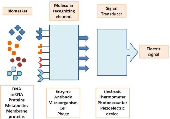
The new strategies for cancer diagnosis at molecular level involve the development of biosensors for biomarker analysis. A biosensor (illustrated on Fig. 2) is, in a traditional sense, a bioanalytical device that incorporates a biological material, which can be microorganisms, enzymes, antibodies, among others, and have a physicochemical transducer, which may be optical, electrochemical, thermometric, piezoelectric or magnetic. The objective of using a biosensor is the production of either discrete or continuous digital electronic signals that can translate qualitative and/or quantitative information about the analyte or group of analytes (Tothill 2009; Soper et al. 2006).
6
Fig. 2 – Schematic representation of a single element biosensor containing a molecular recognizing element, a transducer and the physical output whose magnitude is related with the biomarker concentration.
2.1.
B
IOMARKERSThe discovery of reliable biomarkers that can give trustworthy information about the presence of a tumor, but also the stage of tumorigenesis is the first step to the design of a biosensor (Soper et al. 2006). Biomarkers can be defined as measurable substances that potentially indicate a biological or pathological state. Tumor biomarkers, in particular, can be used to detect the presence of a tumor mass, predict prognosis and the response to a therapeutic agent. In order to have clinical relevance, a biomarker should lead to an improvement in life expectancy or quality of life (Newton et al. 2011; Ludwig & Weinstein 2005; Duffy & Crown 2008). Cancer biomarkers can be DNA, mRNA, proteins, metabolites, whole cells, and even processes like apoptosis, angiogenesis and cell proliferation (Kulasingam & Diamandis 2008). These can be produced by the tumor mass or by other tissues in response to the presence of the foreign mass or associated conditions such as inflammation. Such biomarkers can be found in body fluids like blood, serum or urine, on tissue and cell lines (Even-Desrumeaux et al. 2011). Tumor markers can be divided in some groups according with their specifications and
7 applications. For instance, there are diagnostic, prognostic and predictive biomarkers (Kulasingam & Diamandis 2008). A diagnostic marker is a marker that is used to detect and identify a certain type of cancer in any individual. These kind of markers are expected to have high specificity and sensitivity meaning that their presence on a given sample indicates the existence of cancer, or that cancer will occur with nearly a 100% certainty within a specified time interval (Srivastava et al. 2001). Several criteria must be met before biomarkers can be approved as markers for early detection involving the quality of biomarkers and also the assays for their measurement: (a) the biomarker must be expressed in a different manner in normal, premalignant or high-risk, and tumor tissue; (b) the marker and its assay must provide acceptable predictive accuracy for risk or for the presence of cancer; and (c) the variation of the detection tests and the intra- and inter-laboratory variance must be known (Srivastava et al. 2001). Prognostic biomarkers are expected to predict a likely course of the disease since their status is already established including the probability of recurrence. Hence, a prognostic marker can aid the definition of the therapy to be used (Bendardaf et al. 2004). At last, predictive markers are those that can predict the response to a drug treatment before its application. They allow distinguishing individuals as “responders” and “no responders” to a given treatment (Newton et al. 2011). These biomarkers mainly arise from array-type experiments that make it possible to predict the clinical outcome from the molecular characteristics of a patient’s tumor (Kulasingam & Diamandis 2008).
An ideal cancer biomarker should be measured easily, reliably and cost-effectively by the use of an assay with high analytical sensitivity and specificity (Even-Desrumeaux et al. 2011). Currently, a central concern on the clinical use of biomarkers is that, generally, they lack diagnostic specificity and sensitivity and so they are used as a complement of imaging, biopsy and associated clinic pathological information before a clinical decision is made (Kulasingam & Diamandis 2008).
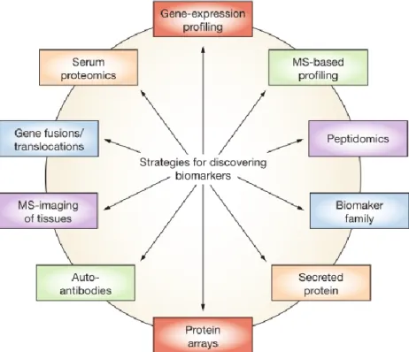
2.1.1. STRATEGIES FOR THE DIS COVE RY OF NOVEL C ANCER BIOMARKERS
The last decade has been marked with an impressive growth in the field of large-scale and high-throughput biology, which contributed to an era of new technological development. The accomplishment of a number of genome-sequencing projects, the discovery of oncogenes and tumor-suppressor genes, and recent advances in genomic and proteomic technologies, jointly with potent bioinformatics tools, are now having a great impact on the cancer biomarkers
8
research field (Kulasingam & Diamandis 2008). Modern technologies enable performing parallel rather than serial analysis, therefore being able to discriminate different patterns and multiple markers simultaneously. Fig. 3 summarizes a number of new strategies that have been using emergent technologies towards tumor markers research.
Fig. 3 – Outline of strategies for biomarker discovery through utilization of emerging technologies. Abbreviation: MS, mass spectrometry [taken from: (Kulasingam & Diamandis 2008)].
2.1.2. DNA AND RNA AS CANCE R BIOMARKERS – GE NE-E XPRESSION PROFILING
On the latest years a new type of tumor classification hypothesis arose. This classification is based on genomic microarrays, a powerful technology for gene-expression studies and it states on the fact that gene-expression patterns identified with DNA microarrays can predict the clinical behavior of tumors (Mohr 2002; Marisa et al. 2013). This method can provide complete gene expression profiles of tumor and normal cells. Comparison of normal and tumor profiles of gene expression (GEPs) can lead to the discovery of different levels of expression of one or more genes, thus indicating a possible DNA or mRNA biomarker (Mohr 2002).
In spite of the positive results of this technology for cancer sub-classification, there are only two multigene panel tests approved by FDA (US Food and Drug Administration), both of them to predict the recurrence of breast cancer (Kulasingam & Diamandis 2008). For CRC,
9 among all molecular markers that have been extensively studied for its characterization and prognosis, microsatellite instability (MSI), caused by defects on the Mismatch Repair System of DNA (MMR), is the only marker that was found to be a significant prognostic factor in early CRC (Marisa et al. 2013). Microarray technology has been used in the recent years to investigate GEPs in CRC, but they are found to be poorly reproducible, possibly because CRC can be developed through multiple pathways being composed of distinct molecular entities. In fact, specific GEP studies, including genetic and epigenetic analysis, have identified at least three distinct molecular subtypes of colon cancer (Shen et al. 2007), proving again that this should no longer be considered an homogenous disease.
2.1.3. PROTEINS AS CANCE R BIOMARKE RS - ONCOPROTE OMICS
Gene expression data gives limited information about the biological processes occurring within a cell since proteins are the main functional units performing all of them. Also, it is known that mRNA suffers post-transcriptional events and proteins, post-translational modifications. Therefore, the direct analysis of proteins, using proteomics has several advantages regardless of requiring more tissue and being more time-consuming (Even-Desrumeaux et al. 2011; Wulfkuhle et al. 2003). For this reason, the use of proteomic patterns for cancer diagnosis and tumor sub-classification seems promising (Kulasingam & Diamandis 2008). The concept of oncoproteomics arises as the study of proteins and their interactions in cancer cells using proteomic technologies. Cells and tissue phenotype is ultimately dependent on which proteins are being expressed and how much of it is being produced at a given time. Hence, all alterations that occur at the cell level during carcinoma process can be monitored evaluating cell protein profiles both qualitatively and quantitatively (Cho 2007). Protein signatures in cancer provide helpful details that may enable more effective diagnosis, prognosis, and response to therapy information.
Several proteomics technologies including 2D-PAGE, Mass Spectrometry (MS), surface enhanced laser desorption/ionization time of flight (SELDI-ToF) (Wulfkuhle et al. 2003), protein arrays and mass-spectrometry techniques such as isotope coded affinity tags (ICAT), iTRAQ and multidimensional protein identification technology (MudPIT) are the approaches being implemented in cancer research to a qualitative analysis (Even-Desrumeaux et al. 2011) aiming at the discovery of novel biomarkers that can be measured using enzyme-linked immunosorbent assay (ELISA) or immunohistochemistry (Cho 2007).
10
ELISA is the most common method used for protein quantification. This system represents the most reliable, sensitive and widely available protein-based methodology for biomedical research and clinical diagnostic (Even-Desrumeaux et al. 2011; Wulfkuhle et al. 2003). ELISA systems can be used for the detection of specific antibodies, soluble antigens adsorbed onto a plastic microtiter plate or cell-surface antigens, which are incubated with reactants covalently coupled to an enzyme. Several washes are done to remove the unbound conjugates and a chromogenic or fluorogenic substrate is added. As the substrate is hydrolyzed by the bound enzyme conjugate, a colored or fluorescent product is generated that can be measured either visually or using a microtiter plate reader (Hornbeck 1991). To test for the presence of disease, these tests requires a scrupulously validated protein biomarker as well as an well-characterized, high-affinity antibody that can detect the protein of interest (Wulfkuhle et al. 2003).
2.2. N
ON-
INVASIVE BIOMARKERS FOR THEE
ARLYD
ETECTION OFCRC
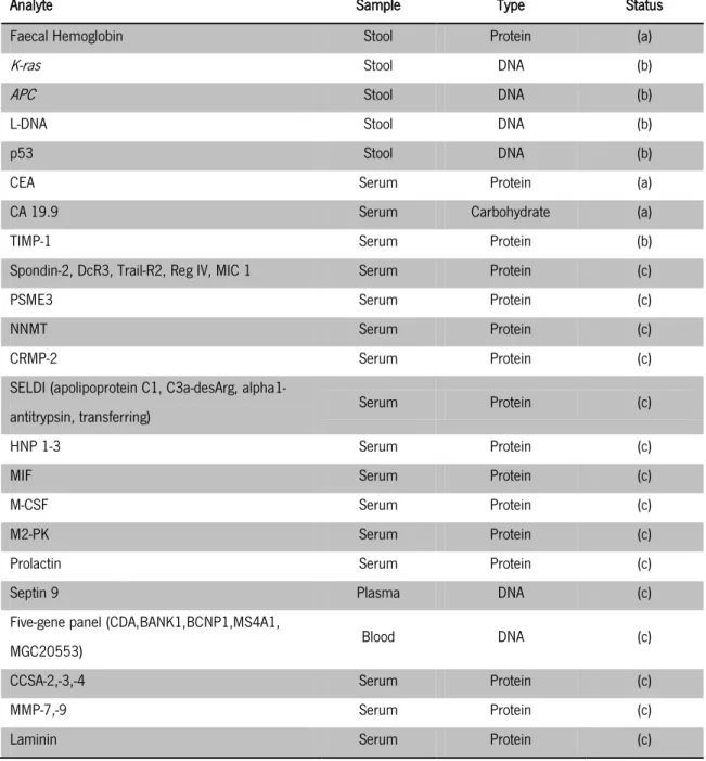
Several markers that include faecal, genetic, epigenetic and serum or blood markers available for the detection of CRC are presented on table 1. These are either already in use for screening of general population (a), in clinical trials (b) or in a preclinical state (c).
Faecal hemoglobin has proved its value as a marker for CRC screening as it is the protein measured on FOBT, i.e. the most widely screening modality available. In addition, DNA stool test, named cologuard test, was approved by FDA on 2014 (Simon 2014), for analysis of cancer faecal colonocytes (colonic epithelium cells). These cells are shed into the faecal stream providing revealing material that can be used to detect mutations on genetic markers and epigenetic markers (Loktionov et al. 1998) as K-ras, p53 (Losi et al. 1996; Shivapurkar et al. 1997), adenomatous polyposis coli (APC) (Smith et al. 1994), microsatellite instability (MSI) or long-form DNA (L-DNA) (Kim et al. 2008). This DNA panel was found to be more sensitive and specific for the detection of CRC than FOBT (Imperiale et al. 2004).
Of all serum or blood markers shown in table 1, the carcinoembryonic antigen (CEA) and carbohydrate antigen 19-9 (CA 19.9) are the only tumor markers that are currently in clinical use. CEA is a high molecular weight glycoprotein that has been used for many years as a biomarker for CRCs and other cancers. This marker is mostly used for monitoring the patient reaction to treatment, such as resection surgery, but does not provide sufficient sensitivity and
11 reliability for early cancer detection (Bates 2008), being instead a prognostic marker since higher levels are indicative of a more aggressive disease and poorer prognosis (Kim et al. 2008). CA 19.9, the second most investigated gastrointestinal tumor marker, is less sensitive than CEA when used as a prognostic marker for CRC patients. This and other carbohydrate antigens have been extensively studied, but due to their low sensitivity, stage dependency and specificity, they are not useful for detection of CRC (Hundt et al. 2007). Moreover, tissue inhibitor of metalloproteinase type 1 (TIMP-1) total levels in patients with CRC have shown to be significantly higher when compared to the ones of healthy blood donors with very narrow range of plasma TIMP-1 levels. More importantly, it has been proven that it can be detected at early stages of CRC (Holten-Andersen et al. 2004). In spite of the promising results, more studies are needed to validate the use of TIMP-1 as a diagnostic and prognostic biomarker for CRC (Kim et al. 2008).
12
Table 1 – Non-invasive molecular markers for the detection of CRC [adapted from: (Kim et al. 2008; Tanaka et al. 2010)]
Analyte Sample Type Status
Faecal Hemoglobin Stool Protein (a)
K-ras Stool DNA (b)
APC Stool DNA (b)
L-DNA Stool DNA (b)
p53 Stool DNA (b)
CEA Serum Protein (a)
CA 19.9 Serum Carbohydrate (a)
TIMP-1 Serum Protein (b)
Spondin-2, DcR3, Trail-R2, Reg IV, MIC 1 Serum Protein (c)
PSME3 Serum Protein (c)
NNMT Serum Protein (c)
CRMP-2 Serum Protein (c)
SELDI (apolipoprotein C1, C3a-desArg,
alpha1-antitrypsin, transferring) Serum Protein (c)
HNP 1-3 Serum Protein (c)
MIF Serum Protein (c)
M-CSF Serum Protein (c)
M2-PK Serum Protein (c)
Prolactin Serum Protein (c)
Septin 9 Plasma DNA (c)
Five-gene panel (CDA,BANK1,BCNP1,MS4A1,
MGC20553) Blood DNA (c)
CCSA-2,-3,-4 Serum Protein (c)
MMP-7,-9 Serum Protein (c)
Laminin Serum Protein (c)
There are evidences that all of the markers mentioned and that are currently in pre-clinical development can be used alone, or in combination with others, for the detection of CRC, since they show a greater level of sensitivity and specificity when compared to CEA. However, large-scale studies are needed to evaluate the potential of using biomarkers that have recently been discovered through genomics and proteomics advances in routine analysis (Tanaka et al. 2010).
13
2.3. M
OLECULAR RECOGNIZING ELEMENTSThe progress of biosensors for the detection of cancer is dependent on the availability of high affinity and specific ligands for the desired cell type and/or biomarker (Mcguire et al. 2009). The ability to recognize a cell or biomarker in a mixed population is viewed as the critical step in any diagnostic assays. The biomarker that can be as complex as a whole cell, or as simple as a single molecule, must be recognized and collected from a heterogeneous population, regardless of the complexity of the sample matrix (Soper et al. 2006).
The most commonly used recognition elements in biosensors are antibodies but, more recently, synthetic based recognition elements such as aptamers, peptides, surface-imprinted polymers, carbohydrates, nucleic acids and molecularly imprinted polymers are being used as a replacement for antibodies (Soper et al. 2006; Petrenko 2008).
For a cell-based recognition, typically very little is known about the specific landscape of the membrane surface making it impossible to design specific cell-targeting ligands, such as specific antibodies. The development of methods to identify specific ligands for tumor biomarkers that can discriminate between normal and cancerous cells is an effective way of cancer cell targeting that can be useful for diagnostic purposes and targeted drug delivery (Zhang et al. 2007), as it has been shown that endothelial cells in the vasculature of tumors differ from normal endothelial cells, and that tumor blood vessels express proteins that are not produced in motionless vascular endothelium (Rasmussen et al. 2002).
The biopanning approach using phage-displayed ligands consists in an unbiased screen technique in which there is no selective pressure towards binding a particular macromolecule (Soper et al. 2006). Phage display screening of random peptide libraries do not require a knowledge of the cell surface nature, and as a result many protocols to identify cell-specific binding peptides have been developed (Barry et al. 1996; Brown 2000; Oyama et al. 2003; McGuire et al. 2004). Peptides selected using biopanning are extremely cell-specific meaning that affinity selection using random peptide libraries can be used to identify different types of tumors that are currently similarly classified (Soper et al. 2006; Oyama et al. 2003). This high discriminating power of the selected phage also suggests that peptides could be used to target cancer cells in vivo, since they can distinguish between normal and cancer cells, being an extremely remarkable characteristic that is useful for target chemotherapy and diagnosis purposes. Also, these peptides can be used for example to drive the delivery of fluorescent nanoparticles, as well as cell capture reagents for cell enrichment, and as antibody replacements
14
for flow cytometry. Another appealing aspect of peptide libraries to screen cancer cells surface is that the peptide found can be characterized as modular, meaning that they retain functionality more or less independently of the protein context. A phage display-derived peptide is, by definition, a peptide that binds to a given target in the context of being coupled to a coat protein of a phage (Voss et al. 2002). A modular peptide is one that was found to be specific to a target in that context, but retains its binding affinity when fused to an heterologous proteins such as glutathione S-transferase, agarose (Frangioni et al. 2000) or a contrast agent. In other words, a specific peptide found using combinatorial phage peptide libraries, can be coupled to a variety of different functional entities. This characteristic has already been demonstrated for target chemotherapy (Pasqualini & Ruoslahti 1996; Arap et al. 1998a; Arap et al. 1998b). Finally, as peptides are amenable to derivatization, it is possible to anticipate that these cell-specific ligands will find utility in a variety of different biosensor platforms (Mcguire et al. 2009).
Synthetic based recognition elements, such as aptamers and peptides, have particular advantages over antibodies for its use as ligands. Aptamers and peptides have robust structures that can be placed in varied conditions without losing their specificity and are easy to modify structurally to support the addition of reporters or immobilization to sensing elements. In addition, their inexpensive chemical synthesis makes their production cost-effective and appropriated for large scale preparation contrarily to the labor-intensive and time-consuming production of antibodies (Soper et al. 2006). In particular, peptides present some advantages especially over antibodies and proteins, namely regarding their use in a clinical context. The small size of peptides makes them very interesting for drug delivery purposes since the degree of penetration of a molecule into a living cell is markedly associated with the size of the compound used for targeting. Moreover, peptides are non-immunogenic, thus promoting their safety profile and making them suitable for clinical uses (Bakhshinejad et al. 2014; Zhang et al. 2007).
Peptides with high affinity for specific tumor antigens like HER2/neu receptor, oligonucleotide receptor and ICAM- 1 have been isolated by phage display (Bélizaire et al. 2003; Karasseva et al. 2002; Schatzlein et al. 2001). Specifically in the case of CRC, peptide libraries have been used successfully for the identification of colon tumor binding peptides (Zhang et al. 2007; Kelly & Jones 2003; Rasmussen et al. 2002).
15
3. S
PECIFIC CANCER CELLS LIGANDS IDENTIFICATION BY
PHAGE DISPLAY TECHNOLOGY
Within the last three decades, biopanning of phage displayed peptide libraries on intact cells proven to be a successful route for the identification of cell-specific ligands (Mcguire et al. 2009). Phage display is a powerful in vitro selection technology that uses bacteriophages (phages) to the display of exogenous (poly)peptides on its surfaces coupled to the phage coat proteins (Huang et al. 2011).
The concept was first introduced in 1985 by George Smith (Smith 1985), who used filamentous f1 phage as an expression vector to insert exogenous DNA sequences into specific sites of its genome where the genes encoding the phage coat proteins are localized, enabling the display of peptides and proteins as a fusion to one of the phage coat proteins. Smith proved that it was possible to introduce such alterations on the phage genome, and consequently on the proteins of the coat, without loss of infectivity capacity (Smith 1985; Smith & Petrenko 1997).
Phage display is a combinatorial technology that has developed enormously in the last decades with the construction of a large number of libraries of peptides, proteins, antibodies, cDNA and mRNA (Bratkovič 2009). It had a major influence on several discoveries in fields like drug discovery (Alirezapour et al. 2013; Christensen et al. 2001), tumor cell-targeting (Rasmussen et al. 2002), molecular imaging and cancer diagnosis (Deutscher 2011), and the identification of several cancer cells specific peptides useful for diagnosis and targeted drug delivery (Oyama et al. 2003; Kelly & Jones 2003; Zhang et al. 2007; Ghosh et al. 2012; Bakhshinejad et al. 2014).
This technology takes advantage of the extensive genetic flexibility of phages to create a huge variety of modifications on their surface, thus allowing the construction of combinatorial libraries composed by phage particles displaying ligands (peptides, proteins, among others) for different targets eliminating the need of genetically or chemically engineer each one of them (Arap 2005). Using standard molecular biology techniques, a phage display library, containing up to 1010 variants can be constructed simultaneously on the surface of phages, and can be used for the selection of any desired target even when the target and its specifications are unknown (Huang et al. 2011). This is the great strength of phage technology; i.e. the ability to identify interactive regions of proteins and other molecules without preexisting notions about the nature of the interaction. For this reason, different screening methods allowed the isolation and
16
characterization of peptides binding to several molecules in vitro, in the context of living cells, on both animals and humans (Arap 2005).
3.1. F
ILAMENTOUS PHAGES FOR PHAGE DISPLAY:
PHAGE STRUCTURE AND PROPAGATIONA variety of phages, obligate intracellular parasites that use the biosynthetic machinery of the bacterial cell host (gram-negative cells) for reproduction, have been used for phage display purposes. M13, f1 and fd phages from Ff family (usually referred as Ff or M13-like phages) are mostly used for phage display technology (Arap 2005; Russel et al. 2004). Filamentous phages are ideal to use as expression vectors and for display in particular. On phage display expression systems (e.g. on Fig. 4) the foreign DNA sequence is introduced into the gene of one of the phage coat proteins so that the respective amino acid sequence is displayed coupled to the endogenous amino acids of the phage coat protein. The hybrid coat protein is incorporated into the viral particles as they are released from the cell host, yielding a recombinant phage with the foreign peptide or protein domain displayed on the outer surface. (Smith & Petrenko 1997).
Fig. 4 – Phage Display expression system (M13 phage): the ligand-encoding oligonucleotide inserted next to gene III that encodes a coat protein of the phage generates a recombinant phage, presented on the right, with the ligand displayed coupled to the respective coat protein [taken from: (Bakhshinejad et al. 2014)].
Ff phages defining characteristic is a circular single-stranded DNA genome enclosed in a flexible tube composed of thousands of copies of a single major coat protein and few minor proteins at the tips. An important feature for phage display is that genomic DNA of these phages can be obtained both as single-stranded and as double-stranded forms which facilitates cloning and library construction as it can be used as a plasmid-based vector. Additionally, phage’s small
17 genome can tolerate large insertions on non-essential regions with no effect on infection capacity as the enlargement of the genome size is accompanied by an equal increase in the length of the phage particle, as well as in the number of pVIII (major coat protein) copies (Bakhshinejad et al. 2014; Ebrahimizadeh & Rajabibazl 2014).
M13 is an E. coli-specific filamentous bacteriophage and is the most commonly used for phage display purposes. It has a flexible tube shaped structure and a circular genome with 6-8 kb enclosed in a coat composed of five different proteins (Sidhu 2001) (Fig. 5).
Fig. 5 – The general structure of M13 phage particle.
[adapted from: http://www.wwnorton.com/college/biology/microbiology2/ch/11/etopics.aspx, on December 17t h, 2014]
The M13 genome contains 11 genes; five of which encode the coat proteins described below, while the others encode proteins necessary for viral replication and assembly. Several thousand copies of pVIII, the major coat protein, cover the length of the phage with five surface-exposed N-terminal residues. On one side of the phage particle, where phage assembly begins, there are 3-5 copies of proteins pVII and pIX whose absence will make impossible to form a phage particle. On the other end there are about 5 copies of both pIII and pVI that are needed for the phage to detach from the host cell membrane (Russel et al., 2004).
All of the five coat proteins of the phage have been used to the display of foreign peptides and proteins, but pIII and pVIII are the most important and each of them has different features (Bakhshinejad et al. 2014). Protein pIII is more suitable for display of a smaller number of larger proteins, while pVIII is appropriate for the display of more copies of the same small peptide (Willats 2002). PIII libraries display 3-5 copies of each individual peptide, whereas pVIII libraries can display up to 2700 copies of small peptides (Kay et al. 1996). However, this cannot be used as a general assumption for phage display. In fact, choosing the coat protein for displaying
18
depends on the type of ligand and the specificity needed for the screening of the target (Bakhshinejad et al. 2014).
Fd particles are able to infect a variety of gram-negative bacteria. F’ and F+ strains (which have an F pilus) of E. coli are widely used to obtain virus particles for phage display. Filamentous phage infection does not produce lytic infection in E. coli, contrariwise induces a state in which the infected bacteria produces and releases phage particles into the growing medium, without killing the cell (Russel et al. 2004). Possessing a non-lytic cycle propagation mechanism is an important feature for the use of M13-like phages as expression systems as it enables the accumulation of a large number of virus particles in the host cell and growth medium (Bakhshinejad et al. 2014). Fig. 6 represents the infection life cycle of filamentous phage that begins with the infection of bacterial cells when the N-terminus of pIII attaches to the F pilus of male E. coli (Sidhu 2001). After this, the single stranded phage genome enters the bacteria and is converted to the double stranded replicative form (RF) by the host DNA replication machinery. RF genome is the template for the production of viral coat proteins and single stranded DNA progeny. The viral coat proteins encapsulate the single stranded DNA during its extrusion from the host cell (Pande et al. 2010).
Fig. 6 – M13 particles life cycle [taken from: (Wilson & Walker 2010)].
The existence of F pilus is very important for phage infection and propagation as, even though infection may occur without appropriate pili, the process is extremely inefficient. There are only a few F pilus per each bacterial cell and the target size at their ends is small. For that reason, the rate and efficiency of infection of a bacterial culture is improved at high multiplicity
19 phage per cell and high cell density. Nevertheless, high density of bacterial cells has to be achieved during log phase because after this point (at stationary phase) pilus expression is decreased and infectivity is compromised. Therefore, to accomplish a higher titer of virus, it is important to have a well aerated bacterial culture so a high cell density is achieved immediately after entering the log phase. Also, F pilus do not assemble at low temperatures, thus temperatures equal or above 34C are needed to assure plaque formation (Russel et al. 2004).
Under optimal conditions, the first progeny particles appear in the culture supernatant 10 min after infection. Phage number increases exponentially on the first 40 min after the infection, after which the rate of propagation becomes linear. In one generation of bacteria, approximately 1000 phage particles are produced and liberated to the surrounding medium (Wilson & Walker 2010). Under optimal conditions infected cells can continue to grow and divide, as well as producing phages, indefinitely (Russel et al. 2004).
3.2. P
HAGE DISPLAY RANDOM PEPTIDE LIBRARIESRandom peptide libraries are the most common type of combinatorial phage libraries (Pande et al. 2010), which provide a rich source of molecules that can be chemically or genetically synthesized and screened to obtain a desired function or affinity. The first combinatorial library was developed five years after the introduction of the phage display concept (Huang et al. 2011). It was constructed by inserting random oligonucleotide sequences into the gene encoding one of the coat proteins of M13. In a phage library, each phage particle displays a unique ligand, but the entire library encompasses a great variety of billions of peptides. The genetic encoding of a library, meaning the presence of the DNA and the peptide of interest in the same virion makes possible to re-synthesize and re-screen binding ligands of interest. Combinatorial libraries turned phage display into a high throughput system that can be used to screen a large pool of potential binding motifs and select those with highest affinity to the target structure (Bakhshinejad et al. 2014). Also, given that a single event of phage/target binding can be detected, this technology can be used to discover binding peptides when the target is ranging from a purified protein sample to a complex mix of proteins, as in the case of a living cell surface (Voss et al. 2002).
For the construction of pIII random peptide libraries, the random oligonucleotides are inserted between the coding sequence for the signal peptide and the N-terminus of the coat