Research Article
Immunodiagnosis of Canine Visceral Leishmaniasis
Using Mimotope Peptides Selected from Phage Displayed
Combinatorial Libraries
Christina Monerat Toledo-Machado,
1Ricardo Andrez Machado de Avila,
2Christophe NGuyen,
3Claude Granier,
3Lilian Lacerda Bueno,
1Claudia Martins Carneiro,
4Daniel Menezes-Souza,
1Rubens Antonio Carneiro,
5Carlos Chávez-Olórtegui,
2and Ricardo Toshio Fujiwara
11Departamento de Parasitologia, ICB, Universidade Federal de Minas Gerais, CP 486, Belo Horizonte 31270-901, MG, Brazil 2Departamento Bioqu´ımica e Imunologia, ICB, Universidade Federal de Minas Gerais, CP 486, Belo Horizonte 31270-901, MG, Brazil 3SysDiag CNRS-BioRad UMR 3145, Cap Delta/Parc Eurom´edecine, 1682 rue de la Valsi`ere, CS 61003,
34184 Montpellier Cedex 4, France
4Departamento de An´alises Cl´ınicas, Escola de Farm´acia, Universidade Federal de Ouro Preto, Ouro Preto 35400-000, MG, Brazil 5Escola de Veterin´aria, Universidade Federal de Minas Gerais, CP 486, Belo Horizonte 31270-901, MG, Brazil
Correspondence should be addressed to Ricardo Toshio Fujiwara; [email protected]
Received 17 November 2014; Revised 27 December 2014; Accepted 11 January 2015
Academic Editor: Mehdi Chenik
Copyright © 2015 Christina Monerat Toledo-Machado et al. This is an open access article distributed under the Creative Commons Attribution License, which permits unrestricted use, distribution, and reproduction in any medium, provided the original work is properly cited.
ELISA and RIFI are currently used for serodiagnosis of canine visceral leishmaniasis (CVL). The accuracy of these tests is
controversial in endemic areas where canine infections byTrypanosoma cruzimay occur. We evaluated the usefulness of synthetic
peptides that were selected through phage display technique in the serodiagnosis of CVL. Peptides were chosen based on their ability to bind to IgGs purified from infected dogs pooled sera. We selected three phage clones that reacted only with those IgGs. Peptides were synthesized, polymerized with glutaraldehyde, and used as antigens in ELISA assays. Each individual peptide or a mix
of them was reactive with infected dogs serum. The assay was highly sensitive and specific when compared to solubleLeishmania
antigen that showed cross-reactivity with anti-T. cruziIgGs. Our results demonstrate that phage display technique is useful for
selection of peptides that may represent valuable synthetic antigens for an improved serodiagnosis of CVL.
1. Introduction
Canine visceral leishmaniasis (CVL) caused byLeishmania (Leishmania) infantum chagasi is a widespread zoonotic disease of both the Old and the New World [1] leading to a considerable number of deaths. Domestic dogs are considered the main animal reservoir hosts of the disease [2,3]. Most infected dogs do not present clinical signs but are seropositive particularly in endemic areas of CVL in the World [2, 4–7]. The seroprevalence of CVL in areas of endemicity in the Mediterranean Basin and the Middle East, including Iran, has been reported to be 10–37% [6,8].
Euthanasia of seropositive dogs has been adopted as a mainstay control measure in some countries [9].
The Brazilian Ministry of Health recommends the use of an immunoenzymatic assay (ELISA) and an indirect immunofluorescence antibody test (IFAT) for the diagnosis of canine visceral leishmaniasis (CVL), employed as criteria for the culling of seropositive dogs in surveillance and control programs for visceral leishmaniasis (VL) [10]. Both the accuracy of these tests and the process of dog culling promote a controversial impact of the leishmaniasis infection [11,12]. However, the development of an effective diagnosis test can be critical for the control and the possible eradication of VL; Volume 2015, Article ID 401509, 10 pages
more sensitive and specific tests may be especially helpful to achieve this goal [13].
Over the past years, synthetic peptides have been used successfully as antigens for the in vitrodiagnosis of many parasitic diseases [14]. Phage display of random peptides has become an alternative method for the study of molecular interactions in many areas of protein science, including antigen-antibody interactions. It has been shown that linear epitopes, as well as mimotopes that mimic discontinuous epitopes of an antigen, can be identified by the screening of phage libraries with monoclonal or polyclonal antibodies [15, 16]. Phage display in neglected disease research has proven successful not only in mapping the protein-protein interac-tions that are important in the etiologic agent biology, but also in the identification of molecules that might be exploited in the design of therapeutic agents, vaccines, or immunodi-agnostics [17–19].
In order to search for diagnostic epitopes without previ-ous knowledge of protein structure, we tested phage-borne libraries displaying foreign peptides at the surface of the major pVIII coat proteins for their capacity to bind anti-L. infantum chagasiproteinantigen (LiPA) IgGs purified from the sera of dogs with visceral leishmaniasis. Phage clones reactive with anti-LiPA IgGs were also tested for reactivity with IgGs purified from the sera of dogs experimentally infected withT. cruzi, because of the known cross reaction between visceral leishmaniasis antibodies and Chagas’ dis-ease antibodies in dogs [20]. In order to estimate the diagnostic accuracy, we found three peptides that could successfully be used as antigens in ELISA assays for a specific immunodiagnosis of canine visceral leishmaniasis without cross reaction with circulating antibodies ofT. cruzi exper-imentally infected dogs.
2. Material and Methods
2.1. Study Dogs. For biopanning assay thirty-eight sera from L. infantum chagasinaturally infected dogs of both genders were used. For the ELISA assays, thirty-eight sera were obtained from beagle dogs experimentally infected with L. infantum chagasifor leishmaniasis studies.The infection was certified through parasitological tests that were conducted on bone marrow cells examined by optical microscopy and immunological tests (ELISA and RIFI). Additionally, for both assays, thirty-eight sera from uninfected dogs were used as negative controls. In order to evaluate cross-reactivity on ELISA assays we also used fifteen sera from dogs experi-mentally infected with T. cruzi parasite obtained from the serum bank of the Laborat´orio de Imunologia e Genˆomica de Parasitos (UFMG).
Proof of dog infection was attested by a positive immunofluorescence titre (IFAT) at the threshold titer of 1 : 40 serum dilution, a positive reactivity in ELISA, and a par-asitological diagnosis ofLeishmania. Antibodies from sera of 38 healthy dogs displaying negative IFAT and parasitological tests were included as uninfected controls. Briefly, the clinical evaluation assessed typical clinical signs for symptomatic visceral leishmaniasis, including lymphadenopathy, decrease of weight and opaque eye, alopecia, eczema, and skin ulcers.
In order to verify possible infection with different pathogens (Babesia canis,Ehrlichia canis, and Trypanosoma cruzi) in allL. infantum chagasinaturally infected animals, parasitological exams were performed in a private laboratory in Belo Horizonte, Minas Gerais, Brazil.
All dogs were maintained in a kennel of Institute of Biological Sciences in the Universidade Federal de Minas Gerais, Belo Horizonte, Brazil, according to university’s ethic committee for clinical research (CETEA) protocol 122/2009.
2.2. Production of L. infantum chagasi Protein Antigen (LiPA). L. infantum chagasi (MHOM/BR/1975/BH46) was grown at 24∘C in Schneider’s medium (Sigma, St. Louis, MO, USA) supplemented with 20% heat-inactivated fetal bovine serum (FBS; Sigma), 200 U/mL penicillin, and 100𝜇g/mL streptomycin, pH 7.2. Protein antigens ofL. infantum chagasi (LiPA) were prepared from stationary phase promastigotes, submitted to 7 cycles of freezing (liquid nitrogen) and thaw-ing (42∘C), followed by ultrasonication (Ultrasonic processor, GEX600) with cycles of 10 sec for 2 min at 35 MHz. The extracts were then submitted to centrifugation at 8,000×g for 20 min at 4∘C. The supernatant was collected and stored at −70∘C. The protein concentration was estimated by the Bradford method [21].
2.3. Preparation of Antibodies for Biopanning. Antibodies used for biopanning (i.e., immunocapture of phages binding to target antibodies) were initially purified from 38 sera from L. infantum chagasinaturally infected dogs of both genders. Fifteen serum samples of dogs experimentally infected with Trypanosoma cruziparasite were also collected.
Polyclonal IgGs to LiPA (anti-LiPA IgGs) used for biopanning were purified from a pool of dogs with VL, using ammonium sulfate precipitation and filtration through Protein A-Sepharose 4B column [22]. Following elution and neutralization using NaOH 0.1 M, the IgG fraction was dialyzed against PBS 1x and the protein concentration was determined by the Bradford method [21]. IgGs from uninfected controls (normal IgG) were also fractionated as described before. IgGs from T. cruzi infected dogs were obtained. The reactivity against LiPA of anti-LiPA IgGs and normal IgGs was confirmed by indirect ELISA.
2.4. Ethics Statement. All sera samples were obtained from the Veterinary Hospital of the Federal University of Minas Gerais (UFMG) and the experiments were performed in compliance with the university’s ethic committee for clinical research (CETEA), protocol 122/2009. All sera were stored at −20∘C until use. All dog owners gave permission to have their
animals sampled.
5𝜇g/mL for the first two rounds of panning and a concentra-tion of 0.5𝜇g/mL for the last panning in 100 mM NaHCO3, pH 8.6, on a shaking platform. For the first panning, 5× 10¹² transducing units (TU) of each library were incubated with the absorbed IgG. After incubation, unbound phages were washed with TBS (Tris 50 mM, NaCl 150 mM, pH 7.5) containing 0.05% (v/v) Tween 20, and bound phages were removed by elution with 0.1 M glycine, pH 2.2, containing 0.1% BSA. Eluted phages were then used to infectEscherichia coliK91 cells. After three rounds of enrichment, individual phage clones were isolated and further analyzed [24].
2.6. ELISA Analysis after Three Rounds of Panning. ELISA plates (Falcon 3912, Becton Dickinson, Oxnard, CA 93030) were coated with 1𝜇g/well of anti-LiPA IgGs in 100 mM NaHCO3, pH 8.6, and overnight at 4∘C. Plates were washed with PBS 0.1% Tween 20 (v/v) and then blocked with PBS, 0.1% Tween 20 and 2% nonfat dried milk (w/v) for 1 h at 37∘C. 1010TU of phages, eluted after each round of panning, were then added to the plate and incubated for 2 h at 37∘C. Binding was detected using a peroxidase conjugated anti-M13 antibody (Roche Molecular Biochemicals) diluted 1 : 3000 in blocking buffer. After 1 h at 37∘C and washing, the peroxidase substrate was added. The resulting color was measured at 492 nm with an automated microtiter plate reader (Model 450, Bio-Rad).
2.7. Screening. ELISA plates (Falcon 3912, Becton Dickinson, Oxnard, CA 93030) were coated with 1𝜇g/well of anti-LiPA IgGs or anti-T. cruziIgGs in 100 mM NaHCO3, pH 8.6, and overnight at 4∘C. Plates were washed with PBS, 0.1% Tween 20 (v/v), and then blocked with PBS, 0.1% Tween 20, and 2% nonfat dried milk (w/v) for 1 h at 37∘C. 1010TU of individual phages was isolated after third panning and 50𝜇L of blocking buffer was then added to each well. Phage particles were incubated for 2 h at 37∘C. Binding was detected using a peroxidase conjugated anti-M13 antibody (Roche Molecular Biochemicals) diluted 1 : 3000 in blocking buffer. After 1 h at 37∘C and washing, the peroxidase substrate was added. The resulting color was measured at 492 nm with an automated microtiter plate reader (Model 450, Bio-Rad). Afterwards, the twelve clones were checked by ELISA for their ability to bind to antiT. cruzidog IgGs.
2.8. DNA Sequencing, Synthesis, Chromatography, and Mass Spectrometry of Soluble Peptides. Approximately, 9𝜇g of single-stranded DNA was purified using the QIA prep Spin M13 protocol (Qiagen). Sequencing reactions were carried out according to the dideoxy chain termination method [25], using the ABI Prism Kit (PE Applied Biosystems) for the automatic method with ABI PRISM 377 (PerkinElmer). The primer reverse 5-TCGGCAAGCTCTTTTAGG-3was used for sequencing. The sequences obtained were translated.
The peptides TTDDDKLKKTLTYRS, KCPSIPGAVLCV, and ICARQDPAGNCS were synthesized in ResPep SL Synthesizer by Fmoc chemistry [26]. After synthesis, the peptides were deprotected and released from the resin by trifluoroacetic acid (TFA) treatment in the presence of
the appropriate scavengers. The peptides were lyophilized and their purity was assessed by HPLC and their mass was confirmed by mass spectrometry according to de Avila et al. (2011) [27].
2.9. Glutaraldehyde Link Reaction. Individual peptides and the combination of three peptides (pool) were polymerized using glutaraldehyde as cross-linking reagent [28]. Briefly, 10𝜇moles of each peptide was diluted in 1 mL of PBS and an equal volume of 1% GLUT (20 mM) in PBS was then slowly added to the solution over the course of 1 hour under constant stirring at 4∘C. The reaction was allowed to proceed for an additional hour and the free aldehyde coupling groups and the Schiff ’s base intermediates were reduced by the addition of NaBH4(10 mg/mL). At the end of the reaction, the solution containing polymerized peptides was dialyzed overnight against PBS.
2.10. Diagnostic ELISA for Canine Sera. ELISA was carried out to detect circulating antibodies that recognized polymer-ized peptide in canine sera. ELISA assays microtiter plates were coated with 50𝜇L/well of each polymerized peptide or with the polymerized mixture of the three peptides or withL. infantum chagasitotal protein antigen (2.5 mg/mL) in Milli-Q water. The wells were blocked with 200𝜇L/well of 5% BSA in PBS at 37∘C for 1 h. After the wells had been washed twice with PBS containing 0.05% Tween 20 (PBS-T), 100𝜇L/well of L. infantum chagasiinfected dog serum; healthy dog serum; and Ehrlichia canis, Babesia canis, and T. cruziinfected dog serum diluted 1 : 100 in PBS- BSA 2.5% was added and incubated at 37∘C for 1 h. The wells were washed six times with PBS-T, incubated with 100𝜇L/well of anti-dog IgG conjugated with peroxidase (Sigma), diluted 1 : 2000 in PBS-BSA 2.5% at 37∘C for 1 h and washed six times with PBST. After incubation with 100𝜇L/well of o-phenylenediamine solution (0.33 mg/mL in citrate buffer, pH 5.2, in the presence of 0.04% hydrogen peroxide) for 15 min at room temperature, the optical density at 492 nm of each well was determined. The cut-off was determined according to ROC curve. The results of the ELISA peptides as antigens in dogs were compared with the immunoassay EIE-LVC kit (FIOCRUZ-Bio-Manguinhos, Brazil), which is the test currently recommended by the Brazilian Ministry of Health for screening seroreactive animals [29]. The assays were conducted according to the manufacturer’s instructions.
0.6
0.4
0.2
0.0
1.00 0.50 0.25 0.12 0.06
Ab
s
492
nm
IgG concentration (𝜇g/well)
IgG from infected dogs IgG from negative dogs
Figure 1: Reactivity of purified IgG antibodies againstL. infantum
chagasi antigen (LiPA). 10𝜇g of affinity-purified IgGs from CVL
dogs (e) and negative dogs (◼) was added to each well (from 10𝜇g
to 0.625𝜇g/well) of microtiter plates coated with 50𝜇g/mL LiPA.
The reaction was detected using a peroxidase conjugated anti-dog IgG antibody (1 : 2000). Absorbance values at 492 nm were means of duplicates.
The degree of agreement between ELISA assays using peptides, L. infantum chagasi antigen, and EIE-LVC Bio-Manguinhos kit test was determined by the Kappa index (𝜅) values with 95% confidence intervals and interpreted accord-ing to the followaccord-ing Fleiss scale: 0.00–0.20, poor; 0.21–0.40, fair; 0.41–0.60, moderate; 0.61–0.80, good; 0.81–0.99, very good; and 1.00, perfect [30]. All of the statistical analyses were performed using GraphPad Prism (version 5.0) and Graph-Pad QuickCals (http://www.graphpad.com/quickcalcs/).
3. Results
3.1. Selection of Phages Recognized by Anti-LiPA IgGs and Mimotope Peptides Synthesis. For the screening of phage-borne peptides from libraries, IgGs purified from sera of thirty-eight naturally infected and sixteen uninfected dogs were used. Levels of anti-LiPA antibodies were previously examined in sera samples collected one week before phage display experiments (data not shown). After fractionation of the pool of sera of infected or noninfected animal through Protein A-Sepharose 4B column, a dose-dependent reactivity of the purified IgGs towards LiPA was observed in an ELISA format (Figure 1).
In order to identify peptides that bind to anti-LiPA antibodies, four different phage libraries were screened. A significant enrichment of phage binding to the target antibod-ies was obtained after three rounds of panning (Figure 2(a)). One hundred and ninety-eight phage clones were randomly picked from the third round of selection, and twelve clones were selected based on their reactivity (absorbance at 492 nm ≥1.0) against IgGs fromL. infantum chagasiinfected dogs (data not shown). Afterwards, the twelve clones were checked by ELISA for their ability to bind to anti-T. cruzi dog
IgGs. Clones 5, 6, and 11 were further selected due their high reactivity againstLeishmania-specific IgGs and without cross-reactivity with IgGs from dogs with Chagas disease (Figure 2(b)).
The DNA sequences of clones 5, 6, and 11 were translated and the amino acid sequences of the peptides were deduced (Figure 2(c)). One peptide (peptide 11) was selected from the 𝑋15 library expressing 15-mer linear peptides, whereas the
other two peptides were selected from theXCX8CXlibrary of constrained 12-mer peptides. The corresponding synthetic peptides, TTDDDKLKKTLTYRS, KCPSIPGAVLCV, and ICARQDPAGNCS, were chemically synthesized and purified by reverse phase chromatography and their correct molec-ular weights were confirmed by mass spectrometry (data not shown).
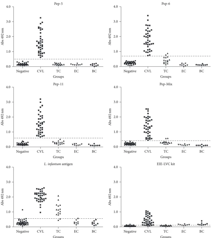
3.2. Sensitivity and Specificity of ELISA Tests with Synthetic Peptides for Serodiagnosis of CVL. In order to evaluate the antigen-specific dog antibody response against synthetic peptides, an ELISA was optimized to obtain the best signal-to-noise ratio and to develop a reproducible and robust assay capable of capturing antibodies over a biologically relevant assay range. The EIE-LVC kit was used as a reference. ELISA cut-off values for peptides 5, 6, and 11 and pooled peptides (peptides 5+6+11) as antigens were 0.488, 0.698, 0.594, and 0.410, respectively. In similar conditions, the cut-off values were 0.546 and 0.104, forL. infantum chagasi antigen and EIE-LVC kit, respectively. The peptides were initially tested on sera of 38 dogs withL. infantum chagasi(CVL) (Figure 3, Table 1). Antibodies against peptides 5, 6, and 11 and pooled peptides were detected in 100% of the serum samples from these dogs. In order to evaluate specificity (Sp), serum samples from negative control dogs and samples from dogs infected by E. canis, B. canis, and T. cruzi were tested. Peptides 5 and 11 demonstrated an excellent specificity value (100.00%) slightly better than the 97.10% obtained for peptide 6 and pooled peptides, which showed 2 false positive results in the T. cruzi group (Figure 3 and Table 1). Comparative analysis using either the crude antigen or the reference EIE-LVC ELISA demonstrated also high sensitivity (100.00% and 94.74%, resp.). However, these tests showed a poorer specificity (76.81% and 68.12%, resp.) due to the large number of false negative results in negative andT. cruzigroup.
3.3. Measures of ELISA Performance and Analysis of Agree-ment between the Different Antigens Used in the Diagnosis of CVL. Measures of performance for the different tests evaluated are shown inTable 1. Maximum positive predictive value (PPV) was achieved by peptides 5 and 11 (100.00%) followed by peptide 6 and pool (95.00% for both). Excellent negative predictive values (NPV) were observed for all three peptides and their mix (100.00%).
2.0
1.5
1.0
0.5
0.0
Ab
s
4
9
2
n
m
R0 R1 R2 R3
Round of panning
(a)
2.0
1.5
1.0
0.5
0.0
Ab
s
492
nm
1 2 3 4 5 6 7 8 9 10 11 12
Clones
IgG anti-L. infantum
IgG anti-T. cruzi
(b)
Peptide5-ICARQDPAGNCS
Peptide6-KCPSIPGAVLCV
Peptide11-TTDDDKLKKTLTYRS
(c)
Figure 2: (a) Enrichment of phage binding after three rounds of panning. 50𝜇L of a 1010transduction unit phage suspension collected after
each round of panning was added to wells of microtiter plates coated with 5𝜇g/mL anti-LiPA IgG. Phages were detected using a peroxidase
conjugated anti-M13 antibody (1 : 3000). Values of absorbance at 492 nm are means of duplicates. (b) Reactivity of individual clones isolated
after panning 3. 50𝜇L of a 1010transduction unit phage suspension isolated after the third round of panning was added to each well of
microtiter plates coated with 1𝜇g/well of anti-L. infantum chagasiIgGs (e) and anti-T. cruziIgGs (◼). Phages were detected using a peroxidase
conjugated anti-M13 antibody (1 : 3000). Values of absorbance at 492 nm are means of duplicates. (c) The aminoacid sequences of the three selected peptides.
Table 1: Diagnostic performance of synthetic peptides,L.infantum chagasiantigen, and EIE-LVC Kit in sera of dogs.
Diagnostic test FN FP Se % Sp % PPV % NPV % AUC AC
5∗ 0/38 0/69 100.00 100.00 100.00 100.00 1.0000 1.0000
6∗ 0/38 2/69 100.00 97.10 95.00 100.00 0.9985 0.9813
11∗ 0/38 0/69 100.00 100.00 100.00 100.00 1.0000 1.0000
Pep-Mix∗ 0/38 2/69 100.00 97.10 95.00 100.00 0.9968 0.9813
Ag∗ 0/38 16/69 100.00 76.81 70.37 100.00 0.9851 0.8505
EIE-LVC# 2/38 22/69 94.74 68.12 62.07 95.92 NA 0.7757
Samples from healthy dogs and dogs with canine visceral leishmaniasis,T.cruzi,E.canis,orB.canis.
∗Cut-off obtained by ROC curve.
#
Cut-off obtained according to the manufacturer.
NA: not applicable; FN: false negative; FP: false positive; Se: sensitivity; Sp: specificity; AUC: area under curve; PPV: positive predictive value; NPV: negative predictive value; AC: accuracy.
chagasiantigen and EIE-LVC kit showed a lower accuracy value (0.8352 and 0.8791, resp.). The agreement between the serological tests using the Kappa Index is shown inTable 2. All peptides showed moderate agreement with EIE-LVC∗and good agreement with crude leishmanial antigen.
4. Discussion
Accurate diagnosis of canine leishmaniasis is essential towards a more efficient control of this zoonosis in endemic
areas where it occurs. Conventional parasitological tech-niques are highly specific but still represent time-consuming and invasive methods that are not appropriate for epidemi-ological surveillance or the daily routine of protocols for control of CVL. On the other hand, the use of rapid and reliable methods such serological assays for diagnosis of visceral leishmaniasis in dogs has been largely hampered by cross-reactivity with other canine pathogens [20, 32], especially when crude antigens are employed [33].
4.0
3.0
2.0
1.0
0.0
Negative CVL TC EC BC
Groups
Ab
s
492
nm
Pep-5 Pep-6
Pep-11 Pep-Mix
EIE-LVC kit
4.0
3.0
2.0
1.0
0.0
Negative CVL TC EC BC
Groups
Ab
s
492
nm
4.0
3.0
2.0
1.0
0.0
Negative CVL TC EC BC
Groups
Ab
s
492
nm
4.0
3.0
2.0
1.0
0.0
Negative CVL TC EC BC
Groups
Ab
s
492
nm
4.0
3.0
2.0
1.0
0.0
Negative CVL TC EC BC
Groups
Ab
s
492
nm
4.0
3.0
2.0
1.0
0.0
Negative CVL TC EC BC
Groups
Ab
s
492
nm
L. infantumantigen
Figure 3: Comparison of ELISA reactivity of canine sera against individual phage-display selected peptides, the pool of peptides, theL.
infantumchagasi antigen (LiPA), and reactivity in the EIE-LVC kit. ELISA was performed in different groups of dogs (negative/control group,
CVL group, and TC/T. cruzigroup). Cut-off was determined according to ROC curves.
extracts, which often do not provide reproducible results due to the variable nature of the protein content, thus contributing to the required standardization of the assay. Moreover, the chemical synthesis of the peptides is relatively simple and does not require the manipulation of pathogenic organisms. In the current study, we assessed the potential of phage dis-play technology [15] to select specific peptide sequences and
100
80
60
40
20
0
0 20 40 60 80 100
S
ens
it
iv
it
y (
%
)
Pep-5 Pep-6
Pep-11 Pep-Mix
100 −specificity (%)
100
80
60
40
20
0
0 20 40 60 80 100
S
ens
it
iv
it
y (
%
)
100 −specificity (%)
100
80
60
40
20
0
0 20 40 60 80 100
S
ens
it
iv
it
y (
%
)
100 −specificity (%) 100
80
60
40
20
0
0 20 40 60 80 100
S
ens
it
iv
it
y (
%
)
100 −specificity (%)
100
80
60
40
20
0
0 20 40 60 80 100
S
ens
it
iv
it
y (
%
)
100 −specificity (%)
L. infantumantigen
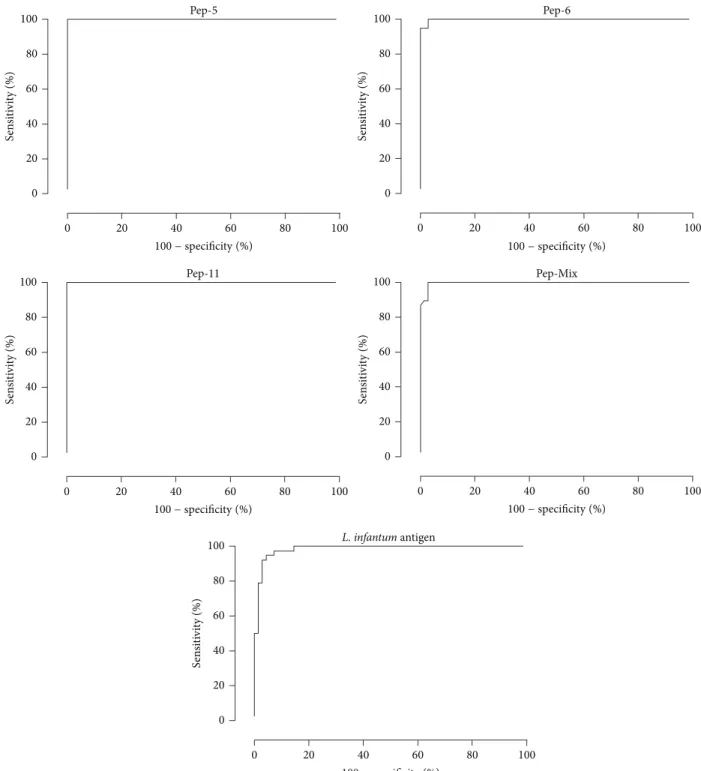
Figure 4: ROC curves obtained from all tests. The curves were used to determine ELISA cut-off, sensitivity, specificity, and AUC.
of three peptides sequence (TTDDDKLKKTLTYRS, pep 11; KCPSIPGAVLCV, pep 6; and ICARQDPAGNCS, pep 5) from theX15andXCX8CXlibraries. The comparison of the sequences of the selected peptides with two GenBank peptide sequence databases did not reveal any significant similarity with amino acid sequences of antigens previously charac-terized from L. infantum chagasi, suggesting that selected peptides might correspond either to yet unknown proteins or to conformational epitopes derived from tertiary/quaternary
structures ofL. infantum chagasiproteins. Of note, the two peptides with a disulfide bridge shared a similar motif (PGA for peptide 6 and PAG for peptide 5), which might correspond to a single epitope of an unknownLeishmaniaprotein or a mimotope [29].
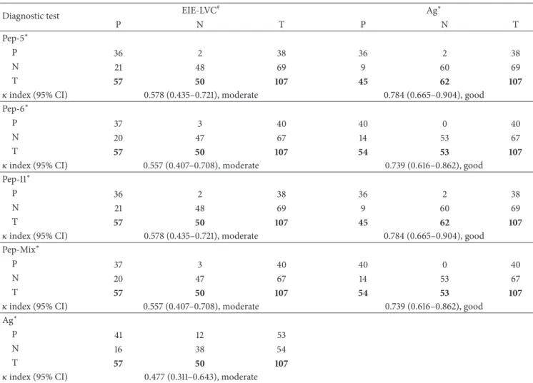
Table 2: Kappa index (𝜅) between paired results of diagnostic tests using peptides,L. infantum chagasiantigen, and EIE-LVC kit.
Diagnostic test EIE-LVC
# Ag∗
P N T P N T
Pep-5∗
P 36 2 38 36 2 38
N 21 48 69 9 60 69
T 57 50 107 45 62 107
𝜅index (95% CI) 0.578 (0.435–0.721), moderate 0.784 (0.665–0.904), good
Pep-6∗
P 37 3 40 40 0 40
N 20 47 67 14 53 67
T 57 50 107 54 53 107
𝜅index (95% CI) 0.557 (0.407–0.708), moderate 0.739 (0.616–0.862), good
Pep-11∗
P 36 2 38 36 2 38
N 21 48 69 9 60 69
T 57 50 107 45 62 107
𝜅index (95% CI) 0.578 (0.435–0.721), moderate 0.784 (0.665–0.904), good
Pep-Mix∗
P 37 3 40 40 0 40
N 20 47 67 14 53 67
T 57 50 107 54 53 107
𝜅index (95% CI) 0.557 (0.407–0.708), moderate 0.739 (0.616–0.862), good
Ag∗
P 41 12 53
N 16 38 54
T 57 50 107
𝜅index (95% CI) 0.477 (0.311–0.643), moderate
Samples from healthy dogs and dogs with canine visceral leishmaniasis,T.cruzi,E.canis,orB.canis.
∗Cut-off obtained by ROC curve.
#
Cut-off obtained according to the manufacturer. P: positive; N: negative; T: total; CI: confidence interval.
large number of sera from dog affected by Chagas disease. In contrast, no positive response was observed when the same sera samples were tested using peptide-based (peptides 5 and 11) ELISA. Noteworthy, the ELISA carried out using synthetic peptides 5 and 11 presented a specificity of 100%, while peptide 6 or the pool of the three peptides rendered a specificity of considerable magnitude (97.10%). Nonetheless, the three peptides were able to detect allLeishmaniapositive samples (100% of sensitivity). These results using peptides selected by phage display for LVC immunodiagnosis tests showed better performance than other studies using crude parasite extracts [20,32,33,38], recombinant antigens [35– 37, 39], and even synthetic peptides [34]. Of note, in order to provide a better binding of the selected peptides to ELISA plates and enhance their antigenic properties, the mimotopes were covalently polymerized using glutaraldehyde (GLUT) as linker [40].
In summary, our data suggest that the three mimotope peptides identified by phage display would represent highly potential antigens to identify canine visceral leishmaniasis.
Such antigens could be used alone or in combination with promising recombinant antigens in order to maximize the performance of the serological assays. Further studies using large cohorts of negative and positive individuals from endemic areas are still required to determine the use of these antigens (alone or in combination with different recombinant proteins) for control of CVL in endemic areas. Finally, our study suggests that mimotope-based ELISA strategy may be useful for the development of a sensitive and highly specific serodiagnosis for CVL or other parasitic diseases.
Disclosure
Claudia Martins Carneiro, Carlos Ch´avez-Ol´ortegui, and Ricardo Toshio Fujiwara are fellows from CNPq/Brazil.
Conflict of Interests
Acknowledgments
The authors thank Dr. J. Scott for the gift of phage libraries.
References
[1] M. Gramiccia and L. Gradoni, “The current status of zoonotic
leishmaniases and approaches to disease control,”International
Journal for Parasitology, vol. 35, no. 11-12, pp. 1169–1180, 2005. [2] J. Moreno and J. Alvar, “Canine leishmaniasis: epidemiological
risk and the experimental model,”Trends in Parasitology, vol. 18,
no. 9, pp. 399–405, 2002.
[3] P. Desjeux, “Focus: leishmaniasis,”Nature Reviews Microbiology,
vol. 2, no. 9, pp. 692–693, 2004.
[4] F. Mancianti, F. Pedonese, and A. Poli, “Evaluation of dot enzyme-linked immunosorbent assay (dot-ELISA) for the sero-diagnosis of canine leishmaniosis as compared with indirect
immunofluorescence assay,”Veterinary Parasitology, vol. 65, no.
1-2, pp. 1–9, 1996.
[5] M. Cabral, J. E. O’Grady, S. Gomes, J. C. Sousa, H. Thompson, and J. Alexander, “The immunology of canine leishmaniosis: strong evidence for a developing disease spectrum from
asymp-tomatic dogs,”Veterinary Parasitology, vol. 76, no. 3, pp. 173–180,
1998.
[6] V. Sideris, G. Papadopoulou, E. Dotsika, and E. Karagouni, “Asymptomatic canine leishmaniasis in Greater Athens area,
Greece,”European Journal of Epidemiology, vol. 15, no. 3, pp.
271–276, 1999.
[7] A. Moshfe, M. Mohebali, E. Afshoun et al., “Canine visceral leishmaniasis in boyer ahmad district, kohgiluyeh & boyer
ahmad province, Southwest of Iran,”Iranian Journal of
Para-sitology, vol. 7, no. 4, pp. 75–81, 2012.
[8] R. Fisa, C. Riera, M. G´allego, J. Manubens, and M. Port´us, “Nested PCR for diagnosis of canine leishmaniosis in peripheral
blood, lymph node and bone marrow aspirates,”Veterinary
Parasitology, vol. 99, no. 2, pp. 105–111, 2001.
[9] R. B. Tesh, “Control of zoonotic visceral leishmaniasis: is it time
to change strategies?”American Journal of Tropical Medicine
and Hygiene, vol. 52, no. 3, pp. 287–292, 1995.
[10] Minist´erio da Sa´ude, Manual de Vigilˆancia e Controle da
Leishmaniose Visceral, Minist´erio da Sa´ude, S˜ao Paulo, Brazil, 2006.
[11] W. A. Alves and P. D. Bevilacqua, “Quality of diagnosis of canine visceral leishmaniasis in epidemiological surveys: an epidemic
in Belo Horizonte, Minas Gerais, Brazil, 1993–1997,”Cadernos
de Sa´ude P´ublica, vol. 20, no. 1, pp. 259–265, 2004.
[12] E. S. Silva, C. M. F. Gontijo, and M. N. Melo, “Contribution of molecular techniques to the epidemiology of neotropical
Leishmaniaspecies,”Trends in Parasitology, vol. 21, no. 12, pp. 550–552, 2005.
[13] G. Matlashewski, B. Arana, A. Kroeger et al., “Visceral
leish-maniasis: elimination with existing interventions,”The Lancet
Infectious Diseases, vol. 11, no. 4, pp. 322–325, 2011.
[14] O. Noya, M. E. Patarroyo, F. Guzm´an, and B. A. de Noya, “Immunodiagnosis of parasitic diseases with synthetic
pep-tides,”Current Protein and Peptide Science, vol. 4, no. 4, pp. 299–
308, 2003.
[15] G. P. Smith, “Filamentous fusion phage: novel expression vectors that display cloned antigens on the virion surface,”
Science, vol. 228, no. 4705, pp. 1315–1317, 1985.
[16] J. K. Scott and G. P. Smith, “Searching for peptide ligands with
an epitope library,”Science, vol. 249, no. 4967, pp. 386–390, 1990.
[17] K. Manoutcharian, A. D´ıaz-Orea, G. Gevorkian et al.,
“Recom-binant bacteriophage-based multiepitope vaccine against
Tae-nia solium pig cysticercosis,” Veterinary Immunology and Immunopathology, vol. 99, no. 1-2, pp. 11–24, 2004.
[18] R. Lanzillotti and T. L. Coetzer, “Phage display: a useful tool for
malaria research?”Trends in Parasitology, vol. 24, no. 1, pp. 18–
23, 2008.
[19] R. C. R. Hell, P. Amim, H. M. de Andrade et al., “Immunodi-agnosis of human neurocysticercosis using a synthetic peptide
selected by phage-display,”Clinical Immunology, vol. 131, no. 1,
pp. 129–138, 2009.
[20] M. Z. Troncarelli, J. B. Camargo, J. G. Machado, S. B. Lucheis,
and H. Langoni, “Leishmaniaspp. and/orTrypanosoma cruzi
diagnosis in dogs from endemic and nonendemic areas for
canine visceral leishmaniasis,”Veterinary Parasitology, vol. 164,
no. 2–4, pp. 118–123, 2009.
[21] M. M. Bradford, “A rapid and sensitive method for the quanti-tation of microgram quantities of protein utilizing the principle
of protein dye binding,”Analytical Biochemistry, vol. 72, no. 1-2,
pp. 248–254, 1976.
[22] P. L. Ey, S. J. Prowse, and C. R. Jenkin, “Isolation of pure IgG1,
IgG2𝑎 and IgG2𝑏immunoglobulins from mouse serum using
protein A-sepharose,”Immunochemistry, vol. 15, no. 7, pp. 429–
436, 1978.
[23] L. L. C. Bonnycastle, J. S. Mehroke, M. Rashed, X. Gong, and J. K. Scott, “Probing the basis of antibody reactivity with a panel of constrained peptide libraries displayed by filamentous phage,”
Journal of Molecular Biology, vol. 258, no. 5, pp. 747–762, 1996. [24] G. Ferri`eres, S. Villard, M. Pugni`ere et al., “Affinity for the
cognate monoclonal antibody of synthetic peptides derived from selection by phage display. Role of sequences flanking the
binding motif,”European Journal of Biochemistry, vol. 267, no.
6, pp. 1819–1829, 2000.
[25] F. Sanger, S. Nicklen, and A. R. Coulson, “DNA sequencing
with chain-terminating inhibitors,”Proceedings of the National
Academy of Sciences of the United States of America, vol. 74, no. 12, pp. 5463–5467, 1977.
[26] H. Gausepohl, C. Boulin, M. Kraft, and R. W. Frank, “Automated
multiple peptide synthesis,”Peptide Research, vol. 5, no. 6, pp.
315–320, 1992.
[27] R. A. M. de Avila, S. Stransky, M. Velloso et al.,
“Mimo-topes of mutalysin-II fromLachesis mutasnake venom induce
hemorrhage inhibitory antibodies upon vaccination of rabbits,”
Peptides, vol. 32, no. 8, pp. 1640–1646, 2011.
[28] E. Harlow and D. Lane,Antibodies: A Laboratory Manual, Cold
Spring Harbor Laboratory, Cold Spring Harbor, NY, USA, 1988. [29] H. M. Geysen, S. J. Rodda, and T. J. Mason, “A priori delineation of a peptide which mimics a discontinuous antigenic
determi-nant,”Molecular Immunology, vol. 23, no. 7, pp. 709–715, 1986.
[30] J. L. Fleiss, R. L. Spitzer, J. Endicott, and J. Cohen,
“Quantifica-tion of agreement in multiple psychiatric diagnosis,”Archives of
General Psychiatry, vol. 26, no. 2, pp. 168–171, 1972.
[31] S. D. Walter, “Properties of the summary receiver operating
characteristic (SROC) curve for diagnostic test data,”Statistics
in Medicine, vol. 21, no. 9, pp. 1237–1256, 2002.
[32] M. F. Znette, V. M. Lima, M. D. Laurenti et al.,
“Serolog-ical cross-reactivity of Trypanosoma cruzi, Ehrlichia canis,
Toxoplasma gondii, Neospora caninum and Babesia canis to
[33] S. Sundar and M. Rai, “Laboratory diagnosis of visceral
leish-maniasis,”Clinical and Diagnostic Laboratory Immunology, vol.
9, no. 5, pp. 951–958, 2002.
[34] M. M. Costa, M. Penido, M. S. Santos et al., “Improved canine and human visceral leishmaniasis immunodiagnosis using combinations of synthetic peptides in enzyme-linked
immuno-sorbent assay,”PLoS Neglected Tropical Diseases, vol. 6, no. 5,
Article ID e1622, 2012.
[35] D. Menezes-Souza, T. A. de Oliveira Mendes, A. C. de Ara´ujo Le˜ao, M. de Souza Gomes, R. T. Fujiwara, and D. C. Bartholomeu, “Linear B-cell epitope mapping of MAPK3 and
MAPK4 fromLeishmania braziliensis: implications for the
sero-diagnosis of human and canine leishmaniasis,”Applied
Micro-biology and Biotechnology, 2014.
[36] D. Menezes-Souza, T. A. Mendes, S. Gomes Mde et al., “Epitope
mapping of the HSP83.1 protein ofLeishmania braziliensis
dis-closes novel targets for immunodiagnosis of tegumentary and
visceral clinical forms of leishmaniasis,”Clinical and Vaccine
Immunology, vol. 21, no. 7, pp. 949–959, 2014.
[37] D. Menezes-Souza, T. A. Mendes, R. A. Nagem et al., “Mapping
B-cell epitopes for the peroxidoxin ofLeishmania (Viannia)
braziliensisand its potential for the clinical diagnosis of
tegu-mentary and visceral leishmaniasis,”PLoS ONE, vol. 9, no. 6,
Article ID e99216, 2014.
[38] D. A. D. Silva, M. D. F. Madeira, T. R. Abrantes, C. J. D. L. B. Filho, and F. B. Figueiredo, “Assessment of serological tests
for the diagnosis of canine visceral leishmaniasis,”Veterinary
Journal, vol. 195, no. 2, pp. 252–253, 2013.
[39] Z. Maia, M. L´ırio, S. Mistro, C. M. C. Mendes, S. R. Mehta, and R. Badaro, “Comparative study of rK39 Leishmania antigen for serodiagnosis of visceral leishmaniasis: systematic review with
meta-analysis,”PLoS Neglected Tropical Diseases, vol. 6, no. 1,
Article ID e1484, 2012.
[40] J. Ramos-Jesus, K. A. Carvalho, R. A. S. Fonseca et al., “A
piezoelectric immunosensor forLeishmania chagasiantibodies
in canine serum,”Analytical and Bioanalytical Chemistry, vol.
Submit your manuscripts at
http://www.hindawi.com
Hindawi Publishing Corporation
http://www.hindawi.com Volume 2014 Anatomy
Research International
Peptides
Hindawi Publishing Corporation
http://www.hindawi.com Volume 2014
Hindawi Publishing Corporation http://www.hindawi.com
International Journal of
Volume 2014
Zoology
Hindawi Publishing Corporation
http://www.hindawi.com Volume 2014
Molecular Biology International
Genomics
International Journal of
Hindawi Publishing Corporation
http://www.hindawi.com Volume 2014
The Scientific
World Journal
Hindawi Publishing Corporation
http://www.hindawi.com Volume 2014
Hindawi Publishing Corporation
http://www.hindawi.com Volume 2014
Bioinformatics
Advances inMarine Biology
Journal ofHindawi Publishing Corporation
http://www.hindawi.com Volume 2014
Hindawi Publishing Corporation
http://www.hindawi.com Volume 2014
Signal Transduction
Journal ofHindawi Publishing Corporation
http://www.hindawi.com Volume 2014
BioMed
Research International
Evolutionary Biology International Journal of
Hindawi Publishing Corporation
http://www.hindawi.com Volume 2014
Hindawi Publishing Corporation
http://www.hindawi.com Volume 2014 Biochemistry Research International
Archaea
Hindawi Publishing Corporation
http://www.hindawi.com Volume 2014
Hindawi Publishing Corporation
http://www.hindawi.com Volume 2014
Genetics
Research International
Hindawi Publishing Corporation
http://www.hindawi.com Volume 2014
Advances in
Virology
Hindawi Publishing Corporation http://www.hindawi.com
Nucleic Acids
Journal ofVolume 2014
Stem Cells
International
Hindawi Publishing Corporation
http://www.hindawi.com Volume 2014
Hindawi Publishing Corporation
http://www.hindawi.com Volume 2014
Enzyme
Research
Hindawi Publishing Corporation
http://www.hindawi.com Volume 2014
International Journal of