© Mary Ann Liebert, Inc. and Korean Society of Food Science and Nutrition DOI: 10.1089/jmf.2007.0676
Brazilian Medicinal Plant Acts on Prostaglandin Level and
Helicobacter pylori
Z.P. Lima,
1T.R. Calvo,
2E.F. Silva,
3C.H. Pellizzon,
4W. Vilegas,
2A.R.M.S. Brito,
3T.M. Bauab,
5and C.A. Hiruma-Lima
11Departamentos de Fisiologia and 4Morfologia, Instituto de Biociências, São Paulo State University,
Botucatu; 2Departamento de Química Orgânica, Instituto de Química, and 5Departamento de Ciências Biológicas, São Paulo State University, Araraquara; and
3Departamento de Fisiologia e Biofísica, Instituto de Biologia,
Campinas State University, Campinas, São Paulo, Brazil
ABSTRACT Among the current treatment strategies for the peptic ulcer patient with Helicobacter pylori infection, the method of choice is triple therapy based on the concurrent use of proton inhibitors and two antibiotics. Alchornea triplinervia is a medicinal plant commonly used by people living in the Cerrado region of Brazil to treat gastrointestinal ulcers. In the present work we proposed therapy based on this medicinal plant that presents effective gastroprotective action with antibiotic effects. Oral pretreatment with methanolic extract (ME) of A. triplinerviain rats and mice decreased the gastric injuries duced by ethanol and HCl/ethanol. Increasing the dose reduced the gastroprotective effects of ME on the gastric lesions in-duced by nonsteroidal anti-inflammatory drug. After pylorus ligature of mice, oral administration of ME inin-duced a decrease not only in total acid but also in the ulcer index. We also observed that ME displayed antibacterial activity against H. pylori. Liquid-liquid separation of ME indicated that active constituents responsible for the gastroprotective action are concentrated in the ethyl acetate fraction (EAF) (50% protection) rather than in the aqueous fraction, which did not induce significant gas-troprotection at the same dose (100 mg/kg). EAF induced an increase of gastric mucosa prostaglandin (PG) E2levels, which remained high even after previous administration of indomethacin. The phytochemical profile of ME revealed that EAF con-tains mainly flavonoids. In conclusion, all these results suggest that ME did not show acute toxicity, but exhibited an antise-cretory property, anti-H. pylorieffect, and gastroprotective action. The observed effect did not involve the participation of ni-tric oxide or endogenous sulfhydryl groups. However, EAF showed a more efficient gastroprotective effect than ME at a lower dose and protected the gastric mucosa by increasing PGE2.
KEY WORDS: • Alchornea triplinervia • flavonoids • gastroprotective action • Helicobacter pylori • prostaglandin E2
701
INTRODUCTION
P
EPTIC ACID ULCERSand diseases have been on the rise intoday’s era of globalization, which is characterized by hurry, worry, and curry.1 This disease is attributed to the
imbalance between aggressive factors (including acid, pepsin, and Helicobacter pyloriinfection) and local mucosal defenses (like secretion of bicarbonate, mucus, and prostaglandins [PGs]).2 The predominant causes of peptic
ulcer disease in the United States are infection with H. py-lori and use of nonsteroidal anti-inflammatory drugs (NSAIDs). H. pylorican be eradicated by triple therapy con-sisting of two antimicrobial agents and a proton pump in-hibitor, such as lansoprazole. However, these drugs are
mainly influenced by bacterial susceptibility and resistance to antimicrobial agents as well as the magnitude of acid in-hibition during the treatment. Patients taking NSAIDs should discontinue their use because of peptic ulcers.3
Despite the progress through conventional chemistry and pharmacology in producing effective drugs, the plant king-dom may provide useful sources of new anti-ulcer com-pounds for development as pharmaceutical entities or, al-ternatively, as simple dietary adjuncts to existing therapies.4
The leaves and aerial parts of Alchornea triplinerviaare commonly used in Brazilian folk medicine in tea form to treat gastric disturbances.5However, there are no
pharma-cological or toxipharma-cological studies on this species. The only report about A. triplinervia describes the isolation of amentoflavone, isocorilagine, gallic acid, and methyl gallate from leaves.6In contrast, other species of this genus already
have been subjects of pharmacological studies: Alchornea cordifoliapresented an anti-inflammatory property, whereas
Alchornea castaneaefoliaand Alchornea glandulosa exhib-ited an anti-ulcer action.7–9Ebi10determined that methano-Manuscript received 21 November 2007. Revision accepted 31 March 2008.
activity against Pseudomonas aeruginosa, Bacillus subtilis, and Escherichia coli.
The present study was carried out to investigate the gas-troprotective and anti-H. pylori effects of ME from A. triplinervia, as well as to evaluate the possible mechanism of action.
MATERIALS AND METHODS
Drugs and chemicals
The chemicals and other solutions used were all of ana-lytical grade. All drugs and reagents were prepared imme-diately before use. The following drugs were used: N
-nitro-L-arginine methyl ester (L-NAME), cimetidine,
N-ethylmaleimide (NEM), indomethacin, and carbenox-olone from Sigma Chemical Co. (St. Louis, MO) and lan-soprazole and piroxicam from Pfizer (São Paulo, Brazil). The extract and fractions were dissolved in saline (0.9% NaCl).
Plant material and preparation of extract and fractions
Leaves of A. triplinerviawere collected at Botucatu, São Paulo State, Brazil, in August 2003, and the vegetal species was identified by Prof. Dr. Jorge Tamashiro from Campinas State University, Campinas, SP, Brazil. A voucher specimen (BOTU number 14873) was deposited at the Herbarium of the São Paulo State University campus in Botucatu, SP, Brazil. The leaves (500 g) of A. triplinervia were air-dried (7 days at 40°C) and powdered. The powdered aerial parts were exhaustively extracted with methanol successively at room temperature (three times, 72 hours for each solvent) to produce the ME with a yield of 15% (75 g). A portion (28 g) of the ME was separated into ethyl acetate/water (1:1, vol/vol), leading to 7.5 g (27%) of the ethyl acetate fraction (EAF) and 15 g (54%) of the aqueous fraction (AqF). Both layers were checked by thin-layer chromatography (silica gel plates, chloroform/methanol/n-propanol/water [5:6:1:4 by volume], organic phase) and visualized with anisalde-hyde sulfuric acid reagent.11Flavonoids (yellow spots) and
gallic acid derivatives (gray spots) were concentrated in the EAF, whereas tannins and free sugars remained in the aque-ous layer.
Analytical and quantitative measurement of total phenolic compounds by high-performance liquid chromatography–ultraviolet (UV)-photodiode array
In the AqF and EAF, the flavonoid concentration was de-termined as follows. An aliquot of each fraction (30 mg) was dissolved in 3 mL of water/methanol (8:2, vol/vol), fil-tered in a Sep-Pak cartridge (C18, 500 mg; Sigma), and
an-alyzed using a Varian (Walnut Creek, CA) ProStar high-per-formance liquid chromatography system equipped with an RP-18 column (250⫻4.60 mm i.d., 5 m, Luna;
Phe-model 7125 sample injector with a 20-L sample loop. The mobile phase was water (A) and acetonitrile (B), both with 0.05% trifluoroacetic acid, in linear gradient elution of 30–70% of B for 60 minutes at a flow rate of 1.0 mL/minute. The effluent was monitored using a ProStar 330 photodiode array UV detection system at 360 nm. Compounds were identified by retention time and characteristic UV spectra and by spiking with standards and isolated compounds from a collection in our laboratory under the same conditions: ellagic acid (1), quercetin-3-O-galactopyranoside (2), quercetin-7-O-glucopyranoside (3), quercetin-3-O -glucopy-ranoside (4), and quercetin-3-O-arabinopyranoside (5). A stock solution (1 mg/mL) of rutin was prepared in methanol. The calibration curve was constructed utilizing seven dif-ferent concentrations (10, 20, 50, 100, 200, 300, and 500
g/mL in rutin) and analyzed in triplicate. The peak areas were correlated with the concentrations according to the cal-ibration curve. A calcal-ibration curve using the external stan-dard rutin was constructed to determine the total concentra-tion of flavonoids. The calibraconcentra-tion curve was linear over the range of 10–500 g/mL with a correlation coefficient of 0.9999. All data are presented as mean⫾standard devia-tion of four independent experiments (n⫽4).
Animals
Male Swiss albino mice (weighing 25–35 g) and male Wistar albino rats (weighing 150–250 g) from the São Paulo State University Central Animal House were used. The an-imals were fed a certified Nuvilab (Nuvital Nutrientes, Colombo, Brazil) diet with free access to tap water under standard conditions of 12 hours dark/12 hours light, hu-midity (60⫾1.0%), and temperature (21⫾1°C). Fasting was used prior to all assays because standard drugs were al-ways administered orally (by gavage) or intraduodenally. Saline solution (10 mL/kg) was always used as the vehicle. Moreover, the animals were kept in cages with raised floors of wide mesh to prevent coprophagy. All experiments were performed in the morning and followed the recommenda-tions of the Canadian Council on Animal Care.12The São
Paulo State University Institutional Animal Care and Use Committee approved all of the protocols used.
Acute toxicity
Gastroprotective activity
HCl/ethanol-induced ulcer. The experiment was
per-formed as described by Mizui and Doteuchi.14Mice were
divided into groups of six or seven animals each that had undergone fasting 24 hours prior to receiving an oral dose of the vehicle (saline), lansoprazole (30 mg/kg), or ME (at a dose of 250, 500, or 1,000 mg/kg of body weight). After 50 minutes, all groups were treated orally with 0.2 mL of a 0.3 M HCl/60% ethanol solution (HCl/ethanol) to induce gastric ulcer. Animals were sacrificed 1 hour after the ad-ministration of ethanol solution, and stomachs were excised. The extent of the lesions was measured, and the lesion in-dex was expressed as the sum of all lesions.15
Ethanol-induced ulcer. Rats were divided into groups of five to seven animals each that had undergone fasting 24 hours prior to receiving an oral dose of the vehicle, lanso-prazole (30 mg/kg), ME (250, 500, or 1,000 mg/kg), EAF (100 mg/kg), or AqF (100 mg/kg). After 60 minutes, all groups were treated orally with 1 mL of absolute ethanol to induce gastric ulcer.16Animals were sacrificed 1 hour after
ethanol administration, and stomachs were excised and gas-tric damage was determined as described above.
NSAID gastric ulcers in mice. In this model, mice were divided into groups of seven animals each, and gastric le-sions were induced with piroxicam (30 mg/kg, s.c.).17ME
(250, 500, or 1,000 mg/kg), cimetidine (100 mg/kg), or saline was administered orally 30 minutes before the in-duction of gastric lesion by NSAID. The animals were sac-rificed 4 hours after treatment, and stomachs were removed and gastric damage was determined as described above.
Shay et al.18 ulcer production method. Mice were ran-domly divided into groups of seven to nine animals each that had undergone fasting for 24 hours with free access to water. Thirty minutes after oral dosing or immediately after intraduodenal administration of a single dose of ME (500 mg/kg), cimetidine (100 mg/kg) as the positive control, or vehicle (saline), pylorus ligature was performed.18 Four
hours later the animals were sacrificed, the abdomen was opened, and another ligature was placed around the esoph-agus close to the diaphragm. The stomach was removed and inspected internally, and its contents were drained into a graduated centrifuge tube and centrifuged at 3,500 gfor 15 minutes. The supernatant volume and pH were recorded with a digital pH meter (PA 200, Marconi SA, Campinas). The total acid content of gastric secretion was also determined by titration to pH 7.0 with 0.01 NNaOH using a digital bu-rette (E.M., Hirschmann Technicolor, Eberstadt, Germany).
Ethanol-induced gastric lesions in L-NAME-pretreated
rats. The rats were divided into six groups of seven rats each that had undergone fasting for 24 hours. The animals were treated with L-NAME (70 mg/kg) or saline intraperitoneally
and 30 minutes later received an oral dose of the vehicle (10
mL/kg), ME (500 mg/kg), or carbenoxolone (100 mg/kg). After 60 minutes, all groups were treated orally with 1 mL of absolute ethanol to induce gastric ulcers.19Animals were
sacrificed 1 hour after ethanol administration, and the stom-achs were excised and gastric damage was determined as described above.
Ethanol-induced gastric lesions in NEM-pretreated rats. Rats were divided into six groups of seven rats each that had undergone fasting for 24 hours. The animals had been previously treated intraperitoneally with NEM (10 mg/kg) or saline and 30 minutes later received an oral dose of the vehicle (10 mL/kg), ME (500 mg/kg), or carbenoxolone (100 mg/kg). After 60 minutes, all groups were treated orally with 1 mL of absolute ethanol to induce gastric ulcers.19Animals
were sacrificed 1 hour after ethanol administration, and the stomachs were excised and gastric damage was determined as described above.
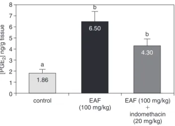
PG synthesis determination. Rats were divided into three groups of six animals each that, after a 24-hour fast, received one of the following solutions: saline (vehicle), EAF (100 mg/kg), or the combination of EAF (100 mg/kg) plus in-domethacin (20 mg/kg, s.c). In this last group, EAF was ad-ministered, followed 30 minutes later by indomethacin (dis-solved in 5% sodium bicarbonate solution). Thirty minutes after treatments, all the animals were sacrificed, and the ab-domen was opened. A sample of the corpus (full thickness) was excised, weighed, and suspended in 1 mL of 1 mM
sodium phosphate buffer, pH 7.4. The tissue was finely minced with scissors and then incubated at 37°C for 20 min-utes. PGE2 in the buffer was measured by enzyme
im-munoassay (Amersham Biosciences, Little Chalfont, Buck-inghamshire, UK), and the absorbance was read at 450 nm.20
Anti-H. pylori activity
The ME was tested to detect anti-H. pyloriactivity.21The
strain of H. pylori (ATCC 43504) had been isolated from patients with duodenal ulcer disease. The frozen H. pylori
isolate was thawed and grown on 5% sheep blood agar plates for 3–4 days at 37°C in 10% CO2and 98% humidity. Each
plate was swabbed with a sterile cotton-tipped applicator, and the cells were suspended in sterile saline to obtain tur-bidity equivalent to a 2.0 McFarland standard. Mueller-Hin-ton broth containing 10% horse serum was added to all wells of a 96-well microtiter plate (Corning, Corning, NY). Each well was incubated with H. pyloriat a final concentration of 1⫻105colony-forming units/mL. The plates were
established by the National Committee for Clinical Labora-tory Standards (Wayne, PA).
Statistical analysis
Results were expressed as mean⫾SE values, and statis-tical significance was determined by one-way analysis of variance followed by Dunnett’s or Tukey’s test with P⬍
.05 defined as significant.
RESULTS AND DISCUSSION
The balance between the therapeutic versus the toxico-logical effects of a drug is an important parameter in as-sessing its applicability as an anti-ulcer or a pharmacologi-cal agent.22As a part of this pharmacological study, ME was
first investigated for acute toxicity in mice. A single oral dose of ME (5 g/kg) did not produce any visible signs or symptoms of toxicity in the treated animals. After 14 days of administration, no animal died, and no significant macro-scopic changes in daily body or organ weights were served (data not shown). Since no acute toxicity was ob-served using ME, we continued our studies evaluating the effect of ME administered to rodents using different stan-dard experimental models of induced gastric ulcer.
cinal plants are generally present at low concentrations. So, in order to establish a general profile of the anti-ulcerogenic activity of the extract, we plotted the dose–response curve using three peroral doses of 250, 500, and 1,000 mg/kg to select the dose that produces the best effect.
Oral administration of absolute ethanol is noxious to the stomach by affecting the gastric mucosa topically by dis-rupting its barrier and provoking pronounced microvascular changes within a few minutes after its application. Konturek
et al.23 reported that the combination of HCl plus ethanol
also promotes stasis in gastric blood flow that contributes to the development of the hemorrhagic and necrotic aspects of tissue injury. Our results obtained with absolute ethanol and HCl/ethanol models gave similar results with 85–86% and 89–90% gastroprotection at the respective ME doses of 500 and 1,000 mg/kg (Table 1). These data suggest that ME displays a gastroprotective effect since it significantly re-duced the occurrence of ethanol-inre-duced ulcers.
It is commonly postulated that a mechanism that causes gastroduodenal damage involves cyclooxygenase-1 inhibi-tion that results in gastric PG suppression.24Deficiency of
endogenous PGs is widely accepted as a major factor in the pathogenesis of gastric lesions caused by NSAIDs.25,26
However, our data showed that in the NSAID-induced gas-tric lesion model, the ME-treated animals exhibited an
un-TABLE 1. GASTROPROTECTIVEEFFECTS OFME, EAF, ORAQF FROMA. TRIPLINERVIAONGASTRIC LESIONSINDUCED BYHCL/ETHANOL, PIROXICAM, ANDABSOLUTEETHANOL INRODENTS
Model (animal) Treatment (dose) Number ULI Inhibition (%)
HCl/ethanol (mice)a Vehicle 7 104.0⫾ 6.3 —
Lansoprazole 7 18.0⫾ 1.9** ⫺83
ME (250 mg/kg) 7 23.2⫾ 2.1** ⫺78
ME (500 mg/kg) 6 14.8⫾ 1.3** ⫺86
ME (1,000 mg/kg) 6 10.8⫾ 1.8** ⫺90
Piroxicam (mice)b Vehicle 7 32.8⫾ 3.3 —
Cimetidine 7 6.7⫾ 1.2** ⫺80
ME (250 mg/kg) 7 23.4⫾ 3.6 —
ME (500 mg/kg) 7 41.0⫾ 4.55 —
ME (1,000 mg/kg) 7 58.0⫾ 4* ⫺77
Ethanol (rats)
ME treatmentc Vehicle 7 65.8⫾ 6.7 —
Lansoprazole 7 29.0⫾ 4.2* ⫺56
ME (250 mg/kg) 7 47.7⫾ 16.6 —
ME (500 mg/kg) 7 10.1⫾ 1.3** ⫺85
ME (1,000 mg/kg) 7 7.0⫾ 1.4** ⫺89
EAF treatmentd Vehicle 5 100.7⫾ 11.9 —
Lansoprazole 6 21.3⫾ 5.6** ⫺79
EAF (100 mg/kg) 6 50.5⫾ 2.5** ⫺50
AqF treatmentd Vehicle 6 92.8⫾ 16.2 —
Lansoprazole 6 31.3⫾ 5.0** ⫺66
AqF (100 mg/kg) 6 71.8⫾ 10.1 —
Data are mean⫾ SE values. ULI, ulcerative lesion index.
For statistical analysis, analysis of variance followed by Dunnett’s test was employed: *P⬍ .05, **P⬍ .01. aF
4; 29⫽138.5. bF
4; 30⫽27.2. cF
4; 30⫽28.6. dF
expected result, i.e., the cytoprotection decreased as the dose was increased (Table 1). In this model, ME showed an ab-sence of gastroprotective effect (P ⬎.05) at doses of 250 mg/kg and 500 mg/kg, while the dose of 1,000 mg/kg in-duced a significant increase of gastric lesions (P⬍.05). It is highly probable that ME contains different active con-stituent(s), i.e., the substance or substances that protected the gastric mucosa against the damage induced by absolute ethanol and HCl/ethanol are different from those that pre-sented cytoprotection against the NSAID-induced gastric le-sions. Among natural substances of plant origin, the litera-ture reports that flavonoids may present this type of activity. Gracioso et al.27 reported that increasing the dose of
flavonoids changes their antioxidant activity to a pro-oxi-dant action, leading to an increase in gastric damage. The literature indicates that other species of the Alchornea gen-era present anti-inflammatory activity.7 Our results agree
closely with those published by Repetto and Llesuy,28which
showed that phenolic compounds have a dual effect on PG biosynthesis, since low concentrations stimulate whereas high concentrations inhibit PGH synthase. Thus, the action mechanism possibly involves the modulation of endogenous PGs by active constituents contained in ME.
With the purpose of investigating the probable gastro-protective mechanisms involved in the action promoted by this extract, we continued our studies using only a single
TABLE2. EFFECTS OFME OFA. TRIPLINERVIAADMINISTEREDORALLY ORINTRADUODENALLY ON GASTRICJUICEPARAMETERS INPYLORUSLIGATURE-INDUCEDGASTRICLESIONS INMICE
Route, Dose Total acid (Eq/mL/ Gastric volume Inhibition
treatment Number (mg/kg) 4 hours) pH (unit) (mL) ULI (%)
Intraduodenal
Control 9 — 32.11⫾ 1.18 2.50⫾ 0.34 0.98⫾ 0.07 49.72⫾ 4.86 — Cimetidine 8 100 18.77⫾ 2.22** 3.90⫾ 0.43* 0.82⫾ 0.06 12.50⫾ 1.95** 75 ME 8 500 29.72⫾ 1.98 1.70⫾ 0.26 1.12⫾ 0.03 19.80⫾ 1.63** 60 Peroral
Control 7 — 46.57⫾ 3.50 3.00⫾ 0.26 0.90⫾ 0.09 46.57⫾ 3.52 — Cimetidine 8 100 16.87⫾ 2.25** 2.50⫾ 0.19 1.09⫾ 0.08 22.14⫾ 3.32** 52 ME 8 500 4.00⫾ 0.50** 3.30⫾ 0.37 1.05⫾ 0.11 9.25⫾ 1.16** 80
Data are mean⫾ SE values. ULI, ulcerative lesion index.
For statistical analysis, analysis of variance followed by Dunnett’s test was employed. By the intraduodenal route: **P⬍ .01 for total acid with F2; 22⫽15.37, *P⬍ .05 for pH with F2; 22⫽9.99; P⬎ .05 for gastric volume with F2; 22⫽13.95, and **P⬍ .01 for ULI with F2; 22⫽ 39.01. By the peroral route: **P⬍ .01 for total acid with F2; 20⫽93.11, *P⬍ .05 for pH with F2; 20⫽2.03, P⬎ .05 for gastric volume with F2; 20⫽0.94, and **P⬍ .01 for ULI F2; 20⫽47.47.
TABLE3. EFFECTS OFME OFA. TRIPLINERVIAONGASTRICLESIONSINDUCED BYETHANOL INRATSPRETREATED WITHNEM OR L-NAME (N⫽7)
Pretreatment Treatment Dose
(intraperitoneal) (peroral) (mg/kg) ULI (% inhibition)
Saline Vehicle — 73.7⫾ 6.7a
Carbenoxolone 100 31.8⫾ 5.9 (57%)**
ME 500 26.6⫾ 2.8 (64%)**b
NEM (10 mg/kg) Vehicle — 148.3⫾ 15.3a
Carbenoxolone 100 106.3⫾ 16.3 (28%)*
ME 500 40.6⫾ 4.5 (73%)**b
Saline Vehicle — 135.5⫾ 18.8c
Carbenoxolone 100 24.0⫾ 5.0 (82%)**
ME 500 20.1⫾ 14.9 (85%)**d
L-NAME (70 mg/kg) Vehicle — 197.7⫾ 15.0c
Carbenoxolone 100 81.7⫾ 20.4 (59%)*
ME 500 41.0⫾ 8.5 (79%)**d
Data are mean⫾ SE values. ULI, ulcerative lesion index.
Analysis of variance followed by Dunnett’s test was used to determine significant differences from the respective con-trol group: *P⬍ .05, **P⬍ .01.
aSaline/vehicle versus NEM/vehicle (P⬍ .05).
bSaline/ME versus NEM/ME (P⬎ .05) by analysis of variance followed by Tukey’s test. cSaline/vehicle versus L-NAME/vehicle (P⬍ .05).
had produced the best results in the previous studies, and no significant differences were observed between groups treated with 500 or 1,000 mg/kg ME.
In pyloric ligation, the digestive effect of accumulated gastric juice (gastric hypersecretion) and the interference of gastric blood circulation are responsible for the induction of ulceration.29,30Animals pretreated orally with ME presented
a decrease of ulcerative lesions and a drop in the total acid value in gastric juice (Table 2). Otherwise, the intraduode-nal administration of ME did not change any gastric juice biochemical parameter but decreased the ulcerative index. Therefore, these data indicate that ME presented antisecre-tory action only when this extract was given orally and that intestinal absorption of ME did not contribute to its antise-cretory effect.
Ethanol-induced gastric damage is associated with a sig-nificant decrease in the mucosal sulfhydryl (SH) level, in-cluding reduced gluthathione, and pretreatment with SH blockers prevents gastroprotection of SH-containing com-pounds.31,32 Our data show that pretreatment with the SH
blocker NEM did not reduce the mucosal protection ob-served with ME treatment. These findings suggest that an increase of endogenous SH is not involved in the gastro-protective effect of ME, as shown in Table 3.
Vascular changes in gastric mucosa appear to be the most pronounced feature of absolute ethanol-induced injury.33
Maintenance of mucosal vasculature and normal blood flow may constitute the major cytoprotective mechanism.34 As
shown in Table 3, L-NAME, a nitric oxide synthase
in-hibitor, also did not attenuate the gastroprotection observed
for ME, suggesting that endogenous nitric oxide did not par-ticipate in the protective effect of ME.
We also evaluated the anti-H. pyloriactivity of ME. In the course of the study, A. triplinerviaME was found to pre-sent antibacterial activity against the standard strain of H.
pylori(ATCC 43504). The results showed that the MIC of
ME against H. pylori was 0.25 mg/mL. The literature re-ported that an MIC of ⬍0.50 mg/mL is considered interest-ing for extracts.21,35We conclude that ME presents
excel-lent antimicrobial action against one of the most important factors that cause gastric ulceration.
In order to better comprehend the effect of ME on gas-tric injuries, we separated ME into ethyl acetate and water, thus obtaining two fractions (EAF and AqF). The pretreat-ments with these fractions were also evaluated against the ethanol-induced gastric lesions that had resulted in the best results with ME. We observed that the active constituents responsible for the protective action are concentrated in the EAF (50% protection), rather than in the AqF, which did not induce significant gastric protection at the same dose (Table 1). Therefore, we can conclude that the active con-stituents responsible for the protective action are concen-trated in the EAF and not in the AqF.
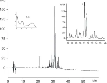
We also determined spectrophotometrically the total con-centration of flavonoids in the A. triplinervia leaves. The flavonoid contents were 19.73% (197.3⫾0.19 mg/g) in the EAF and 1.3% (13.1⫾0.04 mg/g) in the AqF from ME. In agreement with these results, Figures 1 and 2 present the chromatograms and UV spectra of the respective fractions EAF and AqF from ME. The EAF contained primarily five phenolic compounds: ellagic acid, quercetin-3-O -galactopy-ranoside, quercetin-7-O-glucopyranoside, quercetin-3-O
-0 0
10 20 30 40 50 Min
100 200 300 mAU
0
27.5 1
2–4 1
2
3
4 5
Min 35.0 32.5 30.0 100
200 300 mAU
0 0
10 20 30 40 50 Min
25 50 75 100 125 150
0
27 2–3
2
3
Min 35 34 29 30 31 32 33 28
25 100
75
50 125 mAU
FIG. 2. High-performance liquid chromatography profile of AqF from A. triplinervia. Chromatographic conditions were as follows: RP-18 column, 250⫻4.6 mm (i.d.), 5 m; elution with water (0.05% tri-fluoroacetic acid)/acetonitrile (0.05% tritri-fluoroacetic acid), 30% to 70% in 60 minutes; flow rate, 1.0 mL/minute; ⫽360 nm. mAU, milli-absorbance units.
glucopyranoside, and quercetin-3-O-arabinopyranoside. The AqF, which did not exhibit gastroprotective action, con-tained only quercetin-3-O-galactopyranoside and quercetin-7-O-glucopyranoside. So these quantitative (almost 15 times more flavonoid content than found in EAF) and qualitative differences between fractions were essential to the cytopro-tective effect observed with ME.
Although PGs play an important role in modulating gas-tric mucosal integrity and in regulating gasgas-tric acid secre-tion, little is known regarding regulation of PG synthesis by the stomach.20 Arakawa et al.36 suggested that PGs
accel-erate ulcer healing, possibly via angiogenesis, epithelial cell proliferation, reconstruction of extracellular matrices, sup-pression of inflammatory cell infiltration, and production of growth factors such as hepatocyte growth factor and trans-forming growth factor-. In the present work, we observed that treatment with EAF induced a significant jump in PGE2
production to double the basal levels. These results explain the gastroprotective action obtained from ME against ethanol as well as the antisecretory action observed under the pylorus ligature method. It is notable that our previous work with A. castaneaefolia extract also produced an in-crease in PGE2production.8However, more interesting
re-sults were obtained in the group of animals treated with EAF and pretreated with indomethacin (an inhibitor of cy-clooxygenase enzyme). Results displayed in Figure 3 show that EAF was able to maintain a high PGE2 level despite
administration of indomethacin. These results may alter the therapy of gastroprotective drugs associated with NSAIDs because EAF was able to promote a sustained increase of PGE2 levels, which is vital to the integrity of gastric
mu-cosa. Furthermore, the association of EAF and NSAID was not capable of reducing levels of PGE2release in a
signifi-cant manner. Park et al.37 also evaluated flavonoids
origi-nating from Scutellaria baicalensis and observed that this compound provides a cytoprotective effect; they inferred
dual action of wogonin on arachidonic acid metabolism, in-cluding the induction of cyclooxygenase-2 expression.
In conclusion, all these results taken together suggest that ME from the leaves of A. triplinervia did not show acute toxicity and exhibited an antisecretory property, anti-H. py-lorieffect, and gastroprotective action. The gastroprotective effect presented by the EAF was proven more efficient than that shown by the ME, while the gastroprotective action oc-curred by increasing the PGE2level.
ACKNOWLEDGMENTS
We are grateful to the Fundação de Amparo à Pesquisa do Estado de São Paulo (FAPESP), Capes, and CNPq for grants to Z.P.L., W.V., A.R.M.S.B., and C.A.H.-L. and to the Biota-FAPESP Program for funding.
AUTHOR DISCLOSURE STATEMENT
No competing financial interests exist.
REFERENCES
1. Jain KS, Shah AK, Bariwal J, Shelke SM, Kale AM, Jagtap JR, Bhosale AV: Recent advances in proton pump inhibitors and man-agement of acid-peptic disorders. Bioorg Med Chem 2007;15: 1181–1205.
2. Chan FKL, Leung WK: Peptic-ulcer disease. Lancet2002;360: 933–941.
3. Ramakrishnan K, Salinas RC: Peptic ulcer disease. Am Fam Physician2007;76:1005–1012.
4. Schmeda-Hirschmann G, Yesilada E: Traditional medicine and gastroprotective crude drugs. J Ethnopharmacol2005;100:61–66. 5. Silva EM, Hiruma-Lima CA, Lólis SF: Etnobotânica no municí-pio de Porto Nacional [abstract]. In: Symposium of Brazilian Med-icinal Plants. Universidade Federale de Mato Grosso, Cuiabá, Brazil, 2000, p. 106.
6. Braca A, Mendez J, Menichini F, Morelli I: Constituents of Al-chornea triplinervia (Euphorbiaceae). Biochem Syst Ecol 2002;30:1109–1111.
7. Manga MH, Brkic D, Marie DEP, Quetin-Leclerc Q: In vivo anti-inflammatory activity of Alchornea cordifolia(Schumach & Thonn.) Müll. Arg. (Euphorbiaceae). J Ethnopharmacol2004;92:209–214. 8. Hiruma-Lima CA, Calvo TR, Rodrigues CM, Andrade FD, Vile-gas W, Brito AR: Antiulcerogenic activity of Alchornea cas-taneaefolia: effects on somatostatin, gastrin and prostaglandin. J Ethnopharmacol2006;104:215–224.
9. Calvo TR, Lima ZP, Silva JS, Ballesteros KV, Pellizzon CH, Hiruma-Lima CA, Tamashiro J, Brito ARMS, Takahira RK, Vilegas W: Constituents and antiulcer effect of Alchornea glan-dulosa: activation of cell proliferation in gastric mucosa during the healing process. Biol Pharm Bull2007;30:451–459. 10. Ebi GC: Antimicrobial activities of Alchornea cordifolia.
Fi-toterapia2001;72:69–72.
11. Wagner H, Bladt H, Zgainski EM: Plant Drug Analysis. Springer, Berlin, 1984.
12. Olfert ED, Cross BM, McWilliam AA: Guide to the Care and Use of Experimental Animals. Canadian Council on Animal Care, Ottawa, 1993.
control EAF
(100 mg/kg)
EAF (100 mg/kg)
⫹
indomethacin (20 mg/kg) a
b
b
0
[PGE
2
] ng/g tissue
1 3 5 7
2 4 6 8
1.86
6.50
4.30
Editora da Unicamp, Campinas, Brazil, 1995, pp. 15–22. 14. Mizui T, Douteuchi M: Effect of polyamines on acidified
ethanol-induced gastric lesions in rats. Jpn J Pharmacol1983;33:934–945. 15. Szelenyi I, Thiemer K: Distention ulcer as a model for testing of drugs for ulcerogenic side effects. Arch Toxicol1978;41:99–105. 16. Morimoto Y, Shimohara K, Oshima S, Sukamoto T: Effects of the new anti-ulcer agent KB-5492 on experimental gastric mucosal le-sions and gastric mucosal. Jpn J Pharmacol1991;57:595–605. 17. Puscas I, Puscas C, Coltau M, Pasça R, Torres J, Márquez M,
Herrero E, Fillat O, Ortiz J: Comparative study of the safety and efficacy of ebrotidine versus ranitidine and placebo in the pre-vention of piroxicam-induced gastroduodenal lesions. Arzneimit-telforschung1997;47:568–572.
18. Shay H, Komarov SA, Fels SS, Meranze D, Gruenstein M, Siplet H: A simple method for the uniform production of gastric ulcer-ation in the rat. Gastroenterology1945;5:43–61.
19. Arrieta J, Benitez J, Flores E, Castillo C, Navarrete A: Purification of gastroprotective triterpenoids from the stem bark of Amphiptery-gium adstringens; role of prostaglandins, sulfhydryls, nitric oxide and capsaicin-sensitive neurons. Planta Med2003;69:905–909. 20. Curtis GH, MacNaughton WK, Gall DG, Wallace JL:
Intralumi-nal pH modulates gastric prostaglandin synthesis. Can J Physiol Pharmacol1995;73:130–134.
21. Hacem CY, Clarridge JE, Reddy R, Flamm R, Evans DG, Tanaka SK, Graham DY: Antimicrobial susceptibility testing of Heli-cobacter pylori. Diagn Microbiol Infect Dis1996;41:24–37. 22. Ekwall B, Ekwall K: Comments on the use of diverse cell
sys-tems in toxicity testing. Altern Lab Anim1988;15:193–200. 23. Konturek PC, Brzowski T, Sliwoswski Z: Involvement of nitric
oxide and prostaglandin in gastroprotection induced by bacterial lipopolysaccharide. Scand J Gastroenterol1988;33:691–700. 24. Peng S, Duggan A: Gastrointestinal adverse effects of
non-steroidal anti-inflammatory drugs. Expert Opin Drug Safety 2005;4:157–169.
25. Wolfe MM, Lynda S, Sachs G: Proton pump inhibitors and gas-tric acid secretion. Am J Gastroenterol2001;96:3467–3468.
tion. Dig Dis Sci1998;43(Suppl):23S–29S.
27. Gracioso JS, Vilegas W, Hiruma-Lima CA, Souza-Brito ARM: Effects of tea from Turnera ulmifolia L. on mouse gastric mu-cosa support the Turneraceae as a new source of antiulcerogenic drugs. Biol Pharm Bull2002;25:470–491.
28. Repetto MG, Llesuy SF: Antioxidant properties of natural com-pounds used in popular medicine for gastric ulcers. Braz J Med Biol Res2002;35:523–534.
29. Brodie BB, Costa E, Dlabac A, Neff NH, Smookler HH: Appli-cation of steady state kinetics to the estimation of synthesis rate and turnover time of tissue catecholamines. J Pharmacol Exp Ther 1966;154:493–498.
30. Rastogi L, Patnaik GK, Dikshit M: Free radicals and antioxidant status following pylorus ligation induced gastric mucosal injury in rats. Pharmacol Res1998;38:125–132.
31. Szabo S, Trier JS, Frankel PW: Sulfhydryl compounds may me-diate gastric cytoprotection. Science1981;214:200–202. 32. Lopez A, Motilva V, Alarcón de la Lastra C, Martin MJ, La Casa
C: The role of gastric mucosal sulphydryls in the ulcer-protect-ing effects of cisapride. J Pharm Pharmacol1996;48:37–40. 33. Laine L, Weinstein WM: Histology of alcoholic hemorrhagic
“gastritis,” a prospective evaluation. Gastroenterology1988;94: 1254–1262.
34. Szallasi A, Biro T, Modarres S, Garlaschelli L, Petersen M, Klusch A, Vidari G, Jonassohn M, De Rosa S, Sterner O, Blum-berg PM, Krause JE: Dialdehyde sesquiterpenes and other ter-penoids as vanilloids. Eur J Pharmacol1998;356:81–89. 35. Gadhi CA, Benharref A, Jana M, Lozniewski A: Anti-
Heli-cobacter pylorusactivity of Aristolochia paucinervispomel ex-tracts. J Ethnopharmacol2001;75:203–205.
36. Arakawa T, Higuchi K, Fukuda T, Fujiwara Y, Kobayashi K, Kuroki T: Prostaglandins in the stomach: an update. J Clin Gas-troenterol1998;27(Suppl):S1–S11.
1. Jorge M.B. Vítor, Filipa F. Vale. 2011. Alternative therapies for Helicobacter pylori: probiotics and phytomedicine. FEMS Immunology & Medical Microbiology n/a-n/a. [CrossRef]
2. Ariane Leite Rozza, Thiago de Mello Moraes, Hélio Kushima, Alexandre Tanimoto, Márcia Ortiz Mayo Marques, Taís Maria Bauab, Clélia Akiko Hiruma-Lima, Cláudia Helena Pellizzon. 2011. Gastroprotective mechanisms of Citrus lemon (Rutaceae) essential oil and its majority compounds limonene and β-pinene: Involvement of heat-shock protein-70, vasoactive intestinal peptide, glutathione, sulfhydryl compounds, nitric oxide and prostaglandin E2. Chemico-Biological Interactions189:1-2, 82-89. [CrossRef]
3. A. Quílez, B. Berenguer, G. Gilardoni, C. Souccar, S. de Mendonça, L.F.S. Oliveira, M.J. Martín-Calero, G. Vidari. 2010. Anti-secretory, anti-inflammatory and anti-Helicobacter pylori activities of several fractions isolated from Piper carpunya Ruiz & Pav.