1 Recebido para publicação em 12.7.2006 e na forma revisada em 27.2.2007.
2 Eng.-Agrônomo, Doutorando em Fitotecnia da Universidade Federal de Viçosa – DFT/UFV, 36570-000 Viçosa-MG, <[email protected]>; 3D.Sc., Professora do Departamento de Biologia Vegetal – DBV/UFV;4D.Sc., Professor da Universidade do Vale do Rio Doce – UNIVALE, Governador Valadares-MG;5Professor Associado – DFT/UFV.6D.Sc., Professor da Faculdade de Agronomia da Universidade de Rio Verde – FESURV.7Ph.D., Professor associado do Departamento de Biologia Vegetal – DBV/UFV
LEAF
BLADE
QUANTITATIVE
ANATOMY OF
SUGARCANE
CULTIVARS AND
CLONES
1Anatomia Quantitativa da Lâmina Foliar de Genótipos de Cana-de-Açúcar
FERREIRA, E.A.2, VENTRELLA, M.C.3, SANTOS, J.B.4, BARBOSA, M.H.P.5, SILVA, A.A.5, PROCÓPIO, S.O.6and SILVA, E.A.M.7
ABSTRACT- The objective of this study was to make a quantitative assess of the anatomic characteristics of leaf blade of the sugarcane cultivars RB855113, SP80-1842, SP80-1816, RB867515 and clone RB957689 presenting different sensitivity to the mixture of sodium trifloxysulfuron + ametryn herbicides. Compared to the other cultivars assessed, RB855113 cultivar, considered more sensitive to the herbicide mixture, presented relevant differences such as greater proportion of bulliform cells, greater tissue proportion in the transverse section of the leaf blade, greater stomata and trichome density on both surfaces, thinner epidermis on the adaxial surface and length of stomata on both surfaces. The external paraclinal wall of the bulliform cells was thinner than in the common epidermis cells in all the genotypes on the adaxial and abaxial surfaces. Multivariate analysis of the data on the variables considered most relevant to explain the herbicide penetration singled out the sensitive RB855113 from the other materials. Such characteristics can explain the greater penetration, and consequently, greater sensitivity of this cultivar to the sodium trifloxysulfuron + ametryn mixture.
Keywords: leaf anatomy, herbicide,Saccharumsp.
RESUMO-Objetivou-se neste trabalho avaliar, quantitativamente, as características anatômicas da lâmin a foliar dos cultivar es de cana- de-açúcar RB8 551 13, SP80-1 842 , SP80-1 816 e RB867515 e do clone RB957689 com diferentes sensibili dades à mistura dos herbicidas trifloxysulfuron-sodium + ametryn. O cultivar RB855113, considerado mais sensível à mistura de herbicidas, mostrou diferenças relevantes em relação aos demais cultivares avaliados, como: maior proporção de células buliformes, tecido encontrado em maior proporção na seção transversal da lâmina foliar desse cultivar, maior densidade de estômatos e de tricomas em ambas as faces, epiderme menos espessa na face adaxial e estômatos com maior comprimento de ostíolos nas duas faces. Todos os genótipos apresentaram a parede periclinal externa das células buliformes mais delgada do que nas células epidérmicas comuns, nas faces adaxial e abaxial. A análise multivariada dos dados relacionados às variáveis consideradas mais relevantes para explicar a penetração de herbicidas distinguiu o cultivar sensível RB855113 dos demais materiais. Ess as características pod em exp licar a maior pen etraç ão e, con seq üen teme nte, a maior sensibilidade desse cultivar à mistura trifloxysulfuron-sodium + ametryn.
INTRODUCTION
Sug ar can e cul tiv ars and clo nes hav e different sensitivity to herbicides which can cause crop damage, with significant reduction in sugar cane plantation yield (Procópio et al., 2003).
Knowledg e of the mech anisms through which herbicides penetrate the plant tissues is fundamental for the correct use and efficiency of the product. Herbicides can penetrate the plant through the aerial structures (leaves , stems, flowers and fruits) and subterranean structures (roots, rhizomes, tubers, etc.) or, even through seeds during germination and emergence, through the radicle and epicotyl. How ever, th e lea ves are the mai n org ans involved in the penetration of herbicides applied during the post emergence period (Silva et al., 2003).
The quantity of herbicide intercepted and retained in the leaves is mainly influenced by their morphology. However, the anatomical and micro morphol ogical char acte ris tics of the plant determine how easily these products will be absorbed (Hess & Falk, 1990).
Van Dillewijn (1952) reported the work of others noting that the epidermis of sugarcane leaves presents formations such as bulliform cel ls, sto mat a, sil ica cel ls and tri cho mes . Arts chawager (1925) iden tifi ed thre e zones in the upp er epi der mis : 1) a cen tra l zon e consisting of long cells alternated with groups of short cells, presenting trichomes and silica cells; 2) a stomata zone; and 3), a margin zone consisting of long cells alternated with short cell s, also pres enting trichom es and sili ca cells. Inexplicably, Artschwager diagrammed and discussed the role of bulliform cells in the upper epidermis but failed to list these as a fourth zone. He did, however, highlight the impor tance of the epidermis of the abaxia l (lower) leaf surface in identif ying cultivated sug arc ane varie ties, whe re sil ica cel l and stomata distribution were diagnosed.
Pla nt leav es present va ri ous leve ls of trichomes and gland development. Abutilon the ophrasti, for exa mpl e, has simpl e and complex trichomes, whileChenopodium album has a high density of glandular trichomes in th e ad ax ia l ep id er mi s th at can hi de th e
epidermis cells completely (Hess & Falk, 1990). Tri cho mes , esp eci all y the bra nch ed one s, pre sen t on the lea f sur fac e can int erc ept sprayed drops, preventing them from reaching the epidermis. Even when these trichomes are simple and appear in low density, the drops adhe re to the m. The absorpt ion effici ency of he rb icides by the tr ic home s an d th ei r translocation to the epidermis cells are partially understood (Hess & Falk, 1990). However, according to Hull (1970), some absorption of chemical products may occur via the trichomes. Comme lina benghalensis, more sensi tive to glyphosate thanCommelina diffusa, presented a greate r tri chome dens ity than the mor e to le ra nt sp ec ie s (S an to s et al ., 20 01 ). Neve rt he le ss, fe w auth or s st at e th at th e trichomes are a pathway for herbicide entry in the leaves. Generally, a negative relationship seems to exist between herbicide adherence to the trichomes and the efficacy of these products (Hess & Falk, 1990).
According to (Hess & Falk, 1990), most weeds present stomata on the adaxial and abaxi al surfaces (amph isto matic). Procópio et al. (2003) analyz ed 20 weed species and observed that19 were amphistomatic and only one was hypostomatic. Meyer et al. (1973) counted the number of stomata in 39 species, and observed that 16 were amphistomatic and the remaining 23 were hypostomatic. These authors also stated that in the amphistomatic, species,the number of stomata on the adaxial surface was, in most cases, lower than that on the abaxial surface.
The herbicide liquid drop, when sprayed on the leaf surface, can volatilize or be washed away by rain; it can also remain on the surface as a viscous liquid or in the form of crystal; it can penetr ate, but remain absorbed in the lipophylic components of the cuticle; it can penetrate the cuticle and the cell wall and thus trans loc ate bef ore rea chi ng the sym plast. When the product penetrates the cuticle, the cell wall and reaches the cell interior (through the plas malema) , symplas tic tra nslocat ion occurs (Hess, 1995).
Raphanus raphanistrumwere also reported by Ferreira et al. (2003), in quantitative anatomy studies.
The cuticle covers the external paraclinal wall of the epidermis cells forming a covering co ns is ti ng ma in ly of li pi d su bs ta nc es . The cuticle is the main herbicide absorption pa th wa y, so th at un de rs ta nd in g it is fundamental in studies on the absorption of these compounds (Procópio et al., 2003). This covering over the primary celullose wall, from the inside to the outside, consists of secondary cuticle or cutinized wall (cuticle plus wall), pr im ar y cu ti cl e or cu ti cl e it se lf , wh er e embedded wax is found and, on the cuticle covering the most external part, the epicuticle wax (Herédia et al., 1998).
Th e ob je ct iv e of th is st ud y wa s to quantitatively assess the leaf blade anatomic char acterist ics of the sugar cane cultivars RB855113, SP80-1842, SP80-1816, RB867515 and clo ne RB957 689 prese nting dif feren t sensitivities to herbicides.
MATERIAL AND METHODS
The experiment was carried out in the Plant Growth Unit (UCP) of the Department of Plant Biology at the Federal University of Viçosa, in a protected environment. Pots containing six lite rs of Red-Yell ow Argi sol substrat e were used. Two stools were planted and maintained with one shoot eac h. Pot s wer e fert ili zed as rec omm end ed for sug ar cane cropp ing (C FSE MG , 19 99 ). A ra nd om ize d bl oc k ex pe ri me nt al de si gn wa s us ed wi th fi ve replications.
The sugar cane genot ypes used in this ex pe ri me nt ha d be en pr e- se le ct ed in a previous study which analyzed the sensitivity of 15 sugar cane genotypes to the sodium trifloxysulfuron + ametryn herbicide mixture. The following were selected from these cultivars and clones for anatomical studies: RB855113 (sen siti ve to the herb icide mixt ure) , SP80-1842, SP80-1816, (medium sensitivity to the mi xt ur e) an d RB 86 75 15 (t ol er an t to th e mixture) (Ferreira, 2005).
The younge st or uppermost leaf with a visible ligule was collected from five plants of
each genotype. Samples were removed from the mid region of the leaf blade, fixed in FAA50, stored in 70% ethanol (Johansen, 1940) and transported to the Plant Anatomy Laboratory of the Plant Biology Department at the Federal University of Viçosa.
Portions of 1 cm2fixed blade segment were
dehydrated in an ethylic series and blocked in hi st or es in , ac co rd in g to ma nu fa ct ur er ’s re co mm en da ti on s (H is to re si n- Le ic a) , cu t tra nsv ers ely in a rot ating microt ome wit h aut oma tic adv ance, with 8µm thick steel blades. The material was stained with toluidin blue pH 4 (O´Brien et al., 1965) and mounted in synthetic resin (Permount). The epidermis impression with instant glue method was used to observe the leaf surface on both the blade surfaces (Rodella et al., 1993).
To visualize the cuticle, cross sections were made, stained with scarlet red Sudan, and mounted between slides and cover slips with glycerinated gelatin.
Digitalized images of the sections were obta ined with a ligh t microscope attached to a di gi ta l ca mera an d co nn ec te d to a microcomputer. The image Pro plus computer program was used to obtain data on the area and linear means. A light chamber attached to the microscope was used to count.
The fol lowing ana tomic charac ter istics were determined for the transverse section of the leaf blade, using a 10x lens (Figure 1): epide rmis area on the adaxi al and abaxi al surfaces; mesophyll area; bulliform cell area; vascular bundle area; area of bundle sheath cel ls; scl ere nch yma are a; xyl em are a and ph lo em ar ea , ep id er mi s th ic kn es s (b ot h surfaces), external paraclinal wall perimeter of th e bu ll if or m ce ll s an d th e co mm on epidermis cells (both surfaces). The following cha rac ter ist ics were ass ess ed on th e lea f blade surface (Figure 2, A-B) on both surfaces: trichome density, stomata density and length. The area data were transformed in % compared to the total area and the count and linea r measurements were taken from 10 fields per replication.
Figure 1-Cross section of sugar cane leaf blade, RB855113 cultivar.Vascular bundles (VB); bundle sheath cells (BS); schlerenchyma (SC); epidermis (E); bulliform cells (BUL); xylem (X), phloem (P) and mesophyll (M).
Th e ch ar ac te ri st ic s co ns id er ed mo st relevant to herbicide penetration on the axial and abaxial surfaces were selected, such as trichome density , stomata density , stomata length, epidermis thickness, perimeter of the external paraclinal wall of the bulliform cells, and thickness of the external paraclinal wall of the bulliform cells and common epidermis ce ll s (b ot h su rf ac es ). Th es e da ta we re submitte d to stat isti cal anal ysis using the multivariate analysis of the canonic variables method to cluster the treatments according to their degree of similarity.
RESULTS AND DISCUSSION
Univariate analysis
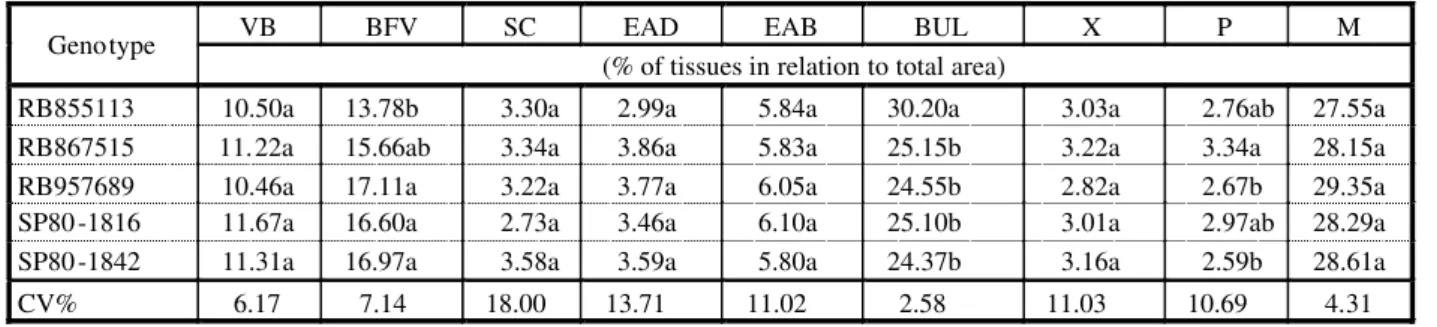
The main cells and tissues present in the suga rcane lea f blad e in cross section and paradermic view are presented in Figures 1 and 2 (A and B). The three tissues/cells that occurred in greatest proportion, in decreasing order, in the cross section of the leaf blade of the RB8 551 13, SP8 0-1 842 , SP80- 181 6, RB867515 cultivars and clone RB957689 were the mesophyll, followed by the bulliform cells and the bundle sheath cells of the vascular bundles. The RB855113 cultivar presented the gre ate st pro por tio n of bul lif orm cel ls, followed by mesophyll and the bundle sheath of the vascular bundle (Table 1, Figure 1). RB855113 presented 30.20% bulliform cells in the cross section of the leaf blade, differing from the others RB867515 (25.15%), SP80-1816 (25.15%), SP80-1842 (24.15%) and clone RB957689 (24.55% ) (Table 1).The bulliform
cells were layers of different widths on the leaf surface (Esau, 1965).
A smaller proportion of bundle sheath cells (13.78%) was detected in B855113, but it did not differ from RB867515 (15.6 6%); however, these cultivars differed from clone RB957689 (17.11%) and from SP80-1816 (16.60%) and SP80-1842 (16.9 7%) for this characterist ic (Table 1).
Cu lt iv ar s RB 95 76 89 an d SP 80 -1 84 2 pr es en te d a lo we r pr op or ti on of ph lo em compared to the other cultivars, respectively, 2.67% and 2.59 %, but did not diff er from RB855113 and SP80-1816 which presented an intermedi ate propo rtion , with 2.76% and 2. 97 % ph lo em , bu t di ff er en t fr om cl on e RB 95 76 89 (3 .3 4% ) (T abl e1 ). A hi ghe r proportio n of phloe m in a cultivar may be related to th e ease of systemic herbicide transportation (Procópio et al., 2003). However, in this case, no rela tionship was obser ved between phloem proportion and sensitivity to herbicide mixture.
Stomata density on the adaxial surface of the sugar can e leaves dif fer ed among the cultivar s. Hig her val ues wer e obs erv ed in RB855113 and clone RB957689 (85.60 and 83. 60 stoma ta mm-2, res pec ti ve ly) , and a
lower value in RB867515 (71.20 stomata mm-2)
(Table 2). RB855113 also presented greater stomata densi ty (163. 40 stomata mm-2) on
th e ab ax ial su rf ac e, di ff er in g fr om th e ot her cu lti var s an d clo ne s as se ss ed : SP80- 184 2 (14 0.8 0 sto mata mm-2), SP8
0-18 16 (1 44 .6 0 st om at a mm-2), RB 86 75 15
(1 44 .0 0 st om at a mm-2) a nd R B9 57 68 9
Table 1 - Tissue proportion in relation to total leaf blade area: Vascular bundles (VB); bundle sheath cells (BS); schlerenchyma (SC); adaxial surface epidermis (EDA) abaxial surface epidermis (EBA); bulliform cells (BUL); xylem (X), phloem (P) and mesophyll (M) in five sugarcane genotypes
VB BFV SC EAD EAB BUL X P M
Genotype
(% of tissues in relation to total area)
RB855113 10.50a 13.78b 3.30a 2.99a 5.84a 30.20a 3.03a 2.76ab 27.55a RB867515 11.22a 15.66ab 3.34a 3.86a 5.83a 25.15b 3.22a 3.34a 28.15a RB957689 10.46a 17.11a 3.22a 3.77a 6.05a 24.55b 2.82a 2.67b 29.35a SP80-1816 11.67a 16.60a 2.73a 3.46a 6.10a 25.10b 3.01a 2.97ab 28.29a SP80-1842 11.31a 16.97a 3.58a 3.59a 5.80a 24.37b 3.16a 2.59b 28.61a
CV% 6.17 7.14 18.00 13.71 11.02 2.58 11.03 10.69 4.31
(139.8 stomata mm-2) (Table 2). It should be
kept in mind that in agric ultural herbicide spraying, it is very difficult for the drops to reach the abaxial surface; consequently, the importance of absorption through the abaxial stomata is thought to be minimal. Another fac tor tha t may red uce the import anc e of herbicide absorption through the stomata may be that they are closed at various times of day and also during the night. However, Taylor et al. (1980) stated that the stomata were the ma in pa th s of be nt az on pe ne tr at io n in Chenopodium album.
Re ga rd in g st om at a si ze , RB 85 51 13 presented longer length, on both surfaces, with a mea n val ue of 23. 41 µm on the adaxia l surface and 26.05 µm on the abaxial surface (Table 3). The stomata cells are covered with a cuti cle that exte nds to the sub-stom ata chambers, and can be comp letely cov ered by wax (Esau, 1982).RB855113 pres ent ed greatest stomata density and greatest length; these characteristics may be important factors in the greater sensitivity of this cultivar to the herbicide mixture.
Regarding the presence of trichomes in the sugar cane leaf limbo, RB855113 presented greatest density on the adaxial and abaxial surfaces (31.28 and 121.04 trichomes mm-2,
respectively, differing from the other cultivars. Lower trichome density was observed in SP80-1816 and SP80-1842 on the adaxial surface. Howev er, in SP80-1816, RB867515 and in clone RB957689, lower trichome density was obse rved on the abaxial surf ace (Table 2). Sargent and Blackman (1962) reported that the cuticle was more permeable on the base of the trichomes ; thus subst ance penetrati on could be facilitated in this region. However, Hess & Falk (1990) reported that the trichomes formed a barr ier to herbici de pene tration. These authors noted that herbicide, adhered to the trichomes without reaching the epidermis. Accordi ng to Motomura et al. (2000), silica deposition is common on grass trichome walls, givin g them great er resistance to herbicide transport.
When the epidermis thickness of the five genotypes was compared, it was observed that RB855113 presented the least thick epidermis (13.09 µm) on the adaxial surface but did not differ from SP80-1842 (14.36 µm) and both
were diffe rent from the other cultivars. No statistical difference was observed among the cult ivars on the abax ial surf ace (Tab le 3). Most of the herbicide applied by spraying is int erc ept ed pre fer abl y by the ada xia l lea f surface, and is not usually deposited on the abaxial surface; thus, the adaxial surface is more important in herbicide interception and penetration (Hess & Falk, 1990).
Analysis of the perimeter of the external paraclinal wall of the bulliform cells showed that SP80-1842 presented greatest perimeter (98.64 µm), this did not differ significantly from RB85511 3 (98.6464 µm) but dif fer ed fro m RB867515 and clone RB957689, which had less bulliform cell perimeter, with mean values of 86.98 and 75.43 µm, respectively.
Table 2 -Mean values of stomata density on the adaxial and abaxial surfaces (DAD and DAB),and trichome density on the adaxial and abaxial surfaces (TAD and TAB) in sugar cane genotypes
Stomata mm-2 Trichomes mm-2
Genotype
DAD DAB TAD TAB
RB855113 85.60a 163.40a 31.25a 121.04a
RB867515 71.20b 144.00b 25.58b 88.44bc
RB957689 83.60a 139.80b 26.30b 85.56bc
SP80-1816 77.00ab 144.60b 19.18c 98.12b
SP80-1842 77.60ab 140.80b 16.84c 82.81c
CV(%) 7.12 3.69 10.85 7.53
Means followed by same letter in the sa me column did not differ by the Tukey test (P<0.05).
Table 3 - Mean stomatal length value on the adaxial and abaxial surfaces (COAD and COAB), epidermis thickness on the adaxial and abaxial surfaces (EEAD and EEAB) and external paraclinal wall perimeter of the bulliform cells (PEB) in sugar cane genotypes
COAD COAB EEAD EEAB PEB
Genotype
(μm)
RB855113 23.42a 26.06a 13.09b 12.22a 98.64ab
RB867515 21.10b 23.49b 15.54a 12.42a 81.95c
RB957689 21.27b 23.86b 15.23a 12.50a 75.43c
SP80-1816 19.76b 22.67bc 15.44a 12.45a 108.29a
SP80-1842 19.58b 21.52c 14.62ab 11.80a 89.99bc
CV(%) 4.52 4.16 5.39 7.39 19.74
No difference was obse rved amon g the cultivars in external bulliform cell paraclinal wall thickness (Table 4). RB867515 presented grea test exte rnal para clin al wall thickness in the epidermis cells on the adaxial surface (4. 71 µm) , dif fer ing fro m clone RB95768 9 and cultivar SP80-1842 (4.06 and 3.97 µm, res pect ive ly). Howe ver , they did not differ from RB85511 3 and SP80 -1816 (4. 42 and 4.14 µm).RB867515 also presented great er extern al par acl in al wa ll th ic knes s of th e epidermis cells on the abaxial surface (4.29 m), differing from clone RB957689 (3.5 µm), SP80-1816 (3.88 µm) and SP80-1842 (3.75 µm), but not from RB855113 (3.85 µm) (Table 4).
Figure 3-Detailof the crosssection of sugarcane leafblade, RB855113, SP80 1842, SP80 1816, RB867515;(A) stained with toluidina blue; (B) stained with scarlet red sudan. E: epidermis, and cell; BUL: bulliform cells; LW: lignified wall; NW non-lignified wall; CUT: cuticle.
Table 4 - Mean values of the external paraclinal wall thickness of the bulliform cells (PBL), on the epidermis cells on the adaxial surface (PAD) and on the abaxial surface (PAB) of the sugar cane genotypes. Means followed by the same lowercase letter in a column did not differ statistically by the Tukey test (P<0.05)
EBL EAD EAB CV (%)
Genotype
(μm)
RB855113 2.38Ab 4.24abA 3.85abA 6.91
RB867515 2.44aB 4.71aA 4.29aA 6.78
RB957689 2.66aC 4.06bA 3.50bB 5.86
SP80-1816 2.78aB 4.14abA 3.88abA 5.00
SP80-1842 2.67aB 3.97bA 3.75abA 6.50
CV(%) 8.18 7.39 7.57
When thicknesses of the external paraclinal wa ll of th e bu ll if or m ce ll s an d co mm on epidermis cells were compared on both surfaces in all the cultivars, the wall was found to be thinner over the former (Table 4). It was also observed that the external paraclinal wall of th e comm on epider mi s cells pr esen te d a greenis h blue colo rin g on the adax ial and abaxial surfaces, indicating the presence of lignification and intense blue coloring over the bullif or m ce ll s th at in dicat ed absen ce of lignification (Figure 3A). According to O’Brien et al. (1964), toluidin blue stain at pH 4 gives di ff er en ti at ed co lo ri ng de pe nd in g on th e chemical composition of the tissue analyzed; the lign ified wall prese nts a greenish blue colori ng due to ligni n, a phenol compoun d pres ent in the wall . The wall wit hout this compound presented an intense blue coloring. The material stained with scarlet red sudan (Figure 3B) revealed the presence of an orange-like color thin cuticle layer over the external paraclinal wall of all the epidermis cells.
As the ext ern al par acl ina l wal l of the bullif orm cells presented cuticle deposition similar to the other epidermis cells (Figure 3B), lig nin absence in the se wal ls (Figur e 3A), combined with reduced thickness, may make this region a preferential path for herbicide penetration.
Multivariate analysis
The canonic variable technique consists of the transforming the original variables into standardi zed and non-correlated vari ables , with the conglomeration process based on th e Ma ha la no bi s di st an ce (M ah al an ob is , 1956, quoted by Cruz, 1990). The new set of variabl es, when rank ed, retains maximum information on total variation. These variables will better explain the variability shown.
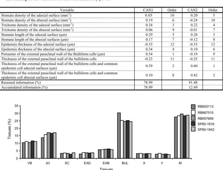
A set of 12 var iables was sel ect ed for multivariate analysis considered most related to herbicide penetration (trichome density on both surfaces, stomata density and length on both surfaces, epidermis thickness on both su rf ac es , exte rn al para cl in al wa ll of th e bulliform cells, perimeter of the bulliform cells, external paraclinal wall of the bulliform cells, thickness of the common epidermis cells and the bulliform cells on both surfaces). Hess
et al. (1990) and Procópio et al. (2003) stated that the main anatomic factors that would in fl uence he rb ic ide penetr at ion we re th e trichomes, stomata, cuticle and wall.
Due to high value of information retained in the CAN1 Canonic variable (78.99%) (Table 5), this first Canonic variable can be considered sufficient to explain the contribution of the original variables analyzed, for the five cultivars in five blocks. For the second Canonic variable (CAN2), the analysis in Table 5 shows that 12.49% of the information was retained. Thus, the first two variables explained 91.48% of the total variation in the original data. According to Regazzi (1998), when the firs t vari ables explain more than 80% of the total variation, divergence study is viable by the dist ance among cultivar s on the dispe rsion graphs, whose coordinates are scores related to the first canonic variables.
Cl us te r an al ys is ai ms to gr ou p th e treatments in various clusters by applying some ranking criteria so that there is homogeneity within the group and heter ogeneity among the groups. Alt er nati vely,c lus ter an alys is techniques aim to divide an original group of observations into various clusters, according to some criteria of similarity or dissimilarity among the treatments (Cruz & Regazzi, 1997). The Tocher optimization method distributed the five cultivars studied into groups, the first formed by RB867515, RB957689, SP80-1842, SP80-1816, and RB867515 and the second by the RB855113 cultivar.
The 12 variables analyzed were efficient in distinguishing two groups of cultivars. The SP80-1842, SP80-1816 culti vars and clone RB957689, presenting medium sensitivity to the sod ium trifl oxy sul furon + ame tryn to herbicide mixture, were placed with RB867515, to le ra nt to he rb ic id e mi xt ur e. RB 85 51 13 cultivar, sensitive to the mixture, was isolated as a separate group (Figure 5).
Figure 4 - Proportion of tissues (%) in relation to total leaf blade area: Vascular bundles (VB); bundle sheath cells (BS); schlerenchyma (SC); adaxial surface epidermis (EDA) abaxial surface epidermis (EBA); bulliform cells (BUL); xylem (X), phloem (P) and mesophyll (M) in five sugar cane genotypes.
Figure 5 - Graphic dispersion of the five sugar cane genotypes (1-RB855113; 2- RB867515; 3- SP957689; 4- 1816; 5- SP80-1842), using the two first Canonic variables (CAN1 and CAN2) for the set of 12 variables (trichome density on both surfaces, stomata density and length on both surfaces, epidermis thickness on the adaxial and abaxial surfaces, external paraclinal wall of the bulliform cells, external paraclinal wall of the common epidermis cells and bulliform cells on both surfaces).
Table 5 - Correlations among the 12 original variables regarding the anatomy of the cross section of sugar cane leaf blade, and the two Canonic variables (CAN1 and CAN2). Percentage of information retained and accumulated in CAN1 and CAN2, and ranking of the variables for their discriminatory power
Variable CAN1 Order CAN2 Order
Stomata density of the adaxial surface (mm-2) 0.05 10 0.20 5
Stomata density of the abaxial surface (mm-2) 0.19 6 -0.24 10
Trichome density of the adaxial surface (mm-2) 0.34 3 0.22 4
Trichome density of the abaxial surface (mm-2) 0.06 9 -0.01 7
Stomata length of the adaxial surface (µm) 0.29 5 0.28 3
Stomata length of the abaxial surfaces (µm) 0.17 7 -0.12 8
Epidermis thickness of the adaxial surface (µm) -0.33 12 -0.33 12
Epidermis thickness of the abaxial surface (µm) 0.34 4 0.10 6
Perimeter of the external paraclinal wall of the Bulliform cells (µm) 0.54 1 -0.19 9
Thickness of the external paraclinal wall of the bulliform cells -0.23 11 -0.25 11
Thickness of the external paraclinal wall of the bulliform cells and common
epidermis cell adaxial surfaces (µm) 0.39 2 0.60 1
Thickness of the external paraclinal wall of the bulliform cells and common
epidermis cell abaxial surfaces (µm) 0.10 8 0.42 2
Retained information (%) 78.99 91.48
Accumulated information (%) 78.99 12.49
0 5 10 15 20 25 30 35
VB BS SC EAD EAB BUL X P M
Tissues
T
issu
es (%)
RB855113
RB867515
RB957689
SP80-1816
surface than the other cultivars. SP80-1816 and RB855113 presented greater perimeter of the external paraclinal wall of the bulliform cells. As already discussed, such characteristics can expla in their greater sensitiv ity to the sodium trifloxysulfuron + ametryn mixture.
Multivariate analysis of the data related to the var iab les con sidere d mos t rel eva nt to explain herbicide penetration distinguished the sensitiv e RB855113 cultivar from the other materials, indicating that the set of 12 variables was efficient to form this clustering, but it did not separate the cultivar groups with medium sensitivity from the cultivars tolerant to the mixture.
LITERATURE CITED
ARTSCHWAGER, H. S. A. Anatomy of the vegetative organs of sugarcane. J. Agric., v. 30, p. 197-221, 1925.
COMISSÃO DE FERTILIDADE DO SOLO DO ESTADO DE MINAS GERAIS – CFSEMG. Recomendações para o uso de corretivos e fertilizantes em Minas Gerais – 5a
aproximação. Viçosa, MG: Universidade Federal de Viçosa, 1999. 359 p.
CRUZ, C. D. Aplicações de algumas técnicas
multivariadas no melhoramento de plantas. Piracicaba: Escola Superior Luiz de Queiroz, 1990. 188 p.
CRUZ, C. D.; REGAZZI, A. J. Modelos biométricos aplicados ao melhoramento genético. Viçosa, MG: Universidade Federal de Viçosa, 1997. 390 p.
ESAU, K. Anatomia de las plantas con semilla. Buenos Aires: Editorial Hemisferio Sur. 1982. 512 p.
FERREIRA, E. A. et al. Estudos anatômicos de folhas de plantas daninhas. I –Nicandra physaloid es,Solanum viarum,Solanum americanumeRaphanus raphanistrum. Planta Daninha, n. 2, p. 159-167, 2002.
FERREIRA, E. A. et al. Sensibilidade de cultivares de cana-de-açúcar à mistura trifloxysulfuron-sodium + ametryn. Planta Daninha, v. 23, n. 1, p. 93-99, 2005.
HEREDIA, A. La cutícula vegetal: estructura y funciones. Ecologia, v. 12, p. 293-305, 1998.
HESS, F. D. Absorption. In:HERBICIDEaction course. West Laffayete: Purdue University, 1995. 785 p.
HESS, F. D.; FALK, R. H. Herbicide deposition on leaf surfaces. Weed Sci., v. 38, p. 280-288, 1990.
HULL, H. M. Leaf structure as related to absorption of pesticides and other compounds. Residue Rev., v. 38, p. 1-155, 1970.
JOHANSEN, D. A. Plant microtechnique. New York: McGraw-Hill Book, 1940. 523 p.
MEYER, B. et al. Introdução à fisiologia vegetal. 2.ed. Lisboa: 1973. 710 p.
PROCÓPIO, S. O. et al. Manejo de plantas daninhas na cultura da cana-de-açúcar. Viçosa, MG: Universidade Federal de Viçosa, 2003. 150 p.
SILVA, A. A.; FERREIRA, F. A.; FERREIRA, L. R. Controle de plantas daninhas. Brasília, DF: ABEAS, 2003. 260 p.
PROCÓPIO, S. O. et al. Anatomia foliar de plantas daninhas do Brasil. Viçosa-MG: Universidade Federal de Viçosa, 2003. 118 p.
RODELLA. R. A.; PIRES, A. I.; MAIMONI-RODELLA, R. C. S. Anatomia comparativa foliar e caulinar de duas espécies daninhas deMerremia(Convolvulaceae). Científica, v. 21, p. 345-353, 1983.
SANTOS, I. C. et al. Características anatômicas de duas espécies de trapoeraba e a eficiência do glyphosate. Planta Daninha, v. 20, p. 1-8, 2001.
SARGENT, J. A.; BLACKMAN, G. E. Studies on foliar penetration. I. Factors controlling the entry of 2,4-dicloroacetic acid. J. Exp. Bot., v. 13, p. 384-368, 1962.
TAYLOR, F. E.; COBB, A. H.; DAVIES, L. G. The effects of bentazon on stomatal behavior inChenopodium albumL. New Phytol., v. 63, p. 369-376, 1980.