Applicatio n o f iso to pe -se le ctive
no n-dispe rsive infrare d spe ctro me try
fo r the e valuatio n o f the
13
C-ure a bre ath
te st: co mpariso n with thre e co nco rdant
m e tho ds
1Serviço de Gastroenterologia, Nutrição, Cirurgia Geral e do Aparelho Digestivo (GEN-CAD), Hospital das Clínicas, Universidade Federal de Minas Gerais, Belo Horizonte, MG, Brasil
2Brisotop Representações Ltda., São Paulo, SP, Brasil L.G.V. Coelho1, M. Reber2,
M.C.F. Passos1, R.O .A. Aguiar1, P.E. Casaes1, M.L. Bueno1, F.R. Yazaki1, F.J. Castro1, W.L.S. Vieira1, J.M.M. Franco1 and L.P. Castro1
Abstract
The aim of this work was to compare the performance of isotope-selective non-dispersive infrared spectrometry (IRIS) for the 13C-urea
breath test with the combination of the 14C-urea breath test (14C-UBT),
urease test and histologic examination for the diagnosis of H. pylori
(HP) infection. Fifty-three duodenal ulcer patients were studied. All patients were submitted to gastroscopy to detect HP by the urease test, histologic examination and 14C-UBT. To be included in the study the
results of the 3 tests had to be concordant. Within one month after admission to the study the patients were submitted to IRIS with breath samples collected before and 30 min after the ingestion of 75 mg 13
C-urea dissolved in 200 ml of orange juice. The samples were mailed and analyzed 11.5 (4-21) days after collection. Data were analyzed statis-tically by the chi-square and Mann-Whitney test and by the Spearman correlation coefficient. Twenty-six patients were HP positive and 27 negative. There was 100% agreement between the IRIS results and the HP status determined by the other three methods. Using a cutoff value of delta-over-baseline (DOB) above 4.0 the IRIS showed a mean value of 19.38 (minimum = 4.2, maximum = 41.3, SD = 10.9) for HP-positive patients and a mean value of 0.88 (minimum = 0.10, maxi-mum = 2.5, SD = 0.71) for negative patients. Using a cutoff value corresponding to 0.800% CO2/weight (kg), the 14C-UBT showed a
mean value of 2.78 (minimum = 0.89, maximum = 5.22, SD = 1.18) in HP-positive patients. HP-negative patients showed a mean value of 0.37 (minimum = 0.13, maximum = 0.77, SD = 0.17). IRIS is a low-cost, easy to manage, highly sensitive and specific test for H. pylori
detection. Storing and mailing the samples did not interfere with the performance of the test.
Co rre spo nde nce
L.G.V. Coelho Rua dos O toni, 705/601 30150-270 Belo Horizonte, MG Brasil
Fax: + 55-31-222-4641 E-mail: lcoelho@ gold.com.br
Research supported by FAPEMIG and CNPq. Publication supported by FAPESP.
Received November 20, 1998 Accepted July 28, 1999
Ke y words
·13C-urea breath test
Intro ductio n
Today, the causal relationship between gastric Helicobacter pylori infection and chronic gastritis and peptic ulcers is well established (1). Recently, this microorgan-ism was considered as a carcinogenic agent (type I) by the World Health Organization for gastric cancer, and studies carried out since 1993 have suggested its role in gastric mucosa-associated lymphoid tissue lym-phoma (2).
The diagnosis of its presence is regularly performed by endoscopic examination with the collection of gastric mucosa fragments for histological examination. This involves the use of various stains such as Giemsa stain, carbolfuchsin and others, microbio-logical tests (Gram and culture smears), or even colorimetric methods like the urease test which rely on the increased production of this enzyme by the microorganism. The diagnosis may even be performed through serological exams or by breath tests employ-ing carbon13- or carbon14- labelled urea. These
tests are based on the elevated production of urease. When the labelled urea is orally ad-ministered, the labelled CO2, originating from
the breakdown of this urea by the urease of the bacteria, can be detected in the air ex-pired by infected individuals. These breath tests, due to their accuracy, simplicity and low cost, are universally accepted today as the gold standard for monitoring patients undergoing anti-H. pylori therapy. The breath tests employing 14C-urea only require a
liq-uid scintillation spectrometer which is easily available at most medium-sized health cen-ters. The tests are inconvenient, however, because of their reliance on the use of a radioactive substance. The substance requires specialized personnel for handling, and should not be used in children and pregnant women. The recent development of instru-ments other than the mass spectrometer, which is of high cost and restricted availabil-ity, for tests using the stable, non-radioactive
isotope 13C has stimulated the wider use of
this methodology in the diagnosis of the pres-ence of H. pylori in the human stomach (3).
The present study aims to compare the
13C-urea breath test with three other methods
(histology examination, urease test, and 14
C-urea breath test) in patients with peptic ulcers.
Patie nts and Me thods
Before participating in the study, all pa-tients gave their written informed consent, and the study was approved by the Research Ethics Committee of UFMG University Hos-pital.
Fifty-three patients (30 men and 23 women) from the Peptic Ulcer Outpatient Clinic of the UFMG University Hospital, Belo Horizonte, were included. All patients had duodenal ulcers, including those admit-ted to the clinic for anti-H. pylori therapy, as well as those already submitted to eradica-tion of the microorganism. All candidates for inclusion in the study underwent upper digestive endoscopy in addition to gastric biopsies to test for H. pylori by both the urease test and histological examination, and the microorganism was detected by hema-toxylin and eosin and modified Giemsa stain-ing. Next, the patients underwent the 14
C-urea breath test as previously described (4), with values above 0.800% CO2/weight (kg)
considered to be positive (5). For the objec-tives of this study, patients were considered to be H. pylori positive when they were positive to the three traditional tests per-formed (urease, histological examination, and
14C-urea breath test), all performed within
13C-ure a bre ath te st
The test was performed using the infra-red isotope analyzer IRIS® (Wagner Analysen
Technik, Bremen, Germany), which allows a precise determination of the two isotopes,
13CO
2 and 12CO2. Two breath samples, taken
respectively before and after the ingestion of
13C-urea, are collected into 1.3-l bags and
are presented together to the instrument. Sam-pling and analysis are fully automated. The following methodology was used: after an overnight fast, a sample of expired CO2 air
was taken, corresponding to time 0 (control), through inflation of a 1.3-l breath bag. Next, patients ingested 75 mg of 13C-urea in 200
ml of orange juice without the addition of water or sugar. Another breath sample was taken 30 min after administration of the tracer. The two-bag samples obtained from each patient were stored at room temperature, packed and later shipped by conventional express air-mail to São Paulo, location of the equipment used in the study, where they were analyzed by one of the authors (MR). The results are presented as delta-over-baseline values (DOB) which indicate the change in the 13CO
2/12CO2 ratio brought about
by the metabolic activity induced by the administration of the labeled urea. Positive test results were those with DOB values above 4‰, as indicated by the manufactur-ers and as recently validated (6).
Statistical analysis
All of the procedures were performed by the same investigators, who were un-aware of the presence or absence of H. pylori in the examined patients. The homo-geneity of the two groups studied was deter-mined by the Mann-Whitney and chi-square tests, with the level of significance set at P<0.05. Spearman’s coefficients were cal-culated to test the correlation between the results of the 13C- and 14C-urea breath
tests.
Re sults
Table 1 shows the demographic charac-teristics of the two groups studied, demon-strating their homogeneity. The median in-terval between performance of the 13C- and 14C-urea breath tests in Belo Horizonte and
their analysis in São Paulo was 11.5 days (minimum = 4, maximum = 21, SD = 5.84, CI 95% = 8-14).
There was 100% agreement between the results of the 13C-urea breath tests and the H.
pylori status determined by the combination of the urease test, histological examination and 14C-urea breath tests.
1
4C
T
e
s
t
v
a
lu
e
s
5.6
4.8
4.0
3.2
2.4
1.6
0.8
0
13C cutoff
14C cutoff
0 4 8 12 16 20 24 28 32 36 40
13C Test values
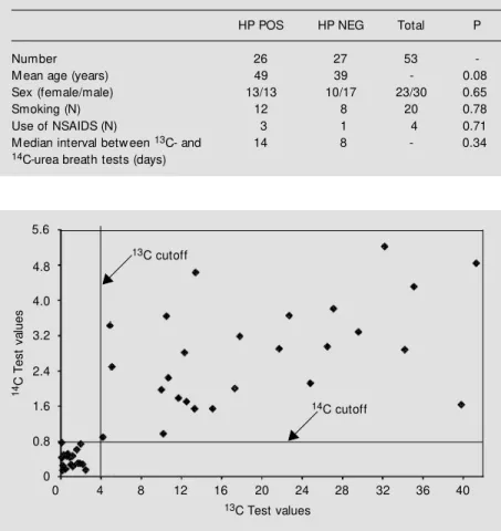
Figure 1 - Comparison of the results of the 13C- and 14C-urea breath tests using a cutoff of
4‰ DOB (delta-over-baseline) for the 13C-urea breath test and a cutoff of 0.800% CO2/
w eight (kg) for the 14C-urea breath test for detection of H. pylori infection.
Table 1 - Demographic characteristics of the 53 patients studied.
HP POS, Helicobacter pylori positive; HP NEG, Helicobacter pylori negative; NSAIDS,
nonsteroidal anti-inflammatory drugs.
HP POS HP NEG Total P
Number 26 27 53
-M ean age (years) 49 39 - 0.08
Sex (female/male) 13/13 10/17 23/30 0.65
Smoking (N) 12 8 20 0.78
Use of NSAIDS (N) 3 1 4 0.71
M edian interval betw een 13C- and 14 8 - 0.34
Figure 1 illustrates the results of the 13
C-and 14C-urea breath tests, showing a 100%
coincidence between the results of the two tests. With a cutoff point corresponding to a DOB value above 4.0, the 13C-urea breath
test had a median value of 19.38 (minimum = 4.2, maximum = 41.3, SD = 10.9, CI 95% = 14.95-23.81) for the positive patients and a median value of 0.88 (minimum = 0.10, maximum = 2.5, SD = 0.71, CI 95% = 0.60-1.17) for the negative patients. With a cutoff point corresponding to a value of 0.800% CO2/weight (kg), the 14C-urea breath test
showed a median value of 2.78 for H. pylori -infected individuals (minimum = 0.89, maxi-mum = 5.22, SD = 1.18, CI 95% = 2.30-3.26). The median value for non-infected individuals was 0.37 (minimum = 0.13, maxi-mum = 0.77, SD = 0.17, CI 95% = 0.30-0.44). There was a significant correlation at the 0.01 level between the results of the two breath tests, with a Spearman’s coefficient of 0.814.
D iscussio n
Various tests have been proposed for the diagnosis of H. pylori infection, all with some limitations. Among the invasive meth-ods, culture of the microorganism, although considered 100% specific, results in up to 20% false-negative results which arise from problems in transport and in the conditions of the culturing environments involved. Moreover, the use of molecular biology tech-niques such as PCR is hampered by the eventual inhibition of Taq polymerase in some cases and the occurrence of false-posi-tive results in others (7). Although useful, the two invasive methods most employed in daily practice, the urease test and histologi-cal examination, may be influenced by a number of variables related to the quality of the samples and the qualifications of the laboratory technician. Thus, the performance of multiple tests to obtain more precise sults is recommended, especially with
re-spect to clinical research.
Among the noninvasive tests, serology has been used, especially in the initial diag-nosis of H. pylori infection in epidemiologi-cal surveys. However, the slow decline of the antibodies after eradication makes it dif-ficult to determine the presence or absence of the microorganism in exams performed up to 6 months after treatment. In addition, it is necessary to collect serum before and after treatment (8). Finally, the 13C- and 14C-urea
breath tests are coming into increasing use, with 90% sensitivity and specificity when compared with the invasive tests already described (1,9).
Our results show that the 13C-urea breath
test is 100% sensitive and specific when compared with the urease test, histological examination, and the 14C-urea breath test,
grouped together to give greater precision to the detection of the presence or absence of H. pylori.
The 13C-urea breath test was described
by Graham et al. (10) using a mass spectrom-eter. The basic principle of this test consists of the administration of 13C-urea followed
by the measurement of the 13CO
2/12CO2 ratio
in the breath. An increase in the proportion of 13CO
2 indicates that the patient is
in-fected. The equipment traditionally used to carry out the breath test with 13C is the costly
and scarcely available mass spectrometer. More recently, other alternative techniques have arisen, such as the non-dispersive in-frared spectrometer (11-14), and in 1997 a laser prototype was described (15). In the non-dispersive infrared spectroscopy the gas sample is fed to the gas analyzer through an autosampler without any sample prepara-tion. The absorption of infrared light in the measurement cell is compared to the specif-ic absorption for 12CO
2 and 13CO2. This is
achieved by using reference gas cells filled with 12CO
2 and 13CO2. After some
correc-tions for cross-sensitivity, the 13C/12C
range of ratios. The non-dispersive infrared spectrometer equipment has the advantage of low cost and easy operation, requiring no helium. However, it requires a larger breath sample (± 500 ml) for analysis, a factor that makes the technique difficult to carry out in the examination of children and when samples have to be shipped over long dis-tances for analysis. In contrast, the two other methods described require small samples (10 ml for the mass spectrometer and 2 ml for the laser analysis), facilitating the ship-ment of samples for analysis by mail. Never-theless, in our study, the greater volume of breath required using the infrared equipment
was not a problem when the samples were shipped to another center located 580 km away. Nor did sample storage for a period of 11.5 days lead to loss of air from the bags. This permitted us to ship the bags in lots containing the exams of several patients in addition to the duplicate analysis of each bag (1300 ml).
With the dissemination of the use of these breath tests in gastroenterology and nutri-tion, and especially in the diagnosis of Heli-cobacter pylori infection, it is hoped that in the short-term, new portable and low-cost equipment is placed on the world market for all to use.
Re fe re nce s
1. M arshall BJ (1994). Helicobacter pylori.
American Journal of Gastroenterology, 89 (Suppl 1): 116-128.
2. Wotherspoon AC, Doglioni C, Diss TC, Pan L, M oschini A, Boni M & Isaacson P (1993). Regression of primary low -grade B-cell gastric lymphoma of mucosa-asso-ciated lymphoid tissue type after
eradica-tion of Helicobacter pylori. Lancet, 342:
575-577.
3. Koletzko S, Haisch M , Seeboth I, Braden B, Hengels K, Koletzko B & Hering P (1995). Isotope-selective non dispersive infrared spectrometry for detection of
Helicobacter pylori infection w ith 13C-urea
breath test. Lancet, 345: 961-962.
4. Coelho LGV, Chausson Y, Passos M CF, Sadala RU, Costa EL, Sabino CVS, Queiroz DM M , M endes EN, Rocha GA, Oliveira CA, Lima Jr GF, Fernandes M LM & Castro LP (1990). Test respirat oire à l’ urée marquée au carbone-14 pour le
diagnos-tic de la colonization gastrique par
Helico-bacter pylori. Gastroenterologie Clinique Biologie, 14: 801-805.
5. Neves OF, Rocha Z, Coelho LGV, Chausson Y, Tóf ani P, Passos M CF, Aguiar ROA & Castro LP (1998). Uso de um método muito simples para a deter-minação e controle do ponto de corte do
teste respiratório com 14C-uréia para a
detecção de Helicobacter pylori no
estô-mago humano. Anais da 7a Semana de
Iniciação Científica da UFM G, 26-31/10/
1998. Pró Reitoria de Pesquisa, UFM G,
Belo Horizonte.
6. Leodolter A, Domínguez-M uñoz JE, Von Arnim U, M anes G & M alferteiner P
(1998). 13C-Urea breath test for the
diag-nosis of Helicobacter pylori infection. A
further simplification for clinical practice.
Canadian Journal of Gastroenterology, 33: 267-270.
7. Lehn N & M égraud F (1996). Diagnosis of
Helicobacter pylori. Current Opinion in Gastroenterology, 12 (Suppl 1): 6-10. 8. Thijs JC, van Zw eet AA, M eyer BC &
Berrelkamp RJP (1994). Serology to
moni-tor the efficacy of anti-Helicobacter pylori
treatment. European Journal of
Gastroen-terology and Hepatology, 6: 579-583. 9. Atherton JC & Spiller RC (1994). The urea
breath test for Helicobacter pylori. Gut,
35: 723-725.
10. Graham DY, Klein PD, Evans DG, Evans Jr DG, Albert LC, Opekun AR & Boutton TW
(1987). Campylobacter pylori detected
noninvasively by the 13C-urea breath test.
Lancet, 1: 1174-1177.
11. Hanauer G, Bethke T, Hermerschmidt M
& Tuch K (1997). Detection of
Helico-bacter mustelae infection in ferrets w ith a
13C-urea breath test using infrared
spec-trometry. Gut, 41 (Suppl 1): 120 (Abstract).
12. Yamamoto S, Kaneko H, Kajiw ara M , Deng W, M ori S, Hayakaw a T, Yamaguchi C, Urum a M , Yam ashit a K, Iyo T, Kusugami K & M itsuma T (1997). Useful-ness of an infrared spectrometer for
moni-toring of Helicobacter pylori infection w ith
13C-urea breath test. Gastroenterology,
112 (Suppl): 335 (Abstract).
13. Kato M , Ohara S, Asaka M & Toyota T (1997). New , on the spot infrared analyser
for 13C-urea breath test: a simple,
easy-to-use method for detecting Helicobacter
pylori in the physician’s office. Gastroen-terology, 112 (Suppl): 169 (Abstract). 14. Hildebrand P & Beglinger C (1997).
Nondispersive infrared spectrometry: a
new method for detection of Helicobacter
pylori infection w ith the 13C-urea breath test. Clinical Infectious Diseases, 25: 1003-1005.
15. van der Hulst RWM (1997). Laser assisted
ratio analyser-13C-urea breath testing, a
novel non-invasive system for the
diagno-sis of H. pylori infection: A prospective
comparative diagnostic multicenter study.