Universidade de Lisboa Faculdade de Ciências Departamento de Biologia Vegetal
Mimicking chromosomal instability observed in cancer – a study
in Mouse Embryonic Fibroblasts
Trabalho orientado por: Drª Rocío Sotillo Profª Drª Rita Zilhão
Rita Gaspar Cabrita
Mestrado em Biologia Molecular e Genética Dissertação
i Acknowledgments
I would like to use this opportunity to express my gratefulness to so many people that deserve it for supporting me through this journey which allowed me to complete this MSc project and writing the report that follows.
First and foremost, I would like to thank my supervisor, Dr Rocío Sotillo, for giving me the opportunity of joining her lab, and also for her advice and continuous support during the course of this MSc project.
Thank you to my internal supervisor, Dr Rita Zilhão, who was always available to guide me and give some words of encouragement.
All the people in the lab: Marion, Charles, Lorena, Kristina, Pino, Tomoko. Thank you for all the good moments we spent together and how much you made me learn with you.
The four special people I had the pleasure to meet and that I never thought that would become such good friends: Tina, Pragya, Joana and Shravan. Thank you for supporting me and for all the moments I will never forget.
Thank you to the rest of my family, my closest friends who always made sure to remind me how much I am loved and were always expecting me at home with their arms wide open.
Moreover, it would be impossible to write these words without mentioning the three most important people in my life. My parents and brother, who are always there for me, with so much love and support to give, no matter how far I am from them. Thank you for never stop believing in me and for always making sure to remind me that my dreams can be a reality.
Last but not least, Humberto, who is unquestionably by my side every step of the way, through ups and downs, even when we are physically so far from each other. Thank you for loving, supporting and reminding me everyday that together we are stronger.
Abstract
MAD21 is a main component of the spindle assembly checkpoint, a pathway that ensures accurate chromosome segregation. This network is responsible for blocking cell cycle progression in case, upon metaphase, the kinetochores do not become correctly attached to the microtuble spindles.
It is known that the overexpression of MAD2 occurs frequently in a number of human cancers. In vitro and also in vivo studies proved that the overexpression of this gene leads to a chromosomal instability (CIN) phenotype and that this phenotype is likely to be responsible for the initiation and progression of different types of cancer.
HER2 is a proto-oncogene which overexpression has been shown to play an important role in the development and progression of certain types of cancer, having also influence in initiating CIN.
In the lab where this work was developed, a Tet-On mouse model is used in order to, by overexpressing certain transgenes, understand the molecular mechanisms that lead to chromosomal instability and the consequences it may have in tumor initiation.
It has been briefly shown before with this model that the concentration of doxycycline commonly used in the experiments in the lab (1000 ng/ml) to activate the system, might not mimic the biological situation, since it induces an overexpression of the transgenes of interest above the overexpression observed in cancer patients.
To better address this matter and attempting to conclude what is the optimal concentration of doxycycline to be used, an in vitro titration of doxycycline in Mouse Embryonic Fibroblasts (MEFs) was done. For this study, both Mad2 and Her2 were overexpressed. This mainly permits to understand if there is a minimum threshold of doxycycline that allows cell survival along with Mad2 and Her2 overexpression.
It was concluded that of the two tested concentrations, 50 ng/ml and 1000 ng/ml, the lower one shows a better correlation between cell viability and the expression of the transgenes of interest, while the higher concentration does not have any advantage, once it has a toxic effect on cells.
Moreover, it was checked what was the phenotype of the cells grown under both concentrations of doxycycline to be able to correlate the concentration of this compound with
1 In this dissertation, the gene and protein nomenclature follows the rules assigned by the HUGO Gene Nomenclature Committee. Human nomenclature is generally used, except when experiments with mice are
iii
the number of cells arrested in mitosis, mitotic errors and also the number of chromosomes. The data collected during the course of this study shows that the concentration of doxycycline of 50ng/ml also induces a CIN phenotype characterized by mitotic errors and a lower number of chromosomes in MEFS.
This corroborates the hypothesis that it is not necessary to compromise the cell viability with a high concentration of doxycycline, when a lower concentration allows having similar results without risking the cell survival.
Resumo
O ciclo celular é o período de vida de uma célula que é definido como uma sequência de etapas bem caracterizadas que levam ao seu crescimento e divisão. Neste rigoroso processo participam inúmeros componentes que asseguram que o mesmo ocorre com a maior acuidade possível.
A proteína MAD22 é um componente principal do checkpoint mitótico, um processo que integra o ciclo celular assegurando a correta segregação de cromossomas durante a mitose. Esta proteína participa numa via responsável por bloquear a progressão do ciclo celular para a anafase caso, aquando da metafase (fase em que os cromossomas estão dispostos na placa equatorial da célula) os cinetocoros não fiquem correctamente ligados aos microtúbulos. Sabe-se que a sobre-expressão de MAD2 ocorre frequentemente em vários tipos de cancro, sendo responsável pela iniciação e progressão tumoral. Estudos in vitro e também in vivo provam que a sobre-expressão deste gene origina um fenótipo de instabilidade cromossómica. Isto ocorre principalmente porque na ausência de um checkpoint mitótico funcional, as células entram prematuramente em anafase, originando erros mitóticos de segregação, como cromossomas lag, pontes em anafase ou micronúcleos. Consequentemente, geram-se células-filhas com mais ou menos cópias de cromossomas do que a célula-mãe, conhecidas como células aneuplóides.
HER2 é um proto-oncogene cuja sobre-expressão já se mostrou ter um papel importante no desenvolvimento e progressão de vários tipos de cancro tendo, tal como no caso de MAD2, influência na iniciação de um fenótipo de instabilidade cromossómica.
No laboratório onde este trabalho foi desenvolvido é usado um modelo animal de roedor em que através do sistema condicional Tet-On se induz, após a adição de doxiciclina, a expressão de transgenes de interesse, como é o caso de Mad2 e Her2. Este sistema auxilia num melhor entendimento dos mecanismos moleculares que levam à instabilidade cromossómica e das consequências desta na iniciação tumoral, particularmente em dois tipos específicos de cancro: de mama e pulmonar.
Dados clínicos mostram que o nível de sobre-expressão em particular de MAD2 varia entre diferentes tipos de cancro e também entre doentes. De forma a garantir que os estudos realizados com o auxílio do sistema Tet-On possam conduzir a conclusões válidas e úteis
2 Nesta dissertação, a nomenclatura de gene e proteína segue as regras estabelecidas pelo HUGO Gene Nomenclature Committee. A nomenclatura humana é a mais frequentemente usada, excepto nos casos de
v
para o futuro, é necessário garantir que a situação biológica é de facto mimetizada e que o sistema está optimizado de forma a recapitular a mesma.
O sistema Tet-On é modulado através da administração de um antibiótico pertencente à classe das tetraciclinas, sendo o mais comum a doxiciclina. O componente mais regulável deste sistema é este composto que é administrado com o objectivo de, no caso do sistema Tet-On, activar a expressão génica do transgene de interesse.
Assim, a melhor forma de optimizar o sistema, para que a expressão do transgene mimetize o que se observa in vivo, é através da regulação da dose de doxiciclina administrada,
Foi previamente mostrado no laboratório que a concentração de doxiciclina normalmente utilizada no modelo animal não mimetiza a situação biológica que ocorre em doentes. Isto porque induz uma expressão demasiado elevada dos transgenes de interesse, não recapitulando o mecanismo de iniciação e progressão tumoral provocado pela aneuploidia. Numa tentativa de concluir qual a concentração ideal de doxiciclina que deve ser usada para que os estudos efectuados sejam recapitulativos, neste estudo foi feita uma titulação de doxiciclina em fibroblastos embriónicos de roedor já contendo as construções genéticas de interesse no seu genótipo, referidas mais adiante neste resumo. Os fibroblastos embriónicos foram escolhidos como modelo celular neste estudo porque para além de serem um modelo clássico para estudar alterações genéticas-chave durante a tumorigénese, oferecem inúmeras vantagens temporais comparativamente com culturas celulares humanas. Estas células foram extraídas de fêmeas cruzadas com machos de forma a originar o genótipo TetO-Mad2/Rosa26-rtTA e TetO-Her2/Rosa26-rtTA. Em cultura celular, após a administração de doxiciclina, os fibroblastos, neste caso, sobre-expressam então Mad2 ou Her2 sob a acção do factor de transcrição rtTA, activado pelo promotor Rosa26.
Neste trabalho, testaram-se duas concentrações distintas: 50 ng/ml e 1000 ng/ml, sendo que a última é a que é normalmente usada em todas as experiências que fazem uso do sistema Tet-On no laboratório.
Observou-se que na concentração mais baixa há uma melhor correlação entre a viablidade celular, comparada através do número de células vivas após vários dias de cultura, e a expressão do transgene de interesse. Por outro lado, a concentração mais elevada e mais comumente utilizada, não terá qualquer vantagem uma vez que tem um efeito tóxico nas células. Para além disto, detetou-se expressão dos transgenes Mad2 e Her2, quando a concentração mais baixa era administrada, o que corrobora a hipótese de que esta concentração é suficiente para activar o sistema Tet-On. Ainda se analisou qual o fenótipo das células plaqueadas com diferentes concentrações de doxiciclina, de forma a que fosse possível
correlacionar a concentração deste composto com o número de células presas/sequestradas em mitose, o número de erros mitóticos e também de cromossomas. Os dados obtidos durante este estudo mostram que uma concentração de 50 ng/ml de doxiciclina, tal como uma concentração elevada do mesmo composto, também é capaz de induzir um fenótipo de instabilidade cromossómica, caracterizado por diferentes erros mitóticos e ganho ou perda de cromossomas.
A administração de diferentes concentrações de doxiciclina, permitiu correlacionar a possível existência de um valor-limite mínimo de doxiciclina que permite a melhor sobrevivência celular associada à sobre-expressão de Mad2 e Her2. A administração deste valor-limite mínimo permitiria realizar todos os estudos futuros nesta área de uma forma mais recapitulativa, mimetizando a situação biológica que ocorre em doentes.
Os resultados deste estudo revelaram que não é necessário comprometer a viabilidade celular utilizando uma concentração elevada de doxiciclina, quando uma concentração mais baixa permite representar uma realidade mais próxima da biológica: ao não ser tão tóxica para as células e ao mesmo tempo, ao recapitular similarmente o que ocorre no organismo de doentes. Este estudo é extremamente útil, na medida em que auxilia na optimização de futuros estudos que façam uso deste sistema induzível por tetraciclina. Particularmente, estudos que visem testar a influência da sobre-expressão de determinados genes, importantes nas mais diversas vias celulares e na iniciação e progressão tumoral.
Table of Contents
Acknowledgments ... i
Abstract ... ii
Resumo ... iv
1. Introduction ... 1
1.1 The eukaryotic cell cycle ... 1
1.1.2 Cell division – Mitosis ... 1
1.2 Controlling the cell cycle – Checkpoints ... 2
1.2.1 The mitotic checkpoint ... 2
1.2.2 Aneuploidy as a consequence of a non-functional mitotic checkpoint ... 3
1.3 Aneuploidy and CIN lead to tumorigenesis ... 4
1.3.1 An over-active SAC is likely responsible for promoting CIN in human tumors ... 4
1.4 The Her2 (human epidermal growth factor receptor 2) oncogene and CIN ... 5
1.5 A CIN mouse model (Tet-On system) ... 5
1.6 Mouse Embryonic Fibroblasts as a useful model to study mitotic checkpoint overexpression ... 6
1.7 Background to project ... 7
1.8 Aims of the project ... 9
2. Materials and Methods ... 10
2.1 Mice ... 10
2.2 Animal Genotyping ... 10
2.3 Mouse Embryonic Fibroblasts (MEFs) ... 11
2.3 Western Blot ... 12
2.4 Immunofluorescence ... 12
2.5 RT-PCR ... 12
2.6 Metaphase Spreads ... 13
3. Results ... 14
3.1 Doxycycline inducible MEFs can be titrated in vitro. ... 14
3.2 The concentration of doxycycline determines the level of expression of Mad2 in MEFs. ... 15
3.4 The concentration of doxycycline affects the number of chromosomes in MEFs expressing Mad2. ... 18
3.5 The Her2 oncogene can be titrated in vitro. Differential outgrowth depends on the expression levels of the oncogene. ... 19
4. Discussion and Conclusions ... 22
1. Introduction
1.1 The eukaryotic cell cycle
1.1.1 Introduction
The eukaryotic cell cycle has four different coordinated phases: cell growth, DNA replication, distribution of the duplicated chromosomes to daughter cells and cell division. This last process is divided into two distinct parts: interphase and mitosis. During interphase, the chromosomes are decondensed and equally distributed in the nucleus. Also, both cell growth and DNA replication occur during this phase, allowing the cell to prepare for its division. Interphase can be divided into four stages: G1, in which the cell is metabolically active and growing; the S-phase, in which DNA replication takes place; and G2, during which cell growth continues and proteins which are indispensable for mitosis are synthesized (Cooper 2003).
A cell has several problems to resolve before the initiation and completion of DNA replication to guarantee that mutations do not arise and more importantly, that every nucleotide is copied once to prevent any damage. The cell cycle is a process that has to be extremely regulated in order to avoid that errors arise.
1.1.2 Cell division – Mitosis
Mitosis is the process by which a eukaryotic nucleus splits in two allowing the division of the parent cell into two daughter cells. The word “mitosis” means “threads” (Nature 2015) and it refers to the specialized network of microtubules, known as the spindle, that extend from structures called centrosomes. These structures attach to the chromosomes and are responsible for pulling one copy of each chromosome to opposite poles of the cell.
Mitosis consists of five distinct phases: prophase, prometaphase, metaphase, anaphase, and telophase.
During prophase, the parent cell chromosomes, duplicated during S phase, condense and become much more compact. In this phase, the mitotic spindle initiates its development as the duplicated centrosomes migrate towards opposite poles of the mother cell. During prometaphase, the nuclear membrane is degraded and the spindle microtubles have direct access to the genetic material of the cell. In metaphase, every chromosome is attached to robust microtubules and aligned in the equatorial plate. During anaphase, enzymatic breakdown of cohesins, leads to the segregation of the chromosome’s sister chromatids and to
2
its separation and movement to opposite poles of the cell. In telophase, the chromosomes are already at opposite poles of the spindle, the chromatin starts to decondense and the nuclear membrane reforms around each group of chromosomes. The whole process ends with the actual division of the cell, the cytokinesis (Cooper 2003).
1.2 Controlling the cell cycle – Checkpoints
During the whole complex process of cell cycle, there are control mechanisms that ensure that chromosomes are intact and also that critical stages of the cell cycle are completed before the initiation of the next stage.
One of the main checkpoints operates during early mitosis to prevent the activation of the Anaphase Promoting Complex (APC). This checkpoint prevents the initiation of anaphase until the mitotic spindle apparatus is completely assembled and all chromosome kinetochores are properly attached to spindle fibers thus, preventing chromosome missegregation.
After this checkpoint, in case the DNA damage is extensive, the tumor suppressor protein p53 activates genes that ultimately induce apoptosis (Lodish et al. 2000).
1.2.1 The mitotic checkpoint
The Spindle Assembly Checkpoint (SAC) or Mitotic Checkpoint is a mechanism that ensures the proper segregation of chromosomes during mitosis. Until all chromosomes achieve bipolar orientation in the mitotic spindle, the SAC proteins are responsible for delaying the transition from metaphase to anaphase (Musacchio & Salmon 2007).
The main target of the SAC machinery is the anaphase promoting complex/cyclosome (APC/C). SAC proteins negatively regulate CDC203, a co-factor of APC/C, which activates the ubiquitination of two proteins: Cyclin B and Securin. Cyclin B is responsible for activating the mitotic kinase CDK1, which promotes exit from mitosis (Coudreuse & Nurse 2010). Securin is an inhibitor of a protease known as Separase, which is responsible for cleaving cohesins that hold sister chromatids together and therefore executing anaphase. Thus by negatively regulating APC/C, the SAC prolongs pro-metaphase until all chromosomes have become bi-orientated on the cellular equatorial axis (Musacchio & Salmon 2007).
3 In this dissertation, the gene and protein nomenclature follows the rules assigned by the HUGO Gene Nomenclature Committee. Human nomenclature is generally used, except when experiments with mice are involved, in which case the mouse nomenclature is preferred.
Figure 1.1: The mitotic checkpoint: a signaling pathway that allows a single unattached kinetochore to inhibit mitotic exit. Left panel: Once an unattached kinetochore is detected, the mitotic checkpoint does not allow mitosis to proceed, by inactivating the APC/C-CDC20 complex. Middle panel: Once all chromosomes are attached and in the cell equatorial plane, the APC/C-CDC20 complex is active and ubiquitinates Securin and Cyclin B. Right panel: Upon anaphase, Securin and Cyclin B have already been degraded, which activates Separase and inactivates CDK1, respectively; allowing the separation of the sister chromatids and the exit from mitosis. (Figure courtesy of Holland & Cleveland 2012).
1.2.2 Aneuploidy as a consequence of a non-functional mitotic checkpoint
The SAC machinery is also known as the Mitotic Checkpoint Complex (MCC) and it contains three main proteins: MAD2, BUBR1 and BUB3. It has been known for almost a decade that MAD2 works as a sensor of the whole mechanism, being closely associated with MAD1. MAD1 and MAD2 accumulate at unattached kinetochores, activating an interaction. MAD2 binds to MAD1, adopting a closed conformation, known as C-MAD2. This conformation allows MAD2 to bind to the CDC20 protein and ultimately inactivate APC/C, which will avoid the transition to late mitotic stages. Once the chromosomes become attached to the mitotic spindle, both MAD1 and MAD2 are removed from the kinetochores. Also, the APC/C complex is activated, thus allowing Securin and Cyclin B to be targeted for degradation and mitosis to proceed. MAD2 converts from the open conformation (MAD2(O)) to the closed conformation (MAD2(C)), which amplifies the unoccupied kinetochore signal. MAD2(C) binds to CDC20 and delivers it to degradation, preventing it from binding to the APC/C complex (Schvartzman et al. 2010).
4
All this happens in normal conditions, when the process is functional. However, every biological process can fail.
1.3 Aneuploidy and CIN lead to tumorigenesis
Aneuploidy is the second major category of chromosome mutations in which chromosome number is abnormal (Griffiths et al. 2000). It is also a common feature of human cancers, although it is still not fully understood whether it is a cause or a consequence of this disease (Sen 2000). There are innumerous studies that have shown that many tumor types are aneuploid. One of the several mechanisms by which aneuploidy is thought to drive tumorigenesis is through a process called chromosome instability (CIN) (Nam et al. 2015). CIN can be divided in two major conditions: the gain and loss of whole chromosomes, also known as whole chromosome instability (W-CIN) and the occurrence of rearrangements in chromosome structure, such as translocations, inversions or amplifications, known as segmental or structural chromosome instability (S-CIN) (Geigl et al. 2008).
1.3.1 An over-active SAC is likely responsible for promoting CIN in human tumors
The deregulation of the mitotic checkpoint can lead to aneuploidy. However, this deregulation can happen through two different processes: the downregulation or overactivation of the SAC machinery. In order to better comprehend tumorigenesis in aneuploid tumors, it is important to understand which process led to it. It is already known that there are few cancer-associated mutations in mitotic genes, which compose the SAC machinery (Perez de Castro et al. 2006). Regarding MAD2, it is known that the partial loss of its function leads to premature degradation of Securin and the separation of the sister chromatids (Michel et al. 2001). Its complete loss leads to chromosome missegregation events, a profound cell death and early embryonic lethality (Dobles et al. 2000). This might explain the fact that to date, tumor cells displaying partial loss of MAD2 have been observed but tumors with a complete loss of this gene have not. However, overexpression of this gene is more commonly observed in many types of human cancers. This can be explained by the fact that when MAD2 is overexpressed, the cells are blocked in mitosis as a result of an over-active SAC. As cells adapt to this status, Securin and Cyclin B are stabilized, leading to the inhibition of Separase activity and of cytokinesis. Once a cell has acquired this CIN phenotype, it is thought that there is an optimal chromosome loss rate (Komarova & Wordaz 2004) that will maximize the loss of tumor suppressor genes and expansion of transformed clones, leading to tumourigenesis (Sotillo et al. 2007).
1.4 The Her2 (human epidermal growth factor receptor 2) oncogene and CIN
The HER2 protein, also known as NEU in rodents, is encoded by the ERBB2 gene and it is a member of the EGFR (epidermal growth factor receptor) tyrosine/kinase transmembrane receptor family. HER2 is expressed at low physiological levels on the surface of epithelial cells and it has an important role in the correct development of the mammary gland (Browne et al. 2009). 20-30% of malignant forms of breast cancer are characterized by amplification or overexpression of HER2 (Slamon et al. 1987).
Overexpression of this proto-oncogene is also known to occur in ovarian, stomach, and aggressive forms of uterine cancer (Santin et al. 2008). In the past, HER2 positive breast cancers have had one of the worst prognosis (Paik & Liu 2000). This is the main reason why there have been innumerous studies in order to develop a potential targeted therapy against the overexpression of the HER2 receptor (Yarden & Pines 2012).
In 2002, the correlation between a higher expression of HER2 oncoprotein and aneuploid tumors was discovered. The fact that this protein is overexpressed in these types of tumors suggests that the increased proliferative activity of aneuploid carcinomas is influenced by the activity of this oncoprotein, which favors a more aggressive biological behavior (Val et al. 2002). Nevertheless, this new development allows research to be targeted in a different way, increasing the chances of development of a more efficient treatment.
1.5 A CIN mouse model (Tet-On system)
There has been an effort over the past few decades to correlate the knowledge about the human genome with the molecular mechanisms responsible not only for standard biological processes, as well as those responsible for pathologies. One interesting and useful approach is the construction of genetically engineered mice in which the role of genes can be explored in vivo. The Tetracycline (Tet)-based system was developed to allow the inducible temporal and spatial regulation of gene expression. In fact, these systems permit the investigators to rapidly and reversibly switch the transgene expression on and off, in the desired cells or tissues and at any time point during development.
Because of this fact, these systems have dramatically changed not only the transgenic mouse modeling field, but also they have opened the doors to even bigger range of possibilities to do research in many other scientific fields (Sun et al. 2007).
6
In order to be fully functional, these Tet-based Systems require two main building blocks: the ligand-dependent transactivator tTA or retrotransactivator (rtTA) as effectors, and a Ptet promoter cassette regulating the expression of the transgene as the responder. Ptet is a synthetic promoter the activity of which depends on the binding of tTA (in Tet-Off systems) or rtTA (in Tet-On systems). The original version of this promoter consists of a CMV minimal promoter fused to an array of seven tetO sequences. While the rtTA requires tetracyclines for binding to tetO region (TRE – Tetracyclin Response Element), the tTA is repressed by the same tetracyclines, making it incabaple of binding to tetO region. This is why specific tetracycline derivates like doxycycline (Dox) are indispensable components of the system. An excellent medical safety record and well-characterized pharmacological properties, such as excellent tissue penetration and low toxicity in eukaryotes make doxycycline the most preferable effector substance for these systems.
Figure 1.2: The Tet-On tetracycline inducible system. Representation of the dynamics of the Tet-On system, off and on, upon doxycycline addition.
1.6 Mouse Embryonic Fibroblasts as a useful model to study mitotic checkpoint overexpression
Mouse Embryonic Fibroblasts (or MEFs) are a classic model system for studying key genetic alterations during human tumourigenesis. At the same time, this model offers convenient advantages over human cell cultures when exploring basic molecular mechanisms, since these cells divide rapidly with no need of strict conditions (Odell et al. 2010).
As explained before, genetically manipulated transgenic and gene-targeted mouse models are indispensible tools used in defining the role of genes in many biological processes, particularly in mitosis. Similarly, cells isolated from these mice bear the same genetic
alterations and therefore can be extremely useful for studying the molecular and cellular mechanisms and regulatory gene networks involving the mutated gene under well-defined culture conditions (Xu 2005).
1.7 Background to project
Currently it is known, not only that an elevated expression of the mitotic checkpoint gene MAD2 is observed in a number of human cancers, but also that this overexpression promotes aneuploidy (Alizadeh et al. 2000; Chen et al. 2002; Garber et al. 2001). Moreover, despite the fact that the role of this gene in tumor initiation and progression was not known in the past, in 2007, a study elucidated about this molecular processes. This study showed that the overexpression of Mad2 in mice leads to tumor initiation, most likely through the acquisition of a chromosomal instability (CIN) phenotype (Sotillo et al. 2007).
The Tet-On model used in the lab was developed with the objective of recapitulating with the maximum accuracy the influence of various genes in chromosomal instability and its correlation with breast and lung cancer. For that, several models have already been developed. These models were obtained by crossing tetracycline inducible Mad2 mice (Sotillo et al. 2007) with mice that express Her2 (Moody et al. 2002) in a MMTV (Mouse Mammary Tumor Virus) (Gunther et al. 2002) background, generating triple transgenic mice TetO-Her2/TetO-Mad2/MMTV-rtTA as well as double transgenic mice with only one of the transgenes. However, MMTV promoter is mainly mammary gland selective, which makes it an ideal component of the model to study breast cancer, but not if the main goal is to study ubiquitous effects.
As it has been previously published, the mouse Rosa26 locus is a preferred site for the integration of transgenes and various reporter constructs because it can be targeted by homologous recombination very easily, it supports strong, ubiquitous expression of inserted sequences and it is not subject to gene-silencing effects (Belteki 2005). For this reason, instead of MMTV-rtTA, Rosa26-rtTA is a much better model to use when the main goal is to consider ubiquitous effects of transgene overexpression.
In the lab, single transgenic mice TetO-Mad2/Rosa26-rtTA and TetO-Her2/Rosa26-rtTA were obtained by the same method as the mice with the promoter MMTV-rtTA.
Clinical data of OncomineTM shows that in breast and lung cancer the overexpression of MAD2 ranges from 1 to 12 fold-change. Considering that the level of overexpression varies between cancer types and also between patients, it is extremely important to consider if the
8
the biological situation. The major regulatable component of the Tet-On system is the doxycycline, particularly the concentration that is administered. Therefore, it is crucial to predict if the concentration of this drug affects the level of expression of the transgene of interest. Several studies have been done in order to answer this question (Figures 1.3 and 1.4).
Figure 1.3: Dose-response of transgene induction. In mice, the more the concentration of doxycycline in the drinking water is increased, the higher is the β-galactosidase activity. Also, there seems to be a limit, where there is a saturation of doxycycline and the β-galactosidase activity decreases. (Figure courtesy of Gunther et al. 2002).
Figure 1.4: Doxycycline dose-response curve for a Tet Control cell line. Upon an increase in the doxycycline concentration, the Luciferase activity also increases. When a concentration of 1000 ng/ml of doxycycline is administered, the Luciferase activity is extremely high. Surprisingly, with 10ng/ml of doxycycline, despite the fact that it is a very low concentration, there is already some Luciferase activity (Figure courtesy of Clontech Laboratories Inc 2012). With these studies, it is easy to conclude that one of the major advantages of this system is that it permits the transgene expression to be titrated to a desired level, with a range of levels of doxycycline concentration. Considering that, the concentration of doxycycline commonly
used in the lab is 1000 ng/ml, it is easy to conclude that the expression that is being induced represents an overexpression of around 1000 fold-change. It is clear that this concentration does not recapitulate the real situation, since it represents an unreal overexpression of the transgenes.
As the concentration of Dox is very important to optimize the system, a brief study of titration of Dox in mouse embryonic fibroblasts (MEFs) was previously performed in the lab, in order to take practical conclusions about the optimal concentration to be used in every study. Based on the evidences of Figure 1.4 that the expression of the transgene is induced from 10 ng/ml of doxycycline, it was decided that five different concentrations of doxycycline should be tested: 1, 10, 50, 500 and 1000 ng/ml. These concentrations were tested performing a Western Blot, which showed that 50 ng/ml of doxycycline was sufficient to activate the system in order for the Mad2 expression to be detectable in MEFs. There was, thus, some evidence that 50 ng/ml was the minimum concentration needed for the system to be fully activated.
In every scientific study in which the main goal is to address biological mechanisms, it is extremely important that the performed studies recapitulate real situations.
In this field in particular, it is even more important, since the ultimate goal is to develop a treatment against one of the worst diseases of the century, cancer. This is the primary reason to pursue this project.
1.8 Aims of the project
The main aims of this project were (i) to investigate whether there is a minimum threshold of doxycycline that allows cell survival along with the overexpression of Mad2 and Her2, (ii) to understand what is the phenotype that arises in cells grown under different concentrations of doxycycline and (iii) to investigate whether a phenotype of chromosomal instability can be mimicked in Mad2 overexpressing MEFs when a lower concentration of doxycycline, than the one commonly used, is administered.
To achieve this, the following experiments have been performed:
- An in vitro titration of doxycycline with two concentrations (50 and 1000 ng/ml) in MEFs. - Comparison of the viability of the cells grown under both concentrations of doxycycline. - Confirmation and comparison of the expression levels of the transgenes of interest in MEFs grown under both concentrations of doxycycline.
- Analysis of the phenotype of cells grown under both conditions in order to understand if with the lower concentration the phenotype of chromosomal instability is still induced.
10 2. Materials and Methods
2.1 Mice
All procedures were approved by the European Molecular Biology Laboratory (EMBL) Ethical Comittee (Monterotondo, Italy) and were in accordance with national and european regulation.
Animals were housed in the animal facility in EMBL Monterotondo, in a 12h light, 12h dark cycle with constant ambient temperature (21.5±1 ºC) and humidity (55±8ºC).
Every transgenic mouse lines used in this study were previously generated: TetO-Mad2 (Sotillo et al. 2007), TetO-Her2 (Moody et al. 2002) (the Her2 oncogene is represented in this study as Neu, a form derived from a rodent glioblastoma cell line), Rosa26-rtTA (Beard et al. 2006).
Mice were bred in order to get two combined genotyes: Mad2/Rosa26-rtTA and TetO-Neu/Rosa26-rtTA.
2.2 Animal Genotyping
Genotyping was performed by PCR using genomic DNA from the heads of the embryos after a digestion of 1h with 50mM KCl at 56ºC and 10mM Tris pH 8.0. PCR amplification was used for transgenes detection in somatic DNA. One μl of each sample supernatant was used for PCR reaction. PCR mastermix was as follows: 0.25 pmol/μl FW primer, 0.25 pmol/μl RW primer, 200uM dNTPs, Taq DNA Polimerase 1U/20μl, Dream Taq Green Buffer (Thermo Scientific) 1X. PCR conditions were as follows: 95ºC for 1’, 2x [95ºC for 15’’, 64ºC for 15’’, 72ºC for 1’30’’], 2x [95ºC for 15’’, 61ºC for 15’’, 72ºC for 1’30’’], 20x [95ºC for 15’’, 58ºC for 15’’, 72ºC for 1’30’’], 10x [9ºC for 15’’, 55ºC for 15’’, 72ºC for 1’30’’], 72ºC for 10’. PCR products were runned on a 1.5% agarose (Sigma) gel with ethidium bromide (Sigma) and visualized with Molecular Imager® GelDocTM XR+ instrument (Biorad).
Table 1.1: Primers
Table 1.2: Primers
2.3 Mouse Embryonic Fibroblasts (MEFs)
After confirmation of pregnancy, 13,5 days were counted to sacrifice the pregnant female and harvest the embryos, in order to extract mouse embryonic fibroblasts. After harvested from the pregnat female, the embryos were isolated from the placenta and the yolk salk. The head was kept for genotyping and the rest of the body (without the liver) was digested (overnight incubation with trypsin 0,05% Invitrogen at 4ºC). After incubation, the embryos were digested with the same volume of trypsin and plated in P20 plates (Nunclon TM Delta Surface). Cells were grown for two days and then frozen in a fetal bovine serum (FBS)/DMSO solution. All cultures were maintained in Dulbecco’s modified Eagle’s medium (DMEM; Gibco) supplemented with 1% penicillin/streptomycin, 1% L-glutamine and 10% FBS.
For growth curve assays, 70,000 cells were plated in 6-well plates (Corning Incorporated) and were treated with doxycycline or left untreated. The number of cells was counted for a period of 6 days.
To obtain protein and RNA extracts, cells were plated in P10 plates (100x20mm, Corning Incorporated) with doxycycline or left untreated for 8 hours, 3 and 5 days. After these timepoints, cells were trypsinyzed and centrifuged at 1000 rpm for 5 minutes at room temperature. The obtained cell pellets were kept at -80ºC until protein or RNA extraction protocols were performed.
4 HA is an epitope tag derived from the Human influenza hemagglutinin molecule, attached to Mad2.
FW RW
TetO-HA4
-Mad2 CCATCCACGCTGTTTTGACCTC GGCTTTCTGGGACTTTTCTCTACG
TetO-Neu GACTCTCTCTCCTGCGAAGAATGG CCTCACATTGCCAAAAGACGG
H2B-GFP CAAGGGCGAGGAGCTGTT AAGTCGTGCTGCTTCATGTG
Mouse line Common (HET) WT (Wild-type) MT (Mutant)
Rosa26-rtTA AAAGTCGCTCTGA GTTGTTAT GGAGCGGGAGAAATGG ATATG GCGAAGAGTTTGTCCT CAACC
12 2.3 Western Blot
Protein extracts, separated by a 20% SDS-PAGE and transferred onto PVDF membranes, were probed with antibodies against HA (H-9658, 1:2000, Sigma) or actin (A2066, 1:6000, Sigma). Proteins of interest were detected with HRP-conjugated sheep anti-mouse IgG antibody (1:10000, GE Healthcare, UK Limited) HRP-conjugated donkey anti-rabbi IgG antibody (1:10000, GE Healthcare, UK Limited), and visualized with the ECL Western Blotting Detection Reagents (GE Healthcare, Amersham) according to the provided protocol.
2.4 Immunofluorescence
In each well of the 6-well plates, a coverslip (previously incubated with poly-L-lysine (P4832, Sigma) was placed. The cells fixed in these coverslips were permeabilized with 0,5% Triton X-100 diluted in PBS 1X for 10 minutes at 37ºC. Following blocking with 5% goat serum (Jackson ImmunoResearch) in PBS 1X for 1 hour, the coverslips were incubated with primary antibodies at room temperature for 1 hour and 30 minutes. Coverslips were then washed three times for 5 minutes in 0,15% Triton X-100 diluted in PBS 1X. After washing, the appropriate AlexaFluor® conjugated secondary antibodies (Life Technologies; dilution 1:1000) and DAPI (Life Technologies; concentration 1μg/ml) were incubated for 1 hour at room temperature in the dark. The coverslips were washed again three times for 5 minutes and then mounted in slides with 20% (w/v) Mowiol mounting medium.
2.5 RT-PCR
RNA was extracted using RNeasy Mini Kit Qiagen® protocol.
After the RNA concentration was measured in a Nanodrop (ND8000), 1μg of RNA was used for cDNA synthesis. This reaction was carried out with Quantitect Reverse Transcription Kit (Qiagen).
Quantitative PCR was performed using SYBR Green qPCR mastermix (Applied Biosystems) according to manufacturer’s instructions in Lightcycler® 480 System instrument (Roche).
Table 1.3: Primers
The fold changes in gene expression were calculated using ΔΔCt method.
2.6 Metaphase Spreads
After 2 days of expansion, MEFs were incubated for 3 hours with colcemid (0,1μg/ml). Media was collected into a 14 ml falcon tube and cells were detached from the plate with 0,05% trypsin. Tripsynization was inactivated with DMEM and cells were collected into the same tube. Cells were centrifuged at 1000 rpm for 5 minutes at room temperature. After removing the supernatant, an hypotonic solution (KCl 56%) was added drop by drop, up to 10 ml. Cells were incubated at 37ºC for 20 minutes. Next, 5 drops of fixative solution (metanol/acetic acid 3:1) were added and tubes gently inverted. Cells were centrifuged at 1000 rpm for 5 minutes at room temperature. The supernatant was removed, leaving 1 ml in the tube in order to ressuspend the pellet by flicking the tube. Fixative solution was added up to 10 ml followed by a centrifugation of 1000 rpm for 5 minutes at room temperature. The last two steps (fixation and centrifugation) were repeated twice. After the last centrifugation, 2 ml of supernatant was left on the tube in order to ressuspend the pellet on it.
Slides (Thermo Scientific) were previously immersed in fixative solution and stored at -20ºC overnight. Paper tissues were placed onto a heating block at 80ºC and wet with water, to produce hot vapors. Slides were placed on the top of the heating block and fixative was let to dry partially. Next, cells were splashed onto these slides and let to dry overnight at room temperature. Cells were then stained with a 20% solution of Giemsa (Wright-Giemsa Stain, Modified, Sigma Aldrich, WG16) for 30 minutes and analyzed on a LMD 7000 microscope (Leica).
FW RW
Rat Her2 TGTACCTTGGGACCAGCTCT GGAGCAGGGCCTGATGTGGGTT
14 3. Results
3.1 Doxycycline inducible MEFs can be titrated in vitro.
It has been previously shown that when cultured in the presence of doxycycline, MEFs obtained from TetO-Mad2/CMV-rtTA mice, proliferate much slower than non-transgenic cells or cells maintained in normal media (Sotillo et al. 2007).
To understand if a similar effect would be observed in MEFs with the genotype TetO-Mad2/Rosa26-rtTA, these cells were maintained in media with doxycycline. One of the main objectives of this project was to address whether the administration of a lower concentration of doxycycline than the one that is currently used in the lab (1000 ng/ml) would have a significant effect on the growth of MEFs. Considering this, the cells were maintained in two concentrations of doxycycline: 50 ng/ml and 1000 ng/ml. As mentioned in Materials and Methods section in order to obtain a growth curve cells were counted daily over 6 consecutive days.
The graphs represented in Figure 3.1 show that the growth of the clone in which a concentration of 50 ng/ml of doxycycline was administered is very similar to the wild-type clone to which no doxycycline was added, although it is slightly impaired. On the other hand, the growth of the clone to which a concentration of 1000 ng/ml of doxycycline was administered, is significantly impaired compared to the wild-type control. These results suggest that a lower concentration of doxycycline and consequently, the resulting lower expression of Mad2, are less detrimental for the cells.
Figure 3.1: Growth curve of three different clones of transgenic MEFs with the genotype
TetO-Mad2/Rosa26- ) express the transgene
Mad2, while the control clone, grown in normal media (Off Dox), does not express the transgene.
3.2 The concentration of doxycycline determines the level of expression of Mad2 in MEFs.
In order to make sure if the growth impairment that the clones with 1000 ng/ml showed was due to a higher expression of Mad2, a Western Blot was performed as described in Materials and Methods section.
Figure 3.2 shows that the wild-type clone never expresses Mad2 during the entire duration of the experiment. Also, one can see that the clone treated with doxycycline (both with 50 and 1000 ng/ml) has an increased expression of Mad2 from 8 hours to day 3. Nevertheless, this expression is much more significant in the cells treated with 1000 ng/ml of doxycycline when compared to the cells grown with 50 ng/ml. However, there seems to be a decrease in the level of expression progressing from day 3 to day 5, suggesting that cells with high levels of Mad2 could be dying at later stages. These results confirm the expression of Mad2 in the MEFs used in the first section.
16
Figure 3.2: Western Blot of protein samples of MEFs with TetO-Mad2/Rosa26-rtTA genotype grown with 50 and 1000 ng/ml of doxycycline. Results of a control grown off doxycycline, which does not express the transgenes (wild-type) are also shown. HA antibody was used to check the expression of exogenous Mad2. Expression of actin was tested as a loading control.
3.3 Low levels of Mad2 induce mitotic defects in vitro, similar to high levels. It has been previously shown that Mad2 overexpression can acutely induce genomic instability, which is illustrated by increased occurrence of abnormal mitoses in MEFs (Sotillo et al. 2007).
To better understand if a lower expression of Mad2 in MEFS, which allows cell survival in vitro, would have an effect on the phenotype, immunofluorescence was performed as described in Materials and Methods section. This technique would allow correlating the level of expression of Mad2 with the number of mitotic errors found.
Cells were stained with DAPI, tubulin and HA antibodies. Tubulin allows to distinguish micronuclei, which is a common mitotic defect caused due to a chromosome not being incorporated into one of the daughter cells during cell division.
Figure 3.3: Number of cells in different mitotic stages (prophase, metaphase and anaphase) in the samples used to obtain the growth curves in section 3.1. Images were obtained at 60x magnification. The counting was done in a single field of vision of the image, per unit of arbitary area. In each case, the same number of images was utilized for counting.
Figure 3.4: Immunofluorescence images of MEFs stained with tubulin (green), HA (magenta) and DAPI (blue) after 3 days on doxycycline. A) Control off dox; B) 50 ng/ml; C) 1000 ng/ml.
As shown in Figure 3.3, MEFs expressing high levels of Mad2, arrest in metaphase with a higher frequency than in other mitotic stages. In addition, a lower concentration of doxycycline is sufficient to arrest cells at metaphase and evidence for arrested cells found in the culture over a 5 day period on doxycycline could be seen. This means that the arrest is persistent after 5 days on doxycycline. This effect is different with regards to doxycycline concentration of 1000 ng/ml.
18
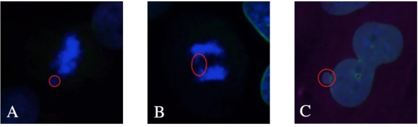
Figure 3.6: Immunofluorescence images of mitotic errors. A) Lagging chromosome in a 50 ng/ml of doxycycline culture at Day 3; B) Anaphase bridge in a 50 ng/ml of doxycycline culture at Day 3; C) Micronuclei in a 1000 ng/ml of doxycycline culture at Day 5.
As observed in Figure 3.5, a doxycycline concentration of 50 ng/ml is sufficient to induce mitotic errors in Mad2 overexpressing MEF cultures.
3.4 The concentration of doxycycline affects the number of chromosomes in MEFs expressing Mad2.
It was previously shown that the overexpression of Mad2 causes gains and losses of chromosomes in vitro (Sotillo et al. 2007).
In order to test if a lower expression of Mad2 could also induce aneuploidy in vitro, metaphase spreads were performed and the number of chromosomes per cell was counted, when treated with two different concentrations of doxycycline for 3 days. This experiment allowed concluding whether there was any clear difference between the concentrations of doxycycline (level of Mad2 overexpression) when it comes to number of chromosomes in each cell. The results obtained not only confirm that the overexpression of the transgene is responsible for the alteration on the number of chromosomes in MEFs, but also show that this alteration occurs in clones grown with both concentrations of doxycycline.
Figure 3.7: Number of chromosomes in TetO-Mad2/Rosa26-rtTA MEFs treated with 50 or 1000 ng/ml of doxycycline. Control cells, off dox, show a normal karyotype, with the majority of cells containing 40 chromosomes. Upon Mad2 overexpression, cells missegregate chromosomes, regardless of the amount of doxycycline. 50 and 1000 ng/ml of doxycycline reached statistical significance (P-value=0,0012 (*) and P-value=0,0244 (**) respectively).
Figure 3.8: Metaphase spreads of MEFs with TetO-Mad2/Rosa26-rtTA genotype. A) Loss of chromosomes in a 50 ng/ml of doxycycline culture; B) Loss of chromosomes in a 1000 ng/ml of doxycycline culture; C) Gain of chromosomes in a 1000 ng/ml of doxycycline culture.
3.5 The Her2 oncogene can be titrated in vitro. Differential outgrowth depends on the expression levels of the oncogene.
Due to the biological relevance of HER2 oncogene and its importance in breast cancer, the growth curve experiment conducted in MEFs expressing Mad2, was conducted in TetO-Neu/Rosa26-rtTA MEFs.
The graphs represented in Figure 3.9 show that the growth of the clone in which a concentration of 50 ng/ml of doxycycline was administered is slightly higher when compared
B C
20
the clone to which a concentration of 1000 ng/ml of doxycycline was administered, is significantly higher compared to the control wild-type.
These results suggest that the concentration of 50 ng/ml is enough to induce an increase in the growth of MEFs that express the Her2 oncogene, once the Tet-On system is activated.
Figure 3.9: Growth curves of three different clones of transgenic MEFs with the genotype
TetO-Neu/Rosa26- ) express the transgene Her2,
while the control clone, grown in normal media (Off Dox), does not express the transgene.
In order to investigate if the more robust increase in growth seen with 1000 ng/ml of doxycycline was due to a higher expression of Her2, the expression levels of this oncogene were tested by RT-PCR as described in Materials and Methods.
Figure 3.10 shows the expression of Her2 is higher in the clone heterozygous for Her2 that was maintained in a media with 1000 ng/ml of doxycycline than in the one that was grown with 50 ng/ml of doxycycline. Nevertheless, the lower concentration still induces an overexpression of the oncogene. Furthermore, these results also show that the expression of
this oncogene decreases throughout the 5 days of the experiment, independent of the doxycycline concentration used.
Figure 3.10: RT-PCR of samples from MEFs from the previous experiment. The graph represents the fold-change of Her2 expression in samples grown with doxycycline compared to a control sample, which was grown with no dox. 8 hours of culture under doxycycline was defined as Day 0.
22 4. Discussion and Conclusions
Cells with altered chromosome numbers are frequently found in numerous human conditions, but mainly in cancerous tissues (Holland & Cleveland 2012); (Nagaoka et al. 2012). Since the beginning of the twentieth century the role of chromosomal instability in cancer biology has been an important topic of study. In the lab where this dissertation was developed, a considerable effort is taken in order to derive conclusions on how aneuploidy and CIN phenotype promote tumor initiation and progression, particularly in breast cancer. Prior evidence supports the use of mouse models harbouring inducible transgenes/oncogenes of interest to study the susceptibility of the mammary gland to tumorigenesis (Gunther et al. 2002). In order to obtain a better understanding of the biological processes that link aneuploidy to tumorigenesis, model in which several transgenes are conditionally overexpressed in mice using tetracycline-inducible and repressible systems is used (Sotillo et al. 2007). In order to achieve reliable results that can have a practical use in the future, all the studies performed need to be as close as possible to the human reality, meaning that every method should recapitulate the biological mechanisms that occur in humans.
The main purpose of this study was to investigate whether the experiments performed in the lab are mimicking the in vivo situation that leads to tumorigenesis in humans. On a broader level, it addressed the extent to which cells can tolerate an extremely harmful condition that is likely to lead to cell death: aneuploidy. With the Tet-On inducible system used in the lab, specific transgenes, Mad2 and Her2 were conditionally expressed in order to promote this state at two different levels, one much higher than the other. To achieve this, two different concentrations of doxycycline were selected. It was decided that the experiments would be performed in mouse embryonic fibroblast taken directly from animals containing the different regulatable transgenes. This MEF model overexpression of the gene(s) of interest is regulatable through the addition of doxycycline to the media resulting in inducible expression. In section 3.1, results obtained by culturing Mad2 inducible MEFs with two concentrations of Dox show that the lower concentration (50 ng/ml) results in growth similar to the no Dox control. Whereas the higher concentration normally used in every experiment in the lab (1000 ng/ml), leads to a significantly impaired growth of the cells. Considering that previous studies have shown that doxycycline is, in fact, toxic for cells and live organisms (as it is discussed further ahead in this section) the question arises, whether this decrease observed with the growth of MEFs is due to the excessive overexpression of Mad2 or the toxic effect of doxycycline itself.
After obtaining the results presented in the growth curves, the aim was to confirm the expression of Mad2 in the cultured MEFs. In fact, as it is shown in Figure 3.2, although 50 ng/ml of Dox is a much lower concentration than 1000 ng/ml, the expression of Mad2 is clear from the 8 hours time point until 5 days of culture. Also, similar to what is seen in Figure 1.3, there seems to be a decrease in the level of expression of clones grown under both concentrations from day 3 to day 5. This might be explained, as it was previously mentioned, by a saturation of doxycycline in the media. Upon this saturation, cells might find a way of excluding this substance, which decreases the level of expression of Mad2, since the activation of the Tet-On system decreases. This need of the cells to expel doxycycline from within might be due to the toxic effect that this substance has on them. However another possibility is that the high Mad2 clones are toxic and therefore actively selected against favouring persistence of lower expressing clones. Nevertheless, these studies might prove to be important, as it is described in the Tet-On® Advanced Inducible Gene Expression Systems User Manual (Clontech Laboratories Inc. 2012) in transgenic organisms, saturation/depletion with doxycycline of different compartments of an organism follows complex pathways which will largely determine the kinetics of activation or inactivation of a gene.
It has been previously shown that overexpression of Mad2 leads to chromosomal instability in MEFs, in which after a karyotype analysis, it was seen that the number of binucleated cells was significantly higher in the Mad2-overexpressing MEFs than in the nontransgenic controls. Also, in this study, overexpression of Mad2 led to a significant increase in the number of chromosomal breaks and fragments, end-to-end fusions (dicentric and acentric chromosomes), as well as chromatid breaks and gaps as compared to wild-type. It was shown that overexpression of Mad2 led to abnormal mitoses, in which errors like lagging chromosomes and chromosome bridges occurred (Sotillo et al. 2007). The phenotype results obtained with the immunofluorescence show that, similar to what was observed by Sotillo et. al, the Mad2-overexpressing MEFs have a significant number of mitotic errors, such as lagging chromosomes and anaphase bridges, when compared to a nontransgenic control. However, as it can be seen in Figure 3.5, MEFs induced with 50 ng/ml of doxycycline, show that this concentration is sufficient to induce mitotic errors.
The confirmation of the effect of the overexpression of Mad2 at the karyotypic level was another aim of this study. As shown in Figure 3.7, Mad2 overexpressing MEFs have different number of chromosomes after division when compared to uninduced clones (Control Off Dox). However, as in the case of cultures with a doxycycline concentration of 1000 ng/ml,
24
transgenic clones grown under a concentration of 50 ng/ml also show gains and losses of chromosomes, although at a lesser frequency.
After assessing the effects of Mad2 overexpression in these cells given the biological relevance of Her2 in cancer, the titration of doxycycline for Her2 in MEFs was repeated. Results show that, similar to what was shown in the case of Mad2, the lower concentration of doxycycline effectively induces Her2 transgene expression. Her2 acts as a networking receptor that mediates signalling to cells, causing them to proliferate (Yarden & Pines 2012). It is observable in Figure 3.9 that, as expected, the overexpression of Her2 increases the growth of MEFs, either in the presence of 1000 ng/ml or of 50 ng/ml of doxycycline. One can see that while the control wild type has a normal growth, the transgenic clones have a significantly higher growth. However, in the case of the clones grown under a concentration of 50 ng/ml of doxycycline, their growth is much more comparable to the wild-type controls. This can be an interesting topic of discussion, since it can be the case that the increased growth induced by the higher concentration does not recapitulate the situation in humans. Nevertheless, this hypothesis would need further research in order to give rise to any conclusion. Concerning the expression of Her2 in this system, the doxycycline concentration of 50 ng/ml induces a much lower expression of the oncogene when compared to the expression induced by a concentration of 1000 ng/ml. Also, contrary to what is seen in Figure 3.2, there seems to be no saturation of the system, since the expression of the oncogene is shown to be constant with both concentrations throughout the 5 days of the experiment. As described in several studies, very little is known about the level of expression of HER2 in human breast cancer data sets. Due to several incongruencies with the technique, usually this expression is not checked by quantitative RT-qPCR but instead by IHC or FISH (Yardley 2015; Cuadros et al. 2010).
Although the exact fold-change of HER2 in human breast cancer data sets is not yet known, one can speculate that the overexpression induced by a concentration of 50 ng/ml might mimic better the situation in humans, just like in the case of Mad2. What little conclusion can be drawn from this observation is that the saturation observed in the Tet system seems to be varied depending upon the transgene induced. Nevertheless, one does not know whether this concentration is already too high. To address this, lower concentrations should be tested under the same conditions conducted in this study.
Overall, the results obtained in this project allow questioning even more whether the concentration of doxycycline normally used in the experiments is the appropriate one. On the other hand, the results conclude that a concentration of 50 ng/ml is sufficient not only to
induce the intended expression of the transgenes of interest (Mad2 and Her2) but also more importantly, to express the phenotype from which the overexpression of these transgenes is responsible for. Despite the fact that further research would be needed to confirm if this level of doxycycline is optimal in vivo, these results aid in the understanding that other solutions are available to activate the system and to make it much more relevant to the human scenario.
In the lab, a state of the art system of three-dimensional (3D) cell culture was adapted to study the behaviour of primary mouse mammary epithelial cells in the response to the conditional expression of oncogenes and other transgenes. This conditional expression was activated by the same Tet-On system used in this project. 3D culture systems are physiologically relevant models that can mimic both the 3D organization and multicellular complexity of an organ but at the same time allow systematic experimental intervention. According to Schmeichel 2003, these culture systems are an important way to study the impact of oncogenic mutations, since they represent the organotypic growth, specialized cell-cell contacts and attachment to an underlying basement membrane – necessary features for the control of cellular proliferation, survival and differentiation (Jechlinger et al. 2009). In order to better understand the effects of the different levels of expression of the several transgenes, similarly to what was done in this project, but in a more biological environment, the use of these 3D systems would be a reliable way to explore it in the future.
Another interesting approach of studying this topic in the future would be by using Live Cell Imaging. Such systems are nowadays widely used by researchers to obtain a better understanding of biological function through the study of cellular dynamics (Baker 2010). These techniques were used before in the lab to examine abnormal mitoses. For that, MEFs infected with a retrovirus expressing histone 2B (H2B)- GFP were used. The method allowed concluding that while nontransgenic or uninduced MEFs underwent a normal and rapid mitosis (59 ± 1.6 min), cells overexpressing Mad2, took an unusually long time to finish the process (89 ± 5 min), showing difficulties in completing cytokinesis and frequent defects in chromosome segregation (Sotillo et al. 2007).
As such, since the technique is already set up, it would also be interesting to follow the differences between cultures with different concentrations of doxycycline making use of a concomitant GFP expression. With the help of such a modern microscopy technique, the conclusions taken would be more accurate and reliable.
26
drinking water with 5% sucrose containing doxycycline. This study could be adapted and a titration of doxycycline with different concentrations in the drinking water could be done. It is known that the expression of the transgenes is seen after 4 days of treatment with doxycycline. This would allow determining the optimum concentration of doxycycline for transgene induction in vivo.
Recent evidences suggest that although the Tet-On/ Tet-Off system provides an excellent flexibility to study gene function, it also has potential detrimental effects, due to the harmful effects of tetracyclines themselves (Brummett & Fox 1989; Mingeot-Leclercq & Tolkens 1999; Selimoglu 2007). These effects deserve to be considered. It has been known since the 1960s that tetracyclines, as well as chloramphenicol, inhibit translation of proteins encoded by mitochondrial DNA (mtDNA) (Clark-Walker & Linnane 1966.). In 2013, Houtkooper et al. showed that this selective inhibition of mitochondrial protein translation by both types of antibiotics leads to a state that impairs mithocondrial proteostasis, called mitonuclear protein imbalance (Houtkooper et al. 2013). In fact, as it was shown in 2015, doxycycline treatment altered the expression of 10% of genes on a cell line and it was severely harmful for homeostasis. At the physiological level of entire organisms, as it was clear in mice, doxycycline treatment impaired the growth, oxygen consumption, among other changes (Moullan et al. 2015). Concluding, some caution should be taken for all research purposes when using these systems, including cancer related, since mitochondria have been implicated in this disease (Andreux et al. 2013). Thus, in the future, the toxicity of doxycycline in MEFs and in mice should be tested in detail, in order to conclude even more precisely what is the adequate concentration to use in the studies performed. This could be achieved culturing MEFs extracted from a non-rtTA animal with doxycycline, since they would never express the transgenes. In this study in particular, this experiment would help concluding, as mentioned before in this section, if the decrease observed with the growth of MEFs is due to the excessive overexpression of Mad2 or the toxic effect of doxycycline itself.
Adding to this, other concentrations should be tested also with other transgenes used in the lab, such as Kras, Myc, PLK1 and Aurora B. This is due to the observation that different transgenes have different effects on cells. For example when Kras protein levels are increased to abnormal levels it can induce growth arrest, apoptosis and replicative senescence (Jančík et al. 2010), meanwhile overexpression of Myc, which binds to target DNA sequences to regulate transcription of genes involved in cell growth and proliferation, can result in proliferation (Dang 2012). On the other hand, PLK1 and Aurora B are two important kinases
that compose SAC. Aurora B is mainly involved in the error correction mechanism, which is required to ensure proper attachment of kinetochore and microtubules. Also, previous studies have revealed that mice expressing Aurora B under a ubiquitous promoter have a higher tumour incidence and these tumours have increased levels of aneuploidy. PLK1 is an early trigger for G2/M transition and it supports the functional maturation of the centrosome and the establishment of the bipolar spindle. Its overexpression is observed in many types of tumours and it can cause aneuploidy due to defects in centrosome amplification, which results in multipolar spindles (Godek et al. 2014).
Considering the wide range of ways through which tumourigenesis can be induced, it is extremely important to hereafter, determine if different transgenes should be induced under different concentrations of doxycycline. If so, these concentrations should be established, in order to make the studies even more reliable. Designing these studies to better mimic the situation in humans will lead to enhanced knowledge and understanding of tumorigenesis which in turn will improve the possibilities to develop more effective therapies.