Bladder cancer
glycome and glycoproteome
microenvironmental
regulation:
the role of oxygen and
glucose
Marta Filipa Relvas dos Santos
Mestrado em Bioquímica
Faculdade de Ciências e Instituto de Ciências Biomédicas Abel Salazar Universidade do Porto
2018
Orientador
José Alexandre Ferreira, Investigador auxiliar, IPO-Porto
Coorientador
André M. N. Silva, Investigador externo, FCUP
Coorientador
Lúcio Lara Santos, Coordenador do Grupo de Patologia e Terapêutica
Experimental, IPO-Porto
Todas as correções determinadas pelo júri, e só essas, foram efetuadas. O Presidente do Júri,
Contribution in the concept and accomplishment of the following research work during the year of development of Master degree’s Thesis:
Scientific Papers
I. Azevedo R, Gaiteiro C, Peixoto A, Relvas-Santos M, Lima L, Santos LL, Ferreira JA. CD44 glycoprotein in cancer: a molecular conundrum hampering clinical applications. Clin Proteomics. 2018; 15:22.
II. Azevedo R, Soares J, Gaiteiro C, Peixoto A, Lima L, Ferreira D, Relvas-Santos M, Fernandes E, Tavares A, Cotton S, Daniel-da-Silva AL, Santos LL, Vitorino R, Amado F, Ferreira JA. Glycan affinity magnetic nanoplatforms for urinary glycobiomarkers discovery in bladder cancer. Talanta. 2018; 184:347-55.
Poster
I. Peixoto A, Fernandes E, Gaiteiro C, Lima L, Azevedo R, Soares J, Cotton S, Parreira B, Neves M, Relvas-Santos M, Amaro T, Tavares A, Teixeira F, Palmeira C, Rangel M, Silva AM, Reis CA, Santos LL, Oliveira MJ, Ferreira JA. Hypoxia enhances the malignant nature of bladder cancer cells and concomitantly antagonizes protein O-glycosylation. Conference: ICS 2018: 29th International Carbohydrate Symposium
Oral communications
I. Peixoto A, Fernandes E, Gaiteiro C, Lima L, Azevedo R, Soares J, Cotton S, Parreira B, Neves M, Relvas-Santos M, Amaro T, Tavares A, Teixeira F, Palmeira C, Rangel M, Silva AM, Reis CA, Santos LL, Oliveira MJ, Ferreira JA.
Hypoxia enhances the malignant nature of bladder cancer cells and concomitantly antagonizes protein O-glycosylation extension. Conference: ICS 2018: 29th International Carbohydrate Symposium
II. Azevedo R, Gaiteiro C, Soares J, Peixoto A, Relvas-Santos M, Ferreira D, Lima L, Santos LL, Ferreira JA. CD44-glycoprofilling: Estabilishing the molecular basis for targeted therapeutics in bladder cancer. Conference: ICS 2018: 29th International Carbohydrate Symposium
Agradecimentos
Na conclusão deste trabalho, gratidão parece-me a palavra certa para exprimir o que sinto. Estou imensamente grata aos meus orientadores por há cerca de ano e meio terem acreditado e apostado em mim. Obrigada por esta oportunidade incrível, por poder aprender tanto todos os dias. Obrigada por toda a ajuda, dedicação, incentivo e amizade.
Ao Doutor José Alexandre Ferreira o meu profundo obrigada pela confiança e pela forma sábia como me ajuda a contruir o meu percurso e a moldar-me como cientista que aspiro um dia ser. É um privilégio poder aprender e fazer Ciência consigo.
Ao Doutor André Silva o meu muito obrigada pela partilha de conhecimento e pela discussão valiosa de abordagens experimentais. Também ele constitui parte muito importante neste percurso e alguém com quem aprendo sempre muito.
Um agradecimento especial ao Professor Doutor Lúcio Lara Santos por me ter permitido integrar tão fantástico grupo.
Ao Grupo de Patologia e Terapêutica Experimental: Andreia, Cristiana, Dylan, Elisabete, Janine, Manuel, Rita, Rui, Sofia e Doutor Luís Lima. Obrigada pela amizade e pela enorme interajuda. O que cresci, aprendi e evoluí, também muito o devo a vocês. Não posso deixar também de agradecer a todos aqueles que na FCUP e no CEMUP de alguma forma contribuíram para que este projeto decorresse ao melhor ritmo.
Aos meus amigos, em particular à Rita e ao Miguel, é muito bom partilhar convosco mais uma etapa.
À minha família, pelas pessoas maravilhosas que são, por todo o apoio que me dão sempre,… Muito obrigada!
Sumário
O tratamento de cancro da bexiga, principalmente dos estadios mais avançados, permanece um desafio, devido à significativa heterogeneidade molecular. Frequentemente, o glicoproteoma da membrana plasmática sofre remodelação, o que contribui para a progressão e disseminação da doença. Além disso, o facto do padrão glicoproteómico ser dependente do contexto em que está inserido pode fornecer informação molecular bastante específica para intervenção dirigida. Alterações na O-glicosilação, uma importante modificação pós-traducional das proteínas à superfície celular, constitui um dos principais eventos moleculares que levam à alteração do glicoproteoma em cancro da bexiga. Contudo, a complexa estrutura dos glicanos e a sua natureza dinâmica, associados à falta de protocolos de glicoproteómica padronizados, têm atrasado o conhecimento compreensivo do glicoma e do glicoproteoma, tendo em vista possíveis biomarcadores para intervenção clínica. Por outro lado, a informação sobre o impacto do microambiente na glicosilação é escassa e, por isso também dificulta estudos funcionais e terapêuticas dirigidas. Assim, este trabalho dedicou-se a padronizar um protocolo para investigação de glicobiomarcadores. De seguida, concentrou-se em explorar as mudanças no glicoma que possam ocorrer em resposta à hipóxia e à privação de glucose, as quais constituem importantes caraterísticas microambientais dos tumores sólidos, devido à ineficiente vasculatura promovida pelo crescimento tumoral rápido e não controlado.
De forma a responder a estes objetivos, este trabalho começa por propor um protocolo apoiado na bioinformática para análise glicómica e glicoproteómica. Resumidamente, três modelos celulares de cancro da bexiga (5637 derivado de grau II, T24 e HT1376 derivados de grau III) foram sujeitos a caraterização O-glicómica, através do método Cellular O-glycome Reporter/Amplification (CORA) e espetrometria de massa (MALDI-TOF-MS e nanoLC-ESI-MS). Estas linhas celulares apresentaram como estruturas dominantes as formas sialiladas do antigénio T, bem como mostraram similaridade a nível do proteoma e do O-glicoma, apesar das suas diferenças moleculares e histológicas. Esta informação permitiu desenhar estratégias adequadas de enriquecimento glicoproteico, o que foi feito através de cromatografia de afinidade com Peanut agglutinin (PNA). A identificação das glicoproteínas foi conseguida através duma estratégia proteómica de bottom up, usando nanoLC-ESI-MS/MS, dados cuidados de ontologia génica para proteínas da membrana plasmática e de previsão de locais de glicosilação, usando o NetOGlyc. As proteínas identificadas foram então integradas com
dados de transcritómica e imunohistoquímica, usando as bases de dados Oncomine e o Human Protein Atlas. Esta abordagem permitiu gerar uma libraria composta por relevantes glicoproteínas, que justificam posteriores estudos clínicos. Além do contributo para um maior conhecimento da glicobiologia do cancro da bexiga, este protocolo pode fomentar novas aplicações clínicas baseadas em glicobiomaracadores. A segunda parte deste trabalho implementou estes avanços analíticos na análise da influência da diminuição de oxigénio e da privação de glucose no O-glicoma. Apesar de explorativo, este trabalho destacou a similaridade e consistência dos padrões de O-glicosilação de todas as linhas celulares em hipóxia, na privação de glucose e quando sujeitas ao efeito sinérgico de ambos os fatores. Resumidamente, a hipóxia teve pouco impacto no O-glicoma, enquanto a privação de glucose originou um impressionante remodelamento. Esta alteração do O-glicoma é caraterizada pela significante diminuição da biossíntese de O-glicanos, pela paragem total da extensão dos glicanos além do core 1, pela troca da fucosilação do core 1 pela sialilação e pela sobreexpressão do sialyl Tn, um glicano bastante associado ao cancro da bexiga. A combinação da hipóxia à privação de glucose, o que frequentemente ocorre in vivo, exacerbou estes eventos. Para conhecimento futuro, este é o primeiro estudo abrangente e compreensivo da plasticidade do O-glicoma, face a estes estímulos microambientais. Além disso, estes resultados, apoiados pela plataforma analítica desenvolvida na primeira parte deste trabalho, ajudam a guiar futuros estudos de glicoproteómica funcional, bem com na descoberta de glicobiomarcadores, especialmente contra os nichos de hipóxia, constituídos por células mais malignas.
Palavras-chave: antigénios T sialilados, cancro da bexiga, glicoproteínas, glicosilação, hipóxia, O-glicanos, privação de glucose
Abstract
Bladder cancer management, especially at advanced stages, remains challenging due to significant molecular heterogeneity. In particular, cancer cells often experience plasma membrane glycoproteome remodelling that contributes to disease progression and dissemination. Moreover, the context-dependent nature of the glycoproteome may provide highly cancer-specific signatures for targeted intervention. Changes in O-glycosylation, a main post-translation modification of cell surface proteins, are amongst the main molecular events driving alteration in the bladder cancer glycoproteome. However, glycans complex structure, non-templated and dynamic nature associated to the lack of standardized glycoproteomics protocols has delayed a comprehensive interrogation of the glycome and glycoproteome for cancer biomarkers capable of aiding clinical intervention. On the other hand, there is little information on the impact of the cancer microenvironment on glycosylation, which has hampered functional studies and targeted therapeutics. As such, this work has devoted to standardizing a workflow to aid glycobiomarker research. It then focused on exploring this knowledge to disclose changes in the glycome in response to hypoxia and glucose deprivation, two salient microenvironmental features of solid tumours due to inefficient vasculature and sustained tumour growth.
Responding to these objectives, this work starts by proposing a bioinformatics-assisted protocol for glycomics and glycoproteomics analysis. Briefly, three of the most studied bladder cancer cell models (5637 from grade II, T24 and HT176 from grade III) were subjected to O-glycome characterization by Cellular O-glycome Reporter/Amplification (CORA) and Mass Spectrometry (MALDI-TOF/TOF-MS and nanoLC-ESI-MS). These cell lines showed some degree of proteome and O-glycome similarity, with sialylated T antigens as dominant glycan species, despite marked molecular and histological differences. This information was latter used to design adequate glycoprotein enrichment strategies by peanut agglutinin (PNA) affinity chromatography. Glycoprotein identification was achieved by bottom-up nanoLC-ESI-MS/MS proteomics and data was curated by gene ontology for plasma membrane proteins and glycosites prediction using NetOGlyc. Identified glycoproteins were comprehensively integrated with transcriptomics and immunohistochemistry data using Oncomine and the Human Protein Atlas databases. This generated a library of potentially relevant glycoproteins that now warrants clinical validation. We anticipate that this
workflow may aid bladder cancer glycobiology interrogation and ultimately foster novel clinical applications based on glycobiomakers.
The second part of this work explored these analytical advances to disclose the influence of oxygen shortage and glucose deprivation in the O-glycome. Despite explorative, it highlighted very similar and consistent patterns for all cell lines in hypoxia, glucose deprivation and under the combination of both factors. Briefly, hypoxia had little impact on the O-glycome. Conversely, glucose deprivation originated a striking glycome remodelling characterized by a significant decrease in O-glycans biosynthesis, complete stop of glycan extension beyond core 1, a shift from fucosylation towards sialylation of core 1 glycans and an overexpression of bladder cancer-associated glycan sialyl-Tn. Such events were reinforced when glucose deprivation was associated with hypoxia, as frequently occurs in vivo. To our knowledge, it is the first comprehensive view on O-glycome plasticity facing these microenvironmental stimuli. Moreover, these findings, supported by the analytical platform developed in the first part of the work, will help guiding functional glycoproteomics and glycobiomarkers discovery, especially against hypoxic niches known to harbour more malignant cells.
Keywords: bladder cancer, glucose deprivation, glycoproteins, glycosylation, hypoxia, O-glycans, sialylated T antigens
Index of contents
Agradecimentos ... iv Sumário ... v Abstract ... vii Index of contents ... ix Index of figures ... xiIndex of tables ... xiii
Abbreviations ... xiv
Chapter I | Introduction ... 1
1. Bladder cancer ... 2
1.1. Epidemiology and risk factors ... 2
1.2. Pathophysiology and disease progression ... 2
1.3. Diagnosis and therapeutics of bladder cancer ... 4
1.4. Characterization of glycoproteome envisaging novel biomarkers and targeted therapeutics... 5
2. Glycosylation and cancer ... 7
2.1. General features and patterns of glycosylation ... 7
2.2. Protein Glycosylation in bladder cancer ... 9
2.3. Expression of short-chain O-glycans in bladder cancer: the disclosed role of the Sialyl-Tn antigen ... 11
3. Microenvironment-induced O-glycome alterations ... 14
3.2. Hypoxia drives metabolic switch and altered glycosylation ... 16
3.3. Hypoxic modulation of O-GalNAc glycans in bladder cancer ... 19
3.4. Hexosamine biosynthesis pathway: linking glucose availability and tumoral aberrant glycosylation ... 19
4. Glycoproteomics and Glycomics: Analytical overview and critical challenges ... 22
4.1. Glycomics ... 22
4.2. Glycoproteomics ... 24
5. Aims and scopes ... 26
Chapter II | A Bioinformatics-Assisted Workflow to address the Plasma Membrane Glycoproteome: Biomarker Discovery Using Bladder Cancer Cell Models ... 27
Chapter III | Bladder cancer O-glycome microenvironmental regulation: the role of oxygen and glucose ... 48
Chapter IV | Concluding remarks ... 62
References ... 66
Index of figures
Chapter I
Figure 1 – Schematic representation of bladder cancer stage and grade 3
Figure 2 – Biosynthesis of core 1 to 4 and sialylated short-chain O-GalNAc glycans 9
Figure 3 – Biosynthesis and interconversion of monosaccharides 21
Chapter II
Scheme 1 – Illustration of the bioinformatics-assisted analytical protocol 35
Figure 1 - Glycome repertoire for 5637, T24 and HT1376 cell lines in bladder cancer 37
Figure 2 - Characterization of bladder cancer cell lines according to GO terms using
STRAP program 40
Figure 3 - String map for glycoproteins that are common to all cell lines and main biological processes, cellular functions and KEGG pathways 41
Figure 4 - Cytoscape analysis highlighting the major biological processes governed by bladder cancer cell lines plasma membrane proteins 42
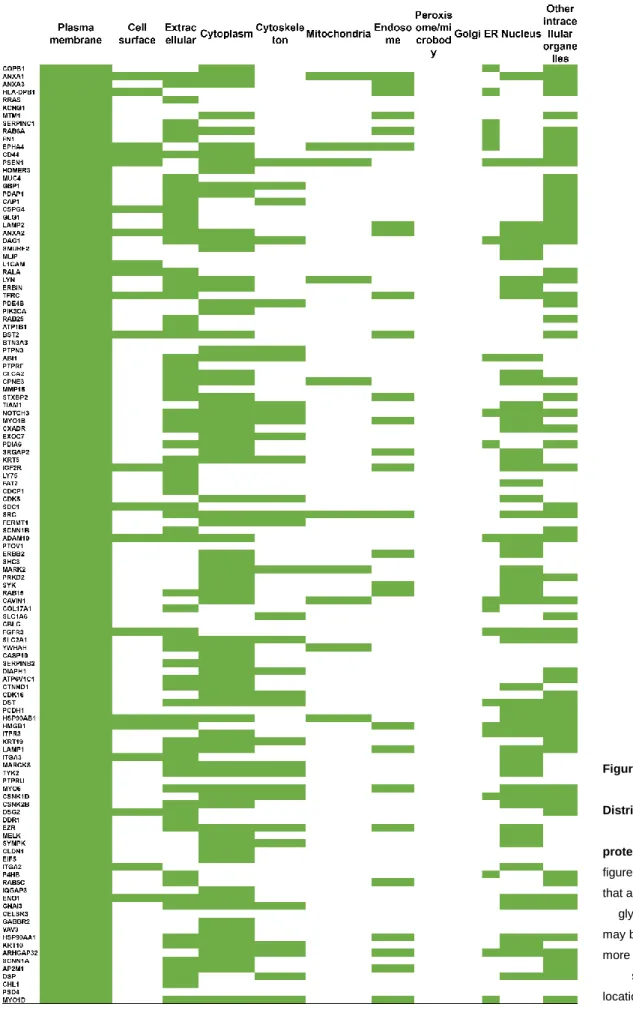
Figure 5 - Cellular Distribution of identified proteins 43
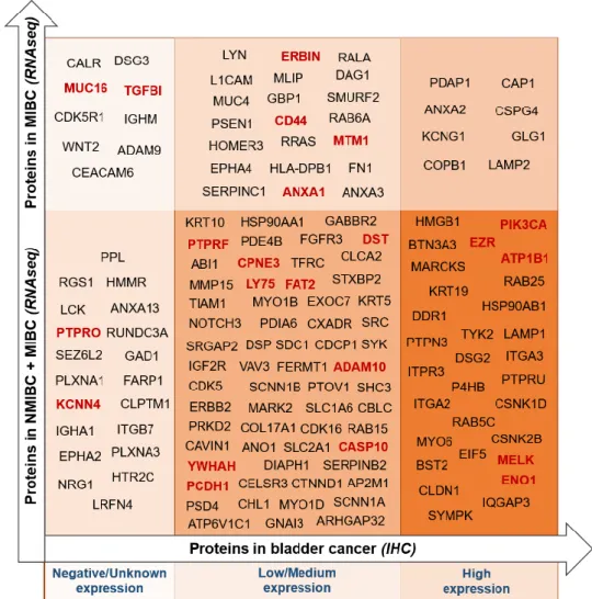
Figure 6 - Oncomine RNAseq cancer database analysis sorting the identified glycoproteins according with its previously reported expression in bladder tumours
Chapter III
Figure 1 – (A) Bladder cancer cell lines expression of HIF-1α hypoxia biomarker over-time (B) Lactate production in bladder cancer cell lines under normoxic and hypoxic
conditions 54
Figure 2 - Impact of hypoxia, glucose deprivation and both factors in in cell proliferation 54
Figure 3 - Relative abundance of O-glycans facing different microenvironmental
challenges 58
Figure 4 - Graphical representation of relative abundance of each O-GalNAc structure under Normoxia, Hypoxia, Normoxia No Glucose and Hypoxia No Glucose for each bladder cancer cell model (A) 5637, (B) T24, (C) HT1376 58
Figure 5 - Comparative relative abundance of O-GalNAc structures in bladder cancer cell lines. (A) Comparison between short-chain and extended (core 2- related structures) O-glycans; (B) Comparison between fucosylated and sialylated structures 60
Index of tables
Appendix
Table S1 – Paired analysis of independent samples comparing MALDI-TOF-MS and
nanoLC-ESI-MS 82
Table S2 – O-glycan structures determined by MALDI-TOF-MS and/or nanoLC-ESI-MS 83
Abbreviations
Asn AsparagineBC Bladder cancer
BCG bacillus Calmette-Guérin
Bn-GalNAc benzyl-α-D-GalNAc or benzyl 2-acetamido2-deoxy-α-D-galactopyranoside CID Collision-induced dissociation
CORA Cellular O-glycome Reporter Amplification DMSO Dimethyl sulfoxide
dST di-Sialyl T DTT Dithiothreitol
ECD Electron capture dissociation EDTA Ethylenediamine tetraacetic acid
EGTA Ethylene glycol-bis(β-aminoethyl ether)-N,N,N′,N′-tetraacetic acid ER Endoplasmic reticulum
ESI Electrospray ionization FBS Fetal bovine serum
FGFR3 Fibroblast Growth Receptor Factor 3 Fuc Fucose G6PD Glucose-6-phosphate dehydrogenase GA Golgi apparatus Gal Galactose GalNAc N-acetylgalactosamine GC Gemcitabine/cisplatin GlcNAc N-acetylglucosamine GLUT Glucose transporter GnT GlcNAc transferase GO Gene ontology
HBP Hexosamine biosynthetic pathway HCD High-energy collision dissociation
HEPES 4-(2-hydroxyethyl)-1-piperazineethanesulfonic acid HIFs Hypoxia-inducible factors
HK Hexokinase
HRAS Harvey rat sarcoma viral oncogene homolog ICORA Isotope-Cellular O-glycome Reporter Amplification
ITGB1 Integrin beta-1 Lea/x Lewis a/x
MALDI Matrix-assisted laser desorption/ionization MIBC Muscle invasive bladder cancer
MS/MS Tandem mass spectrometry nanoLC nano-liquid chromatography Neu5Ac N-acetylneuraminic acid
NMIBC Non-muscle invasive bladder cancer OGA O-GlcNAcase
OGT O-GlcNAc transferase PDH Pyruvate dehydrogenase
PDK Pyruvate dehydrogenase kinase PHDs Prolyl hydroxylase domain enzymes PNA Peanut Agglutinin
PNGase F Peptide-N-Glycosidase F
ppGalNAcT polypeptide GalNAc-transferases PPP Pentose phosphate pathway
PTM Post-translational modification RB Retinoblastoma
RTKs Receptor tyrosine kinases S3T Sialyl-3-T
S6T Sialyl-6-T
SDS Sodium dodecyl sulfate
SDS-PAGE Sodium dodecyl sulfate–polyacrylamide gel electrophoresis Ser Serine
SLea/x Sialyl Lewis a/x
ST Sialyl T
ST3Gal α2-3 sialyltransferase ST6GalNAc α2-6 sialyltransferase STn Sialyl Tn
STRAP Software Tool for Researching Annotations of Proteins STRING Search Tool for the Retrieval of Interacting Genes/Proteins TBS Tris-buffered saline
TCA Tricarboxylic acid cycle TFA Trifluoroacetic acid Thr Threonine
TOF Time-of-flight TUR Tumour resection
UDP-GlcNAc UDP-N-Acetylglucosamine UPR Unfolded protein response
1. Bladder cancer
1.1. Epidemiology and risk factors
Bladder cancer (BC) is the seventh most diagnosed cancer in the male population, dropping to tenth when both sexes are considered (1, 2). Furthermore, BC is the deadliest tumour of the urinary system, with approximately 429,000 new cases diagnosed and 1650,00 deaths estimated worldwide in 2012 (1-3).
There are multiple risk factors identified for BC, which are responsible for variations in incidence and pathophysiology (4). For instance, carcinogenic compounds present in cigarettes, such as aromatic amines, make of tobacco smoking and fume inhalation the main risk factor, being responsible by 50% of diagnosed cases (4-6). Inherited genetic factors represent additional risk by influencing individual susceptibility to external factors, mainly to tobacco smoke. Furthermore, occupational exposure to aromatic amines, polycyclic aromatic hydrocarbons and chlorinated hydrocarbons in industries linked to petroleum, coal, paint and metal, represents an additional risk for BC, despite rising safety measures (4, 5). In line with this, inadvertent consumption of chlorinated and arsenic-contaminated drinking water continues to be a major cause of BC. Other risks factors related to dietary habits as coffee consumption remain controversial (7). In addition, BC can arise from underlying medical conditions such as chronic urinary retention and upper tract dilation, contributing to higher exposure to carcinogens, as well as chronic inflammation or infection with Schistosoma haematobium (4, 5).
1.2. Pathophysiology and disease progression
Urothelial tumours frequently progress along with two molecular pathways, the more common leading to low-grade, multifocal, papillary, non-invasive carcinomas and the other being responsible for high-grade invasive carcinomas (8). The first is thought to arise from nodular hyperplasia and it is known to involve mutations in Fibroblast Growth Receptor Factor 3 (FGFR3), Harvey rat sarcoma viral oncogene homolog (HRAS), as well as phosphatidylinositol 3-kinase (PI3KCA) genes. On the other hand, non-papillary high-grade and invasive tumours should arise from carcinoma in situ or severe dysplasia, harbouring mutations on tumour suppressor genes TP53, p16 and
retinoblastoma (RB) (8-10). Notwithstanding, although papillary tumours tend to recur, the vast majority does not evolve into high-grade and invasive tumours (11).
The vast majority of diagnosed BC cases are urothelial cell carcinomas, followed by carcinomas exhibiting squamous or glandular differentiation and other variants (12). Approximately 75% of newly diagnosed patients present superficial tumours (tumour in situ (Tis), Ta and T1), generally classified as non-muscle invasive bladder cancer (NMIBC). The standard management of these tumours comprises transurethral bladder tumour resection (TUR) and bacillus Calmette-Guérin (BCG) intravesical therapy (2, 13, 14). However, these therapeutic options fail to prevent recurrence and progression; thereby displaying poor disease-specific survival. The risk of progression of T1 tumours at 5 years is superior to 20% and, in case of confirmed progression to muscle invasive bladder cancer (MIBC, T2-T4), the cancer-specific survival drops to 35% after 4 years (Figure 1) (14).
Figure 1 - Schematic representation of bladder cancer stage and grade (15). The stage of the primary tumour (T) is
based on the extent of penetration or invasion into the bladder wall. Regarding tumour grading, bladder lesions can be classified as urothelial papilloma (a benign lesion), papillary urothelial neoplasm of low malignant potential (PUNLMP), low-grade papillary urothelial carcinoma and high grade papillary urothelial carcinoma. Of note, PUNLMP lesions do not have cytological features of malignancy and have a very low risk of progression. Nevertheless, they show high tendency to recur. Tis, Tumour in situ: ‘‘flat tumour’’; Ta, Non-invasive papillary carcinoma; T1, Tumour invades sub-epithelial connective tissue; T2, Tumour invades muscle; T2a, Tumour invades superficial muscle (inner half); T2b, Tumour invades deep muscle (outer half); T3, Tumour invades perivesical tissue; T4, Tumour invades any of the following: prostate, uterus, vagina, pelvic or abdominal wall (15).
1.3. Diagnosis and therapeutics of bladder cancer
The most common BC symptom is painless haematuria, which prompts cystoscopy examination, urine cytology and imaging of the upper urinary tract. The standard therapeutic approach on NMIBC is TUR, which removes all visible lesions while allowing a more precise diagnosis and staging (2). In addition, this surgical procedure is usually followed by adjuvant intravesical BCG immunotherapy, currently the most preventive treatment for NMIBC recurrence in non-immunocompromised patients. In case of BCG treatment failure (1/3 of cases) or in NMIBC patients with high risk of progression, radical cystectomy is the next logical step (2, 16). Radical cystectomy is also the standard treatment for localised MIBC (T2-T4), followed by pelvic lymph node dissection. A viable alternative involves debulking TUR and radiotherapy (13).
Metastatic patients with good renal function may be suitable for systemic
chemotherapy with cisplatin-based combinations as
methotrexate/vinblastine/adriamycin/cisplatin (MVAC) or Gemcitabine/cisplatin (GC) (14). The major difference between the above-mentioned combinations is toxicity. The lower toxicity of GC allied to an increase in cost effectiveness has resulted in it becoming the new standard regimen (17, 18). Carboplatin, an alkylating antineoplastic drug analogue of cisplatin, has been used in urinary and metastatic BC patients with impaired renal function due to its decreased nephrotoxicity compared to cisplatin (19). Even though chemotherapy is efficient against highly proliferative malignant cells that form the tumour bulk, the five-year overall survival does not exceed 25% and many patients die prematurely from adverse drug reactions, urging for effective and safe targeted therapeutics.
Tremendous efforts have been put in the development of biomarker panels for early diagnosis, follow-up, patient stratification, prognosis, treatment selection and development of targeted therapeutics (13). However, the highly heterogeneous molecular nature of bladder tumours has hampered true developments in this field (9) As such, BC remains mostly an “orphan disease” in terms of targeted therapeutics, leading to few improvements in patient’s overall survival over the last decade (10). Importantly, most of this molecular heterogeneity arises from microenvironmental features driving tumour cell adaptation to endogenous stress, such as nutrient deprivation resulting from sustained proliferative signalling and flawed neovascularization, as well as external challenges as chemotherapy. Facing these challenges, glycosylation changes capable of reflecting not only the tumour cells genomic, transcriptomic and metabolomic status but also its microenvironmental context
have major potential for clinical applications. Moreover, glycans and abnormally glycosylated molecules (e.g. proteins and lipids) hold tremendous value for non-invasive cancer detection, while membrane bound glycans may be used to selectively target tumour sites and specific cancer cells.
1.4. Characterization
of
glycoproteome
envisaging
novel
biomarkers and targeted therapeutics
Despite tremendous research efforts, there is still a lack of useful biomarkers to determine prognosis, the most beneficial therapeutic regimen and, in particular, designing novel targeted therapeutics (20, 21). Membrane proteins offer significant potential for the development of targeted therapeutics by being exposed to extracellular ligands such as monoclonal antibodies. In this context, the most explored therapeutic targets in BC are receptor tyrosine kinases (RTKs), including epidermal growth factor receptor (EGFR; Cetuximab, Clinical Phase II), vascular endothelial growth factor receptor (VEGF; Bevacizumab, Clinical Phase II) and human epidermal growth factor receptor 2 (HER2; Trastuzumab, Clinical Phase II) (22). These proteins are essential for communication between cells and their environment and thereby for cell proliferation and growth; however, mutations of RTKs, which are often overexpressed in BC, significantly decrease the efficacy of current immunotherapies targeting these membrane receptors. Other identified potential targets involve signal transduction pathways and include, for example the protein mTOR and the signal transducer and activator of transcription 3 (STAT3) (23, 24). However, few clinical trials have been performed and with limited success. Moreover, many of these membrane glycoproteins are also expressed in many healthy human organs, lacking the necessary cancer-specificity and consequently resulting in relevant toxicity related with off-target effects (22).
Amongst the factors delaying effective targeted therapeutics is the significant molecular microheterogeneity presented by bladder tumours of apparently similar histology (25). This highlights the urgency in the identification of cancer-specific molecular signatures and the development of multi-targeted therapeutics. As such, particular emphasis should be put on screening the membrane proteome of bladder tumour cells for cancer-specific signatures. Moreover, more than 50% of membrane proteins are glycosylated (26) and BC cells are known to present particular glycosylation features (15). Therefore, the identification of cancer-specific glycosylation patterns and protein glycosites may allow narrowing down the specificity of already explored and new
cancer biomarkers. Finally, many biomarker discovery studies disregard the influence of key cancer-associated microenvironment features such as oxygen and nutrient deprivation, which drive key cancer hallmarks. As such, studies envisaging effective therapeutics should also focus on disclosing the impact of these events on the glycome and glycoproteome of cancer cells.
2. Glycosylation and cancer
2.1. General features and patterns of glycosylation
Glycosylation is the most common and structurally diverse post-translational modification (PTM) of membrane-bound and secreted proteins, as well as a common substitution in lipids and intracellular proteins. Accordingly, it has a key role in protein stability, folding, and trafficking, as well as in cell-cell adhesion, differentiation, migration, signalling, host-pathogen interaction and immune recognition (27-29).
This non-templated but highly regulated process is responsive to biological changes and results from the action of several glycosyltransferases, nucleotide sugar transporters and glycan-processing enzymes in the endoplasmic reticulum (ER) and Golgi apparatus (GA) (30, 31). It gives rise to two main classes of glycans found at the cell surface, namely N-glycans and O-glycans. Briefly, N-glycosylation starts with the covalent attachment of a 14-sugar glycan from the lipid precursor Glc3Man9GlcNAc2
-P-P-Dol to Asparagine (Asn) residues in Asn-X-Ser/Thr sequons (X denotes any amino acid except proline) of newly synthetized peptides (32, 33). The next processing steps comprise sugar moiety elongation and maturation at the GA to originate hybrid, complex and oligomannose type N-glycans through the action of glycosyltransferases and glycosidases. Branching of N-glycans results from the action of different GlcNAc transferases (GnT-III-VI), and additional elongation might occur through the addition of galactose, fucose, sialic acid or N-acetyllactosamine (LacNAc, Galβ1-4GlcNAc) sugars (34-36). Frequently, N-glycans exhibit Lewis blood group related antigens (Lea, Lex, Leb,
Ley) or their sialylated forms, as well as ABO blood group antigens as terminal epitopes
(15). As such, glycosylation increases the glycoproteome complexity not only by the vast carbohydrate repertoire but also by the possibility of several glycosidic linkages and isomer forms (37).
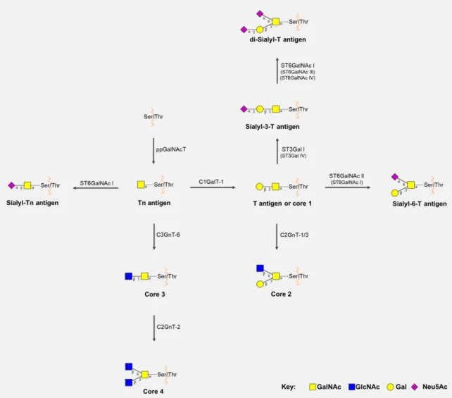
Previously N-glycosylated glycoproteins are subsequently O-glycosylated through the action of polypeptide GalNAc-transferases (ppGalNAcT) which α-link GalNAc moieties to serine (Ser) or threonine (Thr) residues, forming the simplest O-glycan Tn antigen. Subsequently, the attachment of Gal to Tn by C1GALT-1 (T synthase) originates the core 1 T antigen in a chaperone COSMC-dependent manner (38, 39). O-glycan extension beyond the Tn or T antigens can be blocked by sialyltransferases, giving rise to sialyl-Tn (STn), sialyl-T (ST) or di-sialyl-T (dST) antigens. Of note, α2-6 sialyltransferase ST6GalNAc I shows preferred affinity to the Tn antigen, also acting
upon T and sialyl-3-T (S3T) antigen, while ST6GalNAc II acts preferentially on T antigen (40, 41). α2-3 sialyltransferases (ST3Gal), namely ST3Gal I and ST3Gal IV, are also involved in sialylation of T antigen, originating the S3T antigen (39, 41). Furthermore, the synthesis of di-sialylated T antigen involves the conversion of T antigen into ST antigen, by core 1 α2-3 sialyltransferases (ST3Gal I and IV), followed by addition of another sialic acid via ST6GalNAc I, III or IV (41, 42). O-GalNAc glycans can be extended to form core 1 to 4, which are the most common in humans. Namely, Core 2 is formed by the addition of GlcNAc β1-6 branch to core 1, in a reaction catalysed by core 2 β1-6 N-acetylglucosaminyltransferases 1 or 3 (C2GnT-1/3). Moreover, core 3 is formed by core 3 β1-3 N-acetylglucosaminyltransferase 6 (C3GnT-6) which adds a GlcNAc moiety to the Tn antigen. This structure can be converted into core 4 by core 2/4 β1-6 N-acetylglucosaminyltransferase 2 (C2GnT-2) addition of another GlcNAc residue (Figure 3) (39, 43).
In addition to the structural modification of extracellular and cell membrane proteins, intracellular proteins can also be glycosylated, with serious implication to protein function. Glycosylation of intracellular proteins results from the attachment of a N-acetylglucosamine moiety (β-linked GlcNAc) to a Ser or Thr amino acid residue and presents significant differences to other forms of protein glycosylation (44). Namely, it occurs in the cytoplasmic and nuclear compartments, the GlcNAc residue is generally not elongated or modified to generate complex structures and it is a highly dynamic and reversible modification (45) . The dynamic cycling of O-GlcNAcylation is catalysed by two ubiquitously expressed and highly conserved enzymes: uridine diphospho-N-acetylglucosamine:polypeptide β-N-acetylglucosaminyltransferase (O-GlcNAc transferase, OGT), which adds GlcNAc to the hydroxyl side chain of Ser and Thr, and N-acetyl-β-D-glucosaminidase (O-GlcNAcase, OGA), the enzyme that removes O-GlcNAc (46). This post-translational modification has regulatory functions akin to phosphorylation, modulating protein conformation, stability, and reversible multimeric protein assembly (47). Moreover, it functions as a nutrient sensor, providing a biochemical switch to enable the cell to adapt to glucose level alterations and hormonal cues, while regulating a myriad of cellular processes like cellular adhesion, DNA transcription, translation, nuclear transport, and cytoskeletal assembly (48, 49). Interestingly, different isoforms of OGT and OGA vary in length and subcellular localization, suggesting that they target distinct subsets of the proteome (50).
Figure 2 - Biosynthesis of core 1 to 4 and sialylated short-chain O-GalNAc glycans.
2.2. Protein Glycosylation in bladder cancer
The last four decades of glycobiology research have disclosed the existence of profound alterations in glycosylation patterns of proteins, lipids and proteoglycans associated with the neoplastic transformation of the bladder (15). Generally, the most frequently described cancer-related glycosylation modifications include the synthesis of highly branched and heavily sialylated glycans, the premature termination of biosynthesis, resulting in the expression of short-chain forms, and the expression de-novo of glycosidic antigens of foetal type. Herein, protein-associated alterations will be highlighted.
Alterations in N-glycans branching resulting from impaired GnTs expression have been evaluated in the context of BC, being associated with higher disease stage and grade, shorter disease-free survival and recurrence (51). Interestingly, antagonistic effects GnT-III and GnT-V enzymatic activity, producing N-glycans bisection and branching respectively, could be applied to risk stratification in BC, highlighting the importance of GnT-V/III evaluation (52, 53).
Amongst the most common cancer-associated structural features are alterations of terminal glycan epitopes. In fact, the first reported glycosylation alteration in BC was the loss of ABO(H) blood group determinants in advanced stage carcinomas of secretor individuals, as well as changes in Lewis antigens patterns (54). The ABO(H) blood group system refers to terminal oligosaccharides epitopes carried by glycoproteins and glycolipids in hematopoietic and epithelial cells. Their synthesis is controlled by specific glycosyltransferases codified by ABO(H), Se, H, Le and X blood group genes (55). These antigens are present in normal urothelium, but not on some low-grade and early-stage papillary urothelial carcinomas (56). Moreover, loss of these epitopes in initially expressing tumours is associated with local recurrence, progression to invasion, metastization and shorter recurrence-free survival (57). In summary, alterations in ABO(H) genes and epitopes expression accompanying bladder malignant transformation and disease dissemination are well established surrogate markers of poor prognosis.
The ABO(H) determinants have biosynthetic and structural similarities with Lewis antigens a and x (Lea, Lex), which are highly expressed in invasive bladder tumour
compared to healthy urothelium (58). Moreover, Lewis antigens expression was associated with higher tumour grade and shorter recurrence-free survival (58, 59). Their sialylated counterparts SLea and SLex are the molecular ligands of E- and P-selectins in
endothelial cells, actively contributing to early and late stages of the metastatic cascade. First, these sialylated antigens promote anchoring of malignant cells to the activated endothelium, allowing active extravasation into the bloodstream and anchoring of these cells to distant locations (60). As such, overexpression of these glycosidic ligands is associated with the invasive and metastatic potential of primary bladder tumours as well as correlated with decreased survival (61). Importantly, serum overexpression of SLea
(CA19-9 serological marker) was associated with higher stage, grade and invasion, while tissue loss/reduction of SLea expression was associated with higher atypia grade (62,
63). These observations support the need for a comprehensive interrogation of BC cells “sialome” towards understanding tumour progression and dissemination.
Increased expression of complex core-fucosylated N-glycans usually follows the hypersialylation observed in advanced bladder tumours (51). Importantly, changes in BC fucosylation patterns seem to be associated with invasion and progression to metastization in cancer cell lines (61, 64), suggesting that these changes could provide novel strategies for cancer therapy.
Perhaps the most studied glycosylation modification in BC lies in the premature stop of O-glycosylation extension, giving rise to the overexpression of Tn, T, ST, and STn short-chain O-GalNAc glycans. Frequently, these events are associated to an underlying disorganisation of secretory pathway organelles, mutations on Cosmc, a gene encoding the molecular chaperone of T-synthase (65), and absence or altered expression and/or activity of glycosyltransferases (66). The subsequent sections will specially focus on this particular class of O-GalNAc glycans.
2.3. Expression of short-chain O-glycans in bladder cancer: the
disclosed role of the Sialyl-Tn antigen
As previously described, short-chain O-glycans (Tn, T, STn, ST, dST) are the result of a premature stop in O-glycosylation of plasma-membrane and secreted proteins, being expressed in advanced stage bladder tumours (42, 67). Moreover, the fact that these simple glycans are absent, significantly under-expressed or restricted to some cell types in healthy tissues, makes them ideal diagnostic and therapeutic targets for BC therapy (68).
Interestingly, the Tn and T antigens are poorly expressed in bladder tumours in comparison to their sialylated counterparts STn and ST antigens. More importantly, neutral glycoforms are mostly found in high‐grade tumours, irrespective of the degree of invasion (69). Several reports have associated the presence of T antigens with higher grade, stage and poor prognosis in BC (69, 70), suggesting that these antigens may be surrogate markers of profound cellular alterations. Moreover, the expression of T antigen is significantly associated with higher risk for subsequent recurrences with deep muscle invasion and metastatic involvement of regional lymph nodes (70). Contrasting with neutral short‐chain O‐glycans, ST antigens, including mono‐ and/or di-sialyl‐T, are widely detected in bladder tumours irrespective of their grade and degree of invasion (67). Particularly, in opposition to the ubiquitous nature of sialyl-6-T (S6T), the S3T antigen was mostly found in high‐grade NMIBC (67). Nevertheless, many high‐grade tumours co‐express both T sialylated forms. These observations support previous associations
between the overexpression of both sialyl‐T and ST3Gal‐I, the sialyltransferase responsible for T antigen O‐3 sialylation, in high‐grade tumours (71). Moreover, similar to S6T, the S3T antigen was also not detected in the healthy urothelium, reinforcing the cancer‐associated nature of these antigens (67). Glycoproteomic analyses of advanced bladder tumours based on enzymatic treatments, Vicia villosa lectin‐affinity chromatography enrichment and nanoLC‐ESI‐MS/MS analysis resulted in the identification of several key cancer‐associated glycoproteins carrying altered glycosylation, namely MUC16, CD44, and integrins (67).
In turn, the expression of STn in BC has been largely associated with the overexpression of ST6GalNAc I (68). Specifically, the STn antigen is absent in the healthy urothelium, while being present in more than 70% of high-grade NMIBC and MIBC, denoting a cancer specific nature (68). This post-translational modification of cell surface proteins is mostly expressed in non-proliferative tumour areas, known for their high resistance to cytostatic agents currently used to improve the overall survival of advanced stage BC patients (68). Recently, a novel STn-dependent mechanism for chemotherapeutic resistance of gastric cancer cells to cisplatin has been described, in which STn protects cancer cells against chemotherapeutic-induced cell death by decreasing the interaction of cell surface glycan receptors with galectin-3 and increasing its intracellular accumulation (72). Nevertheless, the relationship between chemoresistance and STn overexpression remains to be fully explored in BC. Furthermore, STn expression is significantly higher in MIBC when compared to NMIBC, denoting its association with muscle invasion and poor prognosis (73). Studies in vitro have further demonstrated that this antigen plays an important role in BC cell migration and invasion through mechanisms so far unexplored (68, 74). Recent glycoproteomics studies of BC cell models highlighted that STn was mainly present in integrins and cadherins, further reinforcing a possible role for this glycan in adhesion, cell motility and invasion (74). Moreover, glycoproteomic analysis of advanced-stage bladder tumours has disclosed MUC16 STn+‐glycoforms, characteristic of ovarian cancers, in a subset of
tumours facing the worst prognosis (67). These findings suggest that abnormal MUC16 glycoforms hold potential as surrogate biomarkers of poor prognosis and unique molecular signatures for designing highly specific targeted therapeutics. Also, recent work from our group has demonstrated the presence of STn in MIBC, lymph nodes, circulating tumour cells and in distant metastasis, strengthening the notion that STn expression may influence cancer cell motility and metastization (75, 76). Furthermore, STn-expressing BC cells have shown the ability to induce a tolerogenic microenvironment by impairing dendritic cells maturation, allowing cancer cells to evade
innate and adaptive immune system responses (77). Interestingly, the tolerogenic effect of short-chain O-glycans has also been correlated with bladder tumour metastasis through a mechanism in which MUC1 carrying core 2 O-glycans functions as a molecular shield against NK cells attack, thereby promoting metastization (78). In addition, STn expression in BC tissues has been used in combination with other surrogate markers of tumour aggressiveness envisaging patient stratification regarding disease stage and therapeutic benefit. Specifically, expression of STn and S6T, a sialylated form of T antigen, are independent predictive markers of BCG treatment response and were found useful in the identification of patients who could benefit more from this immunotherapy (79). Moreover, STn was found to be a marker of poor prognosis in BC and, in combination with PI3K/Akt/mTOR pathway evaluation, holds potential to improve disease stage stratification (73).
3. Microenvironment-induced O-glycome
alterations
3.1. Hypoxia: an hallmark of tumour progression
Hypoxia is characterized by a reduction in oxygen tension available to a cell, being characteristic to less vascularized regions of the bone marrow, locales of intense inflammation or necrosis, as well as solid tumours. While oxygen levels of most mammalians tissues vary between 2 and 9%, hypoxia is usually defined as ≤ 2% O2 and
severe hypoxia (or anoxia) is defined as ≤ 0.02% O2 (80). In solid tumours, hypoxia
usually arises from sustained proliferative signalling of tumours cells as well as flawed neoangiogenesis (81). Depending on regional and temporal status of blood flow through leaky vessels, hypoxia can vary from moderate to severe, acute to chronic, and intermittent to persistent. Moreover, both acute and chronic hypoxia co-exist within a tumour, resulting in differential gradients of oxygen consumption and contributing to intra-tumour heterogeneity (82).
The master regulators driving adaptation to O2 deprivation are the
hypoxia-inducible factors (HIFs), heterodimeric transcription factors comprising an α and β subunits. In mammals, there are three isoforms of the HIF-α subunit (HIF-1α, HIF-2α, and HIF-3α). While HIF-1α is ubiquitously expressed, HIF-2α and HIF-3α are tissue-specific (83). Moreover, heterodimers containing HIF-1α or HIF-2α seem to have overlapping but distinct specificities regarding both physiological inducers, target genes and transcriptional co-factors (84). Interestingly, HIF-3α seems to function primarily as a transcriptional inhibitor of HIF-1α. In addition, contrasting with the O2-dependent nature
of the HIF-α subunits, HIF-1β is constitutively expressed and insensitive to changes in O2 levels (85-87).
Regarding the mechanisms of hypoxia sensing, HIF-1α is the major player driving adaptation during acute phases of oxygen shortage (88, 89). Briefly, under normoxia, prolyl hydroxylase domain enzymes (PHDs) hydroxylate two specific proline residues within the O2-dependent degradation (ODD) domain of the α subunit of HIF-1α.
Subsequently, the von Hippel-Lindau (VHL) tumour suppressor E3 ligase complex polyubiquitinates HIF-α and targets it for degradation by the 26S proteasome (84, 90). Concomitantly, factor inhibiting HIF-1 (FIH-1) mediated modification of HIF-1α blocks co-factor binding; thereby inhibiting HIF-1α transcriptional activity (91). Under low O2
tension, HIFs are no longer modified by PHDs, but instead dimerize with ARNT/HIF-1β through HLH and PAS domain interactions, translocate to the nucleus, and recruit co-activators such as CBP/p300. HIF heterodimers bind and recognize hypoxia-response elements (HREs), with the consensus sequence G/ACGTG, within the promoter regions of target genes and drive adaptive gene transcription (92). Through the transcriptional remodelling promoted by HIF, hypoxia selects increasingly aggressive clones endowed with virtually all hallmark capabilities of cancer cells. Namely, by inducing the transcription of mitogenic factors, hypoxia maintains sustained proliferative signalling of tumour cells in a HIF-1α-dependent manner (93). Moreover, HIF-1α can drive antiapoptotic changes, as increase in Bcl-2 and Mcl-1 levels, Bcl-xL induction, and decrease in pro-apoptotic Bid, Bax, and Bak levels, thereby allowing tumour cells to scape programmed cell death (94). In addition, HIF-1α drives a major metabolic adaptation to nutrient shortage, controlling all glucose-dependent biosynthetic pathways, namely glycolysis, hexosamine biosynthetic pathway (HBP), pentose phosphate pathway (PPP), or glycogen synthesis (95). Also, it has been shown that hypoxia selects clones expressing mutant p53, a classical growth suppressor, facilitating the clonal expansion of cells that have a dominant-negative effect on the wild-type cells, thus evading apoptosis (96). The presence of intratumoral hypoxia also promotes genetic instability of tumour cells, leading to altered transcription and translation of several DNA damage response and repair genes, resulting in the inhibition of recombination-mediated repair of DNA double-strand breaks. Moreover, hypoxia increases the rate of mutation, microsatellite and chromosomal instability, driving genetic instability and malignant progression (97). Concomitantly, hypoxia also influences c-Myc activation which is known to transactivate the telomerase reverse transcriptase (TERT) gene, thereby enabling the replicative immortality of tumour cells (98). Tumour hypoxia also promotes vessel growth by upregulating multiple pro-angiogenic pathways that mediate key aspects of endothelial, stromal, and vascular support cell biology, mostly in a HIF-1α-dependent manner (99). Recent studies also show that hypoxia influences additional aspects of angiogenesis, including vessel patterning, maturation, and function (99). Furthermore, hypoxia influences early and late stages of metastasis, mostly in a HIF-dependent manner. Within the primary tumour HIF-HIF-dependent gene expression promotes an immunosuppressive microenvironment, neovascularization, epithelial-to-mesenchymal transition (EMT), and regulates glycosylation in adhesion molecules towards more motile and invasive phenotypes. At a distance, hypoxia contributes to the production of secreted factors and exosomes involved in premetastatic niche formation and regulates metabolic and survival pathways that allow cells to adapt to the
microenvironment of distant tissues while maintaining more aggressive clones (100). Hypoxia also drives cancer-associated inflammation by promoting granulocytes and monocytes/macrophages of the myeloid lineage infiltration and activation in vivo in a HIF-1α-dependent manner (101). Recent findings from our research group showed that hypoxia also regulates cell surface glycosylation towards a simpler phenotype characterized by sialylated short-chain O-glycans. In this context, given the self-like character of these antigens and the immunosupresive role of hypersialylation one can argue that cells might become increasingly less immunogenic, thereby contributing to immune scape.
Overall, these findings reflect the key role of hypoxia in all hallmarks of cancer, highlighting the potential clinical benefit of targeting these particularly aggressive subpopulations of tumour cells.
3.2. Hypoxia drives metabolic switch and altered glycosylation
Hypoxic stress within a tumour leads to a shift from aerobic oxidative phosphorylation to anaerobic glycolysis, with high rates of glucose and glutamine uptake (the Warburg effect) (102, 103). In this context, adaptation to hypoxia and cellular energetic reprograming occurs mostly in a HIF-1α-dependent manner, being frequently accompanied by cell dedifferentiation and acquisition of mesenchymal characteristics (104). Briefly, to compensate the reduction of intracellular ATP levels under hypoxic conditions, HIF-1α upregulates the expression of glucose transporters-1 and 3 (GLUT1, GLUT3), allowing the intracellular uptake of glucose (105). Of note, overexpression of GLUT1/3 is correlated with poor survival in most solid tumours, suggesting that its expression status is a vital prognostic indicator and promising therapeutic target in solid tumours (106-108). Once in the hypoxic cell, glucose is rapidly phosphorylated to glucose-6-phosphate (Glc-6-P), mostly by hexokinase-2 (HK2) and HK1 to a lesser extent, both HIF-1α targets (109). Subsequently, Glc-6-P enters one of several possible biosynthetic pathways, namely glycolysis, HBP, PPP, or glycogen synthesis, all of which substantially regulated by HIF-1α. Particularly, HIF- 1α channels glucose into glycolysis by upregulating the expression of glycolytic enzymes (110). Furthermore, to ensure continuous cycles of glycolysis, HIF-1α enables the removal of pyruvate, the end product of the pathway, as well as NAD+ recycling by upregulating lactate dehydrogenases
(LDH), which catalyses the conversion of pyruvate and NADH to lactate and NAD+ (110).
monocarboxylate transporter 4 (MCT4) (111). Simultaneously, to decrease O2
consumption and reactive oxygen species (ROS) generation, HIF-1α downregulates oxidative phosphorylation within the mitochondria by transactivating genes such as pyruvate dehydrogenase kinase 1 (PDK1), inhibiting pyruvate dehydrogenase (PDH) generation of CoA, CO2, and NADH from pyruvate (112). Under a chronic state of
HIF-1α activation, it can reduce the need for oxygen by limiting mitochondrial biogenesis (113), altering the activity of cytochrome c oxidase (COX-4/10), inducing miR-210 transcription, or by decreasing the expression of iron-sulfur cluster assembly proteins (ISCU) (114, 115); thereby disrupting the electron transport chain and the TCA cycle.
By regulating the flux through the HBP and PPP pathways, HIF-1α dramatically affects glycosylation, either by altering precursor production or by governing enzymatic activity. Specifically, HIF-1α has significant impact on the HBP by increasing both mRNA and expression of its rate-limiting enzyme glutamine-fructose-6-phosphate amidotransferase (GFAT) (116). However, this event is not reflected in the intracellular abundance of the glycosylation precursor UDP-N-Acetylglucosamine (UDP-GlcNAc), mostly because HIF-1α induces PDK transcription, thereby inhibiting PDH activity as previously described (117). This process not only inhibits the TCA cycle but also suppresses the addition of an acetyl group to glucosamine, leading to an overall reduction in UDP-GlcNAc production (112, 117). Moreover, during acute hypoxia, the production of ATP, GTP, UTP and CTP nucleotides through the PPP is decreased, compromising the addition of UDP to GlcNAc (117, 118). Another branch of the HBP, namely the CMP-NeuAc nucleotide sugar biosynthesis pathway, is activated under hypoxia through the epimerization of UDP-GlcNAc by UDP-GlcNAc 2-epimerase (GNE), ultimately enabling cell surface sialylation in a HIF-1α-dependent manner (117, 119). Interestingly, while hypoxia causes downregulation of PPP enzymes, such as the rate limiting Glucose-6-phosphate dehydrogenase (G6PD) and 6-phosphogluconate dehydrogenase (6PGD) in several cancers (120), glycosylation activates G6PD activity and increases glucose flux through the PPP, providing precursors for nucleotide and lipid biosynthesis, and reducing equivalents for antioxidant defence. Particularly, G6PD is dynamically modified with an O-GlcNAc sugar in response to hypoxia and blocking glycosylation of G6PD reduces cancer cell proliferation in vitro while impairing tumour growth in vivo (121). Besides regulating glycolytic enzymes, O-GlcNAcylation also regulates transcription factors as carbohydrate-responsive element-binding protein (ChREBP) and Sp1 to modulate metabolic reprogramming towards increased aerobic glycolysis and lipogenesis (122). O-GlcNAcylation also plays a key role in the stabilization of transcription factor c-MYC, that cooperates with other transcription factors
to regulate genes involved in cell proliferation, differentiation, apoptosis, and nucleotide metabolism, as well as with HIF-1α to regulate genes involved in glucose metabolism (123). Of note, it has been reported that elevated O-GlcNAcylation in cancer cells stabilizes HIF-1α in an indirect manner, thereby reinforcing the Warburg effect (124). c-MYC stabilisation by O-GlcNAcylation occurs at Thr58 residue by competing with
phosphorylation at the same site. The subsequent increase in c-MYC levels contributes to a switch towards aerobic glycolysis and upregulation of glutaminase for anaplerotic resupply of tricarboxylic acid intermediates used in biosynthesis (122). Together, these findings suggest that hyper-O-GlcNAcylation may contribute to oncogenicity and cancer metabolic reprograming through glycolytic enzymes activity modulation and stabilization of oncogenic transcription factors. As such, O-glycosylation directly regulates the PPP to confer a selective growth advantage to tumour cells under hypoxia.
Hypoxia also regulates almost all enzymes involved in glycogen metabolism (125). Moreover, it is proposed that a decrease in oxygen tension leads to glycogenesis, preparing the cells for subsequent nutrient depletion, while glycogenolysis is promptly activated upon glucose deprivation (126). Given these insights, one can conclude that during tumorigenesis tumour cells suffer metabolic reprograming starting with increased glycolytic flux through simple processes such as glycolysis, while decreasing the rate of complex pathways like oxidative phosphorylation. As such, oxygen availability appears to determine which catabolic process glucose undertakes even if it is not the more productive. In addition to intracellular glucose metabolism modifications, glycosyltransferases and glycosidases modulation towards the expression of short-chain sialylated O-glycans, decreased 1,2-fucosylation of cell-surface glycans, and galectin overexpression are some consequences of the hypoxic tumour microenvironment (127). Moreover, increased expression of gangliosides carrying N-glycolyl sialic acids can also be significantly affected by hypoxia (127). For all these reasons, it is possible to realize that hypoxia strongly alters glycobiologic events within tumours, resulting in increased O-GlcNAcylation and sialylation; thereby leading to more aggressive phenotypes (127, 128).
Based on these insights, hypoxia is a major driving force of the energetic reprograming of cancer cells, largely affecting glycosylation in a HIF-1α-dependent manner. As such, both O-GlcNAc modifications and HIF-1α transcriptional activity emerge as key metabolic modulators, envisaging tumour survival and growth.
3.3. Hypoxic modulation of O-GalNAc glycans in bladder cancer
Since hypoxia is a salient feature of advanced stage tumours, our group has recently searched into how it influences BC cells glycophenotype, with emphasis on STn expression (74). Accordingly, hypoxia was shown to promote STn antigen overexpression and enhanced the migration and invasion of cells presenting more mesenchymal characteristics, in a HIF-1α-dependent manner. These effects were reversed by reoxygenation, demonstrating that oxygen affects O-glycan extension (74). Glycoproteomics studies highlighted that STn was mainly present in integrins and cadherins, suggesting a possible role for this glycan in adhesion, cell motility and invasion under hypoxia. The association between HIF-1α and STn overexpressions and tumour invasion was further confirmed in BC patient samples (74). In conclusion, STn overexpression may, in part, result from a HIF-1α mediated cell-survival strategy to adapt to the hypoxic challenge, favouring cell invasion. In addition, targeting STn-expressing glycoproteins may offer potential to treat tumour hypoxic niches harbouring more malignant cells.
A more in-depth screening for other cancer associated O-GalNAc short-chain glycoforms by western blot further revealed that BC cells did not express the Tn and the T antigens in any of the studied conditions. In agreement with these observations and as previously described, the Tn and T antigens are also rarely detected in bladder tumours, irrespectively of their stage (67, 73). However, an increase of sialyl-T (ST) under hypoxia was observed. Nevertheless, when compared to STn expression, the ST antigen was mostly present in high molecular weight glycoproteins, denoting a more restricted glycoproteome which warrants validation future studies. Elevations in the ST antigen have been reported in bladder tumours but their contribution to BC progression and dissemination still warrants clarification (71, 79). These results suggest that sialylation, in particular of the Tn antigen, may be amongst the key events driving the premature stop in O-glycosylation extension in a wide number of membrane glycoproteins.
3.4. Hexosamine biosynthesis pathway: linking glucose availability
and tumoral aberrant glycosylation
As previously described, the HBP provides the UDP-GlcNAc, UDP-GalNAc and CMP-Neu5Ac substrates for N-, O-GalNAc, and O-GlcNAc glycosylation (Figure 4).
Moreover, the increased glucose flux through the HBP upon hyperglycaemia often culminates in the overexpression of glucose transporters, as GLUT1, as well as in increased levels of UDP-GlcNAc and UDP-GalNAc precursors and upregulation of biosynthetic GalNAc transferases as ppGalNAc-T6 (129) (130). Concomitantly, these events translate into aberrant levels of the Tn antigen, α2-3- and α2-6-sialylation, fucosylation as well in complex β1,6-branched N-linked glycans (131). Contrastingly, under glucose starvation, UDP-GlcNAc, UDP-Gal, UDP-Glc and GDP-Man levels are negatively affected, while GDP-Fuc and CMP-NeuAc levels do not suffer alterations due to their posterior biosynthesis (130). In addition, under glutamine abundance but not of glucose, UDP-GlcNAc levels also increase (132). This reflects the fact that glutamine is also a substrate of HBP modulating the production of UDP-GlcNAc (133). Moreover, UDP-GlcNAc levels can be rescued under glucose deprivation through the lysosomal degradation of monosaccharides (134). These events may provide an explanation to the enhancement of HBP flux and to the tolerance acquired by glycolytic tumour cells under glucose depletion (133, 135). Tumour cells that are unable to rescue these alternative pathways commonly undergo programmed cell death due to ER stress and unfolded protein response (UPR) activation due to the accumulation of unfolded poorly glycosylated proteins. In line with this, UDP-GlcNAc supplementation rescues HBP and inhibits UPR activation under glucose deprivation (136).
Given these insights, glucose starvation which follows O2 shortage largely
contributes to aberrant glycosylation of tumour cells, mostly by affecting the bioavailability of glycosylation precursors. Moreover, similarly to the hypoxic challenge, glucose starvation acts as a selective pressor for more aggressive clones capable of enduring metabolic reprograming. As such, glycosylation incorporates the response to the microenvironmental challenge of oxygen and glucose deprivation, opening an avenue to target these highly aggressive subpopulations.
Figure 3 - Biosynthesis and interconversion of monosaccharides (137). Rectangles: donors; ovals:
4. Glycoproteomics and Glycomics: Analytical
overview and critical challenges
The cancer membrane glycoproteome bridges molecular features from the glycan and protein moieties, holding great potential for the identification of clinically relevant biomarkers (15). However, its characterization requires a prior knowledge of the repertoire of glycans that a cell or tissue exhibits under specific conditions, which depends not only on cellular molecular background but also on its microenvironment. In fact, glycomics is pivotal for guiding glycoproteomics, which focus on describing how the glycome appears on the cellular proteome. Glycoproteomics provides information on the nature and abundance of glycosylated proteins, as well as on the distribution and composition of glycosites for a given biological milieu. However, glycans are not direct gene products, but rather the results of a highly regulated process mediated by a wide array of glycosyltransferases, sialidases, chaperones and sugar donors along the protein secretory pathways (30, 31, 37). As such, neither the transcriptome nor the proteome can accurately predict the glycome and glycoproteome, making necessary to adapt conventional proteomics strategies to address both these aspects.
4.1. Glycomics
Glycoproteomics requires a prior knowledge of the cell glycome by mass spectrometry and other complementary techniques. Nevertheless, glycan analysis poses a significant analytical challenge due to its non-templated and structurally diverse nature, frequently characterized by the existence of isomeric/isobaric structures in the same sample (31, 138). Typical protocols initiate with the selective release of glycans from glycoproteins by either enzymatic of chemical methods. Generally, N-glycans are released by PnGaseF digestions whereas O-GalNAc glycans are released by reductive β-elimination (139). Subsequently, glycans may be directly analysed by mass spectrometry, which provides an overview on the main classes of glycans in the sample, or resolved by liquid chromatography into isomeric structures (140). N-glycans are also frequently derivatized by reductive amination using labels such as 2-AB (2-aminobenzamide) or 2-AA (2-aminobenzoic acid). Moreover, this enables its fluorescent detection and ultimately the establishment of mass-spectrometry independent analytical
platforms (141). Another widely explored derivatization method, explored for both N- and O-glycan analysis, involves the permethylation of glycans to stabilize labile sugars such as fucose and sialic acids, enabling detection by matrix-assisted laser desorption/ionization (MALDI) and softer ionization methods such as electrospray (ESI) (139). Moreover, all the above-mentioned derivatization methods render glycans more hydrophobic, thus facilitating separation by conventional C18 reverse phase LC columns, ionization and detection by mass spectrometry. Currently, underivatized glycan analysis has been gaining ground with the introduction of porous graphitized liquid chromatography columns that allow a good separation of isomeric structures. However, the standardization and generalization of these protocols has not yet been achieved. As such, permethylation is still by far the most used derivatization method for mass spectrometry analysis of glycans at the micromolar scale. In addition, several libraries exist for interpretation of permethylated glycans LC chromatograms. Moreover, product ion spectra are generally greatly informative by providing ions derived from both glycoside linkages and cross-ring fragmentations. Notwithstanding, the analysis of O-glycans still poses a significant challenge since it relies on chemical methods, namely β-elimination, that often degrades the protein backbone and significantly reduces the sensitivity of the analysis. This limitation has been recently addressed by a novel method based on CORA and, more recently, ICORA (Isotope-Cellular O-glycome Reporter Amplification), a semi-quantitative and qualitative tool for comparative O-glycomics (142, 143). CORA results in an ∼100–1,000-fold increase in sensitivity compared to β-elimination and identifies a more complex repertoire of O-glycans. In this new approach, cells are incubated with the previously peracetylated benzyl-α-D-GalNAc (benzyl 2-acetamido2-deoxy-α-D-galactopyranoside, Bn-GalNAc), a compound usually used in higher concentrations as inhibitor, mimicking the initial O-GalNAc-Ser/Thr. Peracetylation of Bn-GalNAc renders it more hydrophobic to enhance its cellular uptake where it is acted upon by deacetylases to constitute a substrate to glycosyltransferases. Subsequently, extended Bn-O-glycans are secreted into the extracellular medium, purified and permethylated before mass spectrometry analysis (143). In turn, ICORA uses a stable Benzyl-α-D-GalNAcAc3 precursor presenting a deuterated benzyl group,
allowing quantitative glycomics, since the isotope label precursor is 7 Da heavier than its hydraded counterpart (142).