Review
Biodiversity of cyanobacteria and green algae on
monuments in the Mediterranean Basin: an overview
Maria Filomena Macedo,
1Ana Ze´lia Miller,
2Ame´lia Dionı´sio
3and Cesareo Saiz-Jimenez
4Correspondence Maria Filomena Macedo [email protected]
1VICARTE, Departamento de Conservac¸a˜o e Restauro, Faculdade de Cieˆncias e Tecnologia,
Universidade Nova de Lisboa, Monte da Caparica, 2829-516 Caparica, Portugal
2Departamento de Conservac¸a˜o e Restauro, Faculdade de Cieˆncias e Tecnologia, Universidade
Nova de Lisboa, Monte da Caparica, 2829-516 Caparica, Portugal 3
Centro de Petrologia e Geoquı´mica, Departamento de Engenharia de Minas e Georrecursos, Instituto Superior Te´cnico, Av. Rovisco Pais, 1049-001 Lisboa, Portugal
4Instituto de Recursos Naturales y Agrobiologia, CSIC, Apartado 1052, 41080 Sevilla, Spain
The presence and deteriorating action of micro-organisms on monuments and stone works of art have received considerable attention in the last few years. Knowledge of the microbial populations living on stone materials is the starting point for successful conservation treatment and control. This paper reviews the literature on cyanobacteria and chlorophyta that cause deterioration of stone cultural heritage (outdoor monuments and stone works of art) in European countries of the Mediterranean Basin. Some 45 case studies from 32 scientific papers published between 1976 and 2009 were analysed. Six lithotypes were considered: marble, limestone, travertine, dolomite, sandstone and granite. A wide range of stone monuments in the Mediterranean Basin support considerable colonization of cyanobacteria and chlorophyta, showing notable biodiversity. About 172 taxa have been described by different authors, including 37 genera of cyanobacteria and 48 genera of chlorophyta. The most widespread and commonly reported taxa on the stone cultural heritage in the Mediterranean Basin are, among cyanobacteria,Gloeocapsa,Phormidiumand Chroococcusand, among chlorophyta,Chlorella, StichococcusandChlorococcum. The results suggest that cyanobacteria and chlorophyta colonize a wide variety of substrata and that this is related primarily to the physical characteristics of the stone surface, microclimate and
environmental conditions and secondarily to the lithotype.
Introduction
Stone monuments, statues and historic buildings are exposed to the effects of physical, chemical and biological deteriorating factors. This review will focus on the damage caused by micro-organisms, in a process referred to as biodeterioration. Stone works of art can be colonized by different groups of micro-organisms, including bacteria, cyanobacteria, algae and fungi. Microbial populations present in a stone substratum are usually the result of successive colonization by different micro-organisms that has taken place over several years. It is a process that relies upon the capacity of a substratum to provide a protective niche on which micro-organisms can develop. According to several authors, cyanobacteria and chlorophyta (green algae) are considered the pioneering inhabitants in the colonization of stone (Ortega-Calvo et al., 1991; Tiano et al., 1995; Cecchi et al., 2000; Lamenti et al., 2000; Tomaselliet al., 2000b; Crispim & Gaylarde, 2005). Due to their photoautotrophic nature, these micro-organisms develop easily on stone surfaces, giving rise to coloured
patinas and incrustations (Fig. 1) (Tomaselliet al., 2000b). Identifying the micro-organisms involved in biodeteriora-tion is one of the most important steps in the study of the microbial ecology of monumental stones. It can help us to understand the microbial biodiversity, the phases of colonization and the relationship among populations on the surfaces and between micro-organisms and substrata.
Here we review the occurrence of cyanobacteria and green algae identified on stone monuments, statues and historic buildings in European countries of the Mediterranean Basin. Marble, limestone, travertine, dolomite, sandstone and granite were the six lithotypes considered in this work, corresponding to the lithotypes mainly used in the construction of buildings and monuments in this region.
usually, extended periods of sunshine throughout most of the year; temperatures during winter rarely reach freezing (except in areas with a high elevation), and snow is unusual.
This review catalogues the cyanobacteria and green algae occurring on stone monuments from the Mediterranean area, together with the type of mineral substratum, occurrence locations and references. The data gathered together here will be useful for the study of biological colonization in cultural heritage buildings, and should provide a basis for laboratory experiments on stone colonization, allowing for the selection of single species or mixed communities of micro-organisms and/or stone substrata for ecological studies.
Cyanobacteria on monuments
Table 1 lists the monuments, statues and historic buildings reported in the literature covered in this review. Most of these works of art were built in limestone (32 %) and marble (30 %). Travertine (7 %) and dolomite (2 %) were less represented lithotypes (Fig. 2).
Table 2 lists the cyanobacteria detected on monuments and works of art in the Mediterranean Basin, together with the
substratum. The data on cyanobacteria occurring on stone monuments and works of art is rather wide. A total of 96 taxa of cyanobacteria were found. The genus Gloeocapsa (Fig. 3) was the most widespread, occurring on 20 of the 45 monuments reported, on all substrata and represented by eight species. Cyanobacterial species from the genera Phormidium andChroococcuswere able to colonize five of the six stone substrata considered and they were represented by high species diversity: eight species of Phormidiumand four species ofChroococcuswere reported. Chroococcus species were detected colonizing 13 different monuments while Phormidium was found on 15 monu-ments. Pleurocapsa occurred on 13 monuments but only one species was identified, while the genus Scytonema occurred with five species and colonizing eight different monuments. Therefore, we can consider that Gloeocapsa, Phormidium and Chroococcus are the most common cyanobacterial genera on the monuments of the Mediterranean Basin. This is in accordance with Ortega-Calvo et al. (1995), who stated that the most common species found on monuments located in Europe, America and Asia belong to the genera Gloeocapsa, Phormidium, ChroococcusandMicrocoleus. These genera are ubiquitous and therefore their presence is not strictly related to a specific lithic substratum or climate. Tomaselli et al. (2000b) also found that the data reported in the literature did not establish a clear relationship between organisms and the nature of the substratum. Nevertheless, these authors contend that Phormidium tenue, Phormidium autumnale and Microcoleus vaginatus prefer siliceous substrata.
Fig. 4 shows the number of cyanobacterial taxa present on each lithotype. According to the literature reviewed in this study, marble and limestone were colonized by about the same number of taxa (56 and 55, respectively). Travertine was a lithotype present only in 7 % of the monuments reported but it was colonized by a considerable number of taxa (23). Granite was colonized by a very low number of different taxa (only 10) when compared with limestone or marble. According to Ortega-Calvo et al.(1995), granite, with a very low porosity and pH, is an unfavourable substratum for cyanobacteria. In fact, the colonization of stones is closely correlated with porosity, roughness, hygroscopicity and capillary water absorption, which strongly influence water availability for micro-organisms (Urzı` & Realini, 1998; Prieto & Silva, 2005; Miller et al., 2006).
A high number of taxa found on a substratum does not necessarily imply high bioreceptivity of that substratum, since many other environmental parameters play an important role in a successful colonization (solar radiation, temperature, water regime, climate, etc.). In this study we attempted to select monuments subjected to similar climatic conditions (the Mediterranean climate); neverthe-less, specific microclimatic parameters (orientation, expo-sure to shadow, permanent capillary humidity, etc.) are generally missing in the available literature, and the extent Fig. 1. Wall of the Pala´cio Nacional da Pena, Sintra (Portugal),
to which microclimate determines colonization is unknown. The microclimate determines the degree of colonization, the type of community and its specific composition. Monuments can create microclimatic differ-ences between places that are very close. Ortega-Calvoet al.
(1993a) observed that samples taken near ground level were characterized by the absence of cyanobacteria, which were however present in samples taken from places more exposed to sunlight. Detailed observations in different monuments subjected to lower humidity suggested that the
Table 1. Investigated monuments, statues and historic buildings in European countries from the Mediterranean Basin, and their bibliographic references
No. Monument Reference
1 Ajuda National Palace, Lisbon (Portugal) Milleret al.(2009) 2 Bibatauı´n Fountain, Granada (Spain) Zuritaet al.(2005) 3 Boboli Garden statues, Florence (Italy) Lamentiet al.(2000) 4 Brunelleschi Rotunda, Florence (Italy) Tomaselliet al.(2000b) 5 Ca’ d’Oro fac¸ade, Venice (Italy) Salvadoriet al.(1994)
6 Caestia Pyramid, Rome (Italy) Canevaet al.(1992)
7 Carrascosa del Campo church, Cuenca (Spain) Go´mez-Alarco´net al.(1995) 8 Cathedral of Granada, Granada (Spain) Milleret al.(2009) 9 Cathedral of Seville, Seville (Spain) Saiz-Jimenezet al.(1991) 9a Cathedral of Seville, Seville (Spain) Milleret al.(2009)
10 Cathedral, SI (Italy) Tomaselliet al.(2000a)
11 Church of St Cruz, Coimbra (Portugal) Santos (2003)
12 Crypt, Church M. Favana, Lecce (Italy) Tomaselliet al.(2000b) 13 Fontana dei Quattro Fiumi, Rome (Italy) Ricci & Pietrini (1994) 14 Forte Belvedere, Florence (Italy) Tomaselliet al.(2000b)
15 La Citadelle, Blaye (France) Crispimet al.(2003)
16 La Mola quarry, Novelda, Alicante (Spain) Ascasoet al.(2004)
17 Largo da Pac¸o Building, Braga (Portugal) Leite Magalha˜es & Sequeira Braga (2000)
18 Leaning Tower, Pisa (Italy) Tomaselliet al.(2000b)
19 Fountains of the Alhambra, Granada (Spain) Bolı´var & Sa´nchez-Castillo (1997) 19a Lions Fountain at the Alhambra Palace, Granada (Spain) Sarroet al.(2006)
20 Lungotevere walls, Rome (Italy) Bellinzoniet al.(2003) 21 Lutheran church, Florence (Italy) Tomaselliet al.(2000b) 22 Magistral church, Alcala de Henares (Spain) Floreset al.(1997) 23 Michelangelo cloister, National Roman Museum, Rome (Italy) Pietriniet al.(1985) 24 Ordem de Sa˜o Francisco Church, Oporto (Portugal) Pereira de Oliveira (2008)
25 Orologio Tower, Martano (Italy) Milleret al.(2009)
26 Palace of Sts George and Michael, Corfu (Greece) Pantazidou & Theoulakis (1997) 27 Paleolithic sculptures in Angles-sur-l’Anglin (France) Dupuyet al.(1976)
28 Parthenon and Propylaea acropolis, Athens (Greece) Anagnostidiset al.(1991)
29 Parthenon, Athens (Greece) Anagnostidiset al.(1983)
30 Roman monuments in Appia road, Rome (Italy) Bartoliniet al.(2004)
31 Roman Statue, Volterra (Italy) Tomaselliet al.(2000b)
32 Romanic portal of the Sant Quirze de Pedret church, Berga (Spain) Alvarezet al.(1994) 33 Salamanca Cathedral, Salamanca (Spain) Ortega-Calvoet al.(1993a)
34 San Francisco church, Betanzos, La Corun˜a (Spain) Noguerol-Seoane & Rifo´n-Lastra (1996) 35 San Miniato Basilica, Florence (Italy) Tomaselliet al.(2000b)
36 Santa Clara-a-Velha Monastery, Coimbra (Portugal) Milleret al.(2008, 2009)
37 Santiago church, Betanzos, La Corun˜a (Spain) Noguerol-Seoane & Rifo´n-Lastra (1996) 38 Sculptures in Ostia Antica (Italy) Giacconeet al.(1976)
39 St Maria church, Alcala de Henares (Spain) Floreset al.(1997) 40 Tacca’s Fountains, Florence (Italy) Tomaselliet al.(2000a) 41 Temples of Athena, Neptune and Basilica, archaeological area of Paestum,
Salerno (Italy)
Altieriet al.(2000)
42 The Pyramid, Florence (Italy) Tomaselliet al.(2000b)
43 Toledo Cathedral, Toledo (Spain) Ortega-Calvoet al.(1993a)
44 Trajan’s Forum, Rome (Italy) Canevaet al.(1992)
formation of a photosynthetic biofilm developing on the stone surface is related substantially to the length of the period of wetness and the spatial orientation of the substratum (Fig. 5). In addition, the physico-chemical characteristics of the materials favour the establishment of photosynthetic communities at depths that also depend on external environmental factors, especially light, which influences the total biomass of the community (Saiz-Jimenez, 1995). Bellinzoni et al. (2003) assessed the correlation between biological growth and environmental factors on travertine. The biological colonization showed a characteristic trend with the microclimate (orientation and presence or absence of trees).
Under the most extreme terrestrial climates, such as hot and cold deserts, endolithic cyanobacterial growth can occur, and the cyanobacteria commonly inhabit the outer millimetres to inner centimetres of rocks exposed to such environments (Walker et al., 2005). The endolithic microhabitat gives protection from intense solar radiation and desiccation, and it provides mineral nutrients, rock moisture and growth surfaces (Friedmann, 1982; Bell, 1993; Walkeret al., 2005). However, very few studies report the presence of endolithic cyanobacteria in the monuments of the Mediterranean Basin. Pentecost (1992) observed that endolithic growth was often obscured by superficial algal growths, and consequently overlooked. In our study, we noted endolithic growth of Phormidium, Plectonema, Chroococcopsis,SynechocystisandSynechococcus on Ordem de Sa˜o Francisco Church, Portugal. These cyanobacteria were found growing under a black patina in granite (Pereira de Oliveira, 2008; Pereira de Oliveiraet al., 2008). Phormidium was also found growing under a black sulphated crust developed on limestone in the Cathedral of Seville, Spain (Saiz-Jimenez et al., 1991). Endolithic growth of Hyella fontana was also observed in marble statues in Rome, Italy (Giacconeet al., 1976).
Cyanobacteria can live in rock fissures and cracks and in cavities occurring in porous transparent rocks such as sandstones and marble, but not in dense dark volcanic rocks. Chasmo- and endolithic cyanobacteria and
chlor-ophyta were present in all samples examined from the marbles of the Parthenon and Propylaea Acropolis in Athens, Greece (Anagnostidis et al., 1991). Cryptoendolithic cyanobacteria such as Chroococcidiopsis live beneath rock surfaces together with cryptoendolithic lichens, fungi and bacteria. Chroococcidiopsis can survive extreme cold, heat and arid conditions and it may be the single autotrophic organism most tolerant to envir-onmental extremes (Graham & Wilcox, 2000).
The cyanobacterium Borzia periklei, a rare aerophytic species – first described and found on the marbles of the Parthenon, Greece (Anagnostidis & Komarek, 1988) – is of particular interest. The second record was in the stuccos of the Roman town of Baelo Claudia, South Spain (Hernandez-Marine et al., 1997) (Fig. 6). The growth of this cyanobacterium on mortars suggests a good adaptation to carbonate environments. Mortars, not considered in this review, can provide niches which are suitable for relatively rare species or species peculiar to other, very specific, ecological niches. The high porosity and the mobilization of salts, together with the humidity retained in the inner layers of the wall, facilitate the colonization and growth of such organisms.
Most of the cyanobacteria mentioned in this review (e.g. Gloeocapsa, Gloeothece, Phormidium, Chroococcus, Plectonema, Scytonema, Lyngbya and Microcoleus) have a gelatinous sheath that acts as a reservoir of water, where it is bound through strong molecular forces, allowing these cyanobacteria to colonize stone even when dry conditions prevail (Ortega-Calvoet al., 1991). The sheath can also play an important role in adhesion to the substratum (Fig. 7). Sometimes sheaths may be pigmented. This is particularly evident in some cyanobacterial genera such as Gloeocapsa orScytonemathat have thick sheaths with intense colours, being the expression of different ecological stages and environmental adaptations. Cyanobacteria can take on a yellow-brown colour under low-nitrogen conditions due to a reduction in chlorophyll and phycocyanin and an increase in carotenoids. In other cases, it has been shown that pigmentation changes in response to environmental factors including light intensity, light quality, nutrient availability, temperature and the age of cells. Bartoliniet al. (2004), in a study on monuments located on the Appia Antica road (Rome), observed that grey-black patinas were widespread on marble and travertine stoneworks exposed to sun irradiation, and that green patinas were more frequent on tufaceous materials and mortar in shaded areas. These coloured patinas cause aesthetic damage, giving an unsightly appearance of neglect to buildings, statues and monuments.
Green algae on monuments
A considerable number of green algae (chlorophyta) have adapted to life on land. The chlorophyta constitute the most common group of algae colonizing stone cultural heritage (Ortega-Calvoet al., 1993b). In Table 3, 76 taxa of Fig. 2. Percentage of lithotypes present in the monuments,
Table 2. Cyanobacteria reported on stone monuments, statues and historic buildings in European countries from the Mediterranean Basin, on different substrata
Cyanobacterium Substratum Monument no. (Table 1)
Aphanocapsasp. Marble, limestone 26, 27, 28
Aphanocapsa grevillei(Berkeley) Rabenhorst Travertine 13
Aphanocapsa roeseanaDe Bary Marble 6, 44
Aphanothecesp. Sandstone, limestone 27, 32
Aphanothece saxicolaNa¨geli Marble, limestone 19
Borzia perikleiAnagn. Marble 28
Borzia trilocularisCohn ex Gomont Marble 29
Calothrixsp. Sandstone, limestone 26, 32
Calothrix brauniiBornet & Flahault Limestone 38
Calothrix marchicaLemmermann Marble 6, 44
Calothrix parietina(Na¨geli) Thuret Marble 38
Chamaesiphonsp. Limestone 2
Chamaesiphon incrustans Marble, limestone 19
Chlorogloeasp. Limestone, travertine 2, 41
Chlorogloea microcystoidesGeitler Travertine, marble, limestone 19, 41
Chlorogloea purpureaGeitler Marble, limestone 19
Chroococcidiopsissp. Limestone, marble, granite, sandstone 1, 2, 3, 8, 9a, 14, 18, 21, 40, 42
Chroococcopsissp. Granite 24
Chroococcussp. Limestone, travertine, sandstone, dolomite 2, 16, 32, 38, 41
Chroococcus lithophilusErcegovic´ Travertine, marble 20, 30
Chroococcus minor(Ku¨tzing) Na¨geli Marble, limestone 6, 26, 28, 29, 44
Chroococcus minutus(Ku¨tzing) Na¨geli Travertine, limestone 13, 26
Chroococcus tenax(Kirchner) Hieronymus Limestone 38
Cyanosarcinasp. Marble, limestone 26, 28
Cyanosarcina parthenonensisAnagnostidis Marble 28
Cylindrospermopsissp. Limestone 1
Dermocarpa kerneri(Hansgirg) Bourr. Marble 29
Geitlerinemasp. Marble 3, 10, 31, 35, 40
Gloeocapsasp. Limestone, marble, travertine, sandstone, dolomite, granite
2, 3, 5, 11, 15, 16, 27, 32, 35, 41, 42, 45
Gloeocapsa alpina(Na¨geli) Brand Limestone 45
Gloeocapsa biformisErcegovic Travertine, marble 6, 20, 26, 27, 28, 30, 44
Gloeocapsa calcareaTilden Marble 6, 44
Gloeocapsa compactaKu¨tzing Marble, travertine 30
Gloeocapsa decorticans(A. Braun) Richter Marble 29
Gloeocapsa kuetzingianaNa¨geli Limestone 11, 26
Gloeocapsa novacekiiKoma´rek & Anagnostidis Limestone 11
Gloeocapsa sanguinea(Agardh) Ku¨tzing Limestone 26
Gloeothecesp. Sandstone 32
Gomphosphaeriasp. Sandstone 32
Hydrocoleus homeotrichusKu¨tzing ex Gomont Marble 29
Hyella fontanaHuber & Jadini Marble, limestone 19, 38
Hyella fontana var. maximaGeitler Limestone 38
Leptolyngbyasp. Marble, limestone 3, 9a, 18, 26, 28, 35, 36
Leptolyngbya boryanum(Gomont) Anagnostidis & Koma´rek Marble, limestone 26, 28
Lyngbyasp. Travertine, limestone 27, 41
Lyngbya spiralisGeitler Travertine 20
Merismopediasp. Sandstone 32
Microcoleussp. Limestone 1, 2
Microcoleus chthonoplastesGomont Limestone 26
Microcoleus lacustris(Rabenh.) Farlow ex Gomont Marble 29
Microcoleus vaginatus(Vaucher) Gomont Marble, sandstone 7, 29, 33, 43
Microcystissp. Marble, sandstone 6, 32, 44
Myxosarcinasp. Limestone, marble, travertine 5, 10, 11, 19, 26, 41
Myxosarcina chroococcoidesGeitler Limestone, marble 19, 38
chlorophyta found on monuments and works of art in the Mediterranean Basin are listed, together with the sub-stratum on which they occurred. Most of these genera are soil algae. This is predictable, since the main source of biological colonization of stone is the surrounding soil, containing large numbers of many different types of bacteria, algae and fungi, which can contaminate the stone shortly after quarrying. Windblown detritus or rising groundwater infiltration may also be a source of stone inoculation (Koestler, 2000).
From Table 3 we can see that chlorophyta were not found on dolomite. The genus Chlorella was the most widespread, occurring on 20 of the monuments reported, represented by four species and occurring on four different substrata. Stichococcus was found on 17 monuments and was also present on four substrata. Members of the chlorophyte genusChlorococcumwere detected on all the stone substrata considered, with the exception of dolomite, and this genus occurred on 15 distinct monuments. Therefore, we can consider that Chlorella,Stichococcus andChlorococcum are
Table 2.cont.
Cyanobacterium Substratum Monument no. (Table 1)
Myxosarcina spectabilisGeitler Travertine, marble 6, 20, 44
Nodularia harveyana(Thwaites) Thuret Marble 29
Nostocsp. Limestone, marble, travertine 1, 3, 10, 11, 19, 26, 27, 28, 38, 41
Nostoc punctiformeHariot Travertine 30
Oscillatoriasp. Limestone 27
Oscillatoria lacustris(Klebahn) Geitler Travertine 13
Phormidiumsp. Limestone, granite 2, 9, 11, 24
Phormidium ambiguumGomont Limestone, marble 19
Phormidium autumnaleGomont Marble, sandstone, travertine 7, 13, 33, 43
Phormidium favosum(Bory) Gomont Limestone, marble 19
Phormidium foveolarumGomont Marble, travertine, limestone 6, 13, 26, 28, 29, 44
Phormidium fragileGomont Sandstone, granite 33, 34
Phormidium retziiGomont Travertine, limestone, marble 13, 19
Phormidium subfuscumKu¨tzing Marble, sandstone 33, 43
Phormidium tenue(Meneghini) Gomont Marble, granite, sandstone, limestone 7, 26, 43
Plectonemasp. Granite, marble, limestone, sandstone 3, 12, 14, 21, 24, 42, 43
Plectonema boryanumGomont Granite 34, 37, 43
Plectonema boryanumf.hollerbachianumElenk. Marble 29
Plectonema radiosum(Schiedermann) Gomont Marble 6, 44
Pleurocapsasp. Limestone, marble, granite 1, 2, 3, 8, 9a, 10, 14, 18, 25, 28, 35, 36
Pleurocapsa minorHansgirg Limestone, marble 19
Schizothrixsp. Limestone, marble 27, 19
Schizothrix bosniaca(Hansgirg) Geitler emend. Claus Marble 29
Schizothrix coriaceaGomont Limestone 26
Schizothrix tenuisWoronichin Limestone, marble 19
Scytonemasp. Limestone, marble 3, 11, 12, 21, 35
Scytonema crustaceum(Agardh) Bornet & Flahault Limestone 26
Scytonema hofmanniAgardh Limestone 26
Scytonema javanicumBornet Limestone 11
Scytonema julianumMeneghini Travertine 20
Scytonema myochrous(Dillwyn) Agardh ex Bornet & Flahault Limestone, marble and travertine 26, 30
Stigonemasp. Limestone 27
Symplocasp. Limestone 2
Symploca elegansKu¨tzing ex Gomont Limestone, marble 19
Synechococcussp. Marble, granite 3, 6, 18, 24, 44
Synechococcus aeruginosusNa¨geli Travertine 20
Synechococcus elongatus(Na¨geli) Na¨geli Marble 28, 29
Synechocystissp. Sandstone, limestone, granite 7, 15, 20, 24, 30, 37
Synechocystis pevalekiiErcegovic Travertine 20
Tolypothrixsp. Limestone, sandstone 11, 32
the green algal genera most abundant on the monuments of the Mediterranean Basin. This is in fair agreement with Ortega-Calvo et al. (1995), who stated that Chlorella, Chlorococcum Klebsormidium and Trentepohlia can be readily observed in monuments located in Europe, America and Asia. However, it was not possible to establish a correlation between these genera and a specific substratum or climate.
Klebsormidium, Trebouxia and Trentepohlia (Fig. 1) also showed a significant representation among the chlorophyte genera colonizing stone substrata in the Mediterranean Basin. The occurrence of Trebouxia and Trentepohlia indicates that these microalgae could be involved in the lichenization process leading to colonization by lichens. In fact, the genusTrebouxiaoccurs in approximately 20 % of all lichens and has rarely been found free-living. Regarding endolithic growth of green algae,Trentepohlia,Chorellaand Klebsormidiumwere found growing under a black patina, probably a cryptoendolithic niche, in the Ordem de Sa˜o Francisco Church, Portugal (Pereira de Oliveira, 2008; Pereira de Oliveiraet al., 2008). Cryptoendolithic growth ofStichococcus bacillaris was also observed in granite from the Cathedral of Toledo, Spain (Ortega-Calvoet al., 1995).
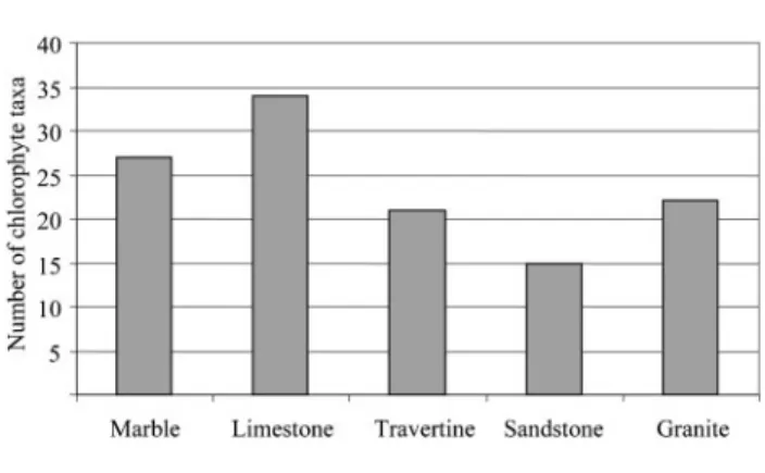
Fig. 8 shows the number of chlorophyta taxa present in each lithotype. Limestone was colonized by the highest number of taxa (34), followed by marble (27). Travertine and granite were colonized by about the same number of taxa (21 and 22, respectively), although the number of monuments built of travertine (7 %) was considerably lower than the number made of granite (18 %). From Table 3 we can see that Oocystis, Cosmarium and Staurastrum species appear almost exclusively on trav-ertine. Nevertheless, the majority of the results suggest that green algae can colonize a wide variety of substrata and this is primarily related to the physical characteristics of the stone surface (porosity, roughness and permeability) and secondarily to the nature of the substratum. This is in accordance with Tianoet al.(1995). These authors carried out a laboratory experiment using two photosynthetic strains: a green alga (Pleurococcus) and a cyanobacterium (Lyngbya), inoculated on 12 different lithotypes exposed to constant climatic conditions. They demonstrated that the Fig. 3. Gloeocapsa sp. observed by optical microscopy. Scale
bar, 20mm.
Fig. 4. Number of cyanobacterial taxa found on each lithotype.
Fig. 5. Klebsormidium flaccidum biofilm on a terracotta statue, Cathedral of Seville.
preferential colonization (percentage of stone surface coverage) was correlated mainly with physical character-istics of the stone (roughness and porosity) while the chemical composition had low influence (Tiano et al., 1995).
Survival strategies of cyanobacteria and algae on stone
As mentioned above, environmental conditions, climate, microclimate and other site-specific characteristics may have a stronger influence on community development than stone lithotype. However, green algal and cyanobacterial diversity as well as abundance is clearly dependent on the availability of water, allowing micro-organisms to form subaerial biofilms on virtually any surface (Gorbushina, 2007). This leads to species characteristic of very different habitats and ecological requirements. Moreover, it was observed that the most representative genera occurring in unfavourable environments develop different strategies to survive in such conditions; these include the production of a sheath composed of extracellular polymeric substances (EPS) outside the cyanobacterial cells (Fig. 7) as a protection against desiccation (Urzı` & Realini, 1998; Tomaselli, 2003). Due to the presence of these colloidal polymeric substances, the biofilm incorporates large amounts of water into its structure, ensuring the maintenance of moisture by balancing changes in humidity and temperature, which permits cyanobacteria and algae to resist drought periods (Go´mez-Alarco´n et al., 1995; Saiz-Jimenez, 1999; Schumannet al., 2005). These biofilms are composed of populations or communities of different micro-organisms (microalgae, cyanobacteria, bacteria and fungi) immobilized on the stone surface (substratum) and frequently embedded in an organic polymer matrix formed by EPS, such as polysaccharides, lipopolysaccharides,
proteins, glycoproteins, lipids, glycolipids, fatty acids and enzymes (Younget al., 2008). EPS are also responsible for binding cells and other particulate matter together (cohesion) and to the substratum (adhesion) (Cecchi et al., 2000; Warscheid & Braams, 2000) (Fig. 9).
The stone surface may have also several characteristics that are important in the attachment process. The extent of microbial colonization appears to increase as the surface roughness increases. This is because shear forces are diminished, and total surface area is higher on rougher surfaces (Mortonet al., 1998; Donlan, 2002). Tomaselliet al. (2000a) showed that high stone porosity and rough surface, together with environmental factors, played a greater role than mineral composition in promoting microbial estab-lishment. The most important of the environmental factors is water availability. Adequate temperature and solar irradiance, and type of atmospheric deposition, are also relevant factors (Tomaselliet al., 2000a).
Endolithic colonization is also a successful survival strategy when surface environmental conditions are adverse for life on stone. The protection provided by the rock leads to the abundance of endolithic micro-organisms in extreme environments, such as cold and hot deserts, semiarid lands and even polar regions (Friedmann, 1982; Bell, 1993; Walkeret al., 2005).
Biodeterioration of stone
The presence of cyanobacterial and algal biofilms on stone surfaces can be considered biodeteriogenic, simply because of the aesthetic damage they cause, producing variously coloured patinas (Ortega-Calvoet al., 1995). These micro-organisms have an important role in the disfigurement of monuments and stone works of art (Figs 1 and 5). Moreover, there are several references in the literature that point to direct decay mechanisms caused by these photosynthetic micro-organisms (Anagnostidis et al., 1991; Griffin et al., 1991; Krumbein & Urzı`, 1991; Ortega-Calvo et al., 1992, 1993b; Wakefield & Jones, 1998; Saiz-Jimenez, 1999; Warscheid & Braams, 2000; Crispim & Gaylarde, 2005; Zurita et al., 2005). In fact, cyanobacteria and green algae living in rocks can enhance soil formation and water retention. It has been estimated that 20–30 % of stone deterioration is a result of biological activity (Wakefield & Jones, 1998). Two types of biodeter-ioration can be considered: biogeophysical and biogeo-chemical. While the effects and extent of biogeochemical deterioration processes are controlled and determined by the chemistry of minerals and the binding cement of each rock, biogeophysical mechanisms are mostly regulated by the porosity and shape of the interior surface of the rocks (Warscheid & Braams, 2000).
Biogeophysical deterioration can be defined as the mechanical damage caused by exerted pressure during biological growth, resulting in surface detachment, super-ficial losses, or penetration and increased porosity (Griffin Fig. 7. SEM micrograph of a biofilm induced in vitro on a
Table 3. Chlorophyta reported on stone monuments, statues and historic buildings in European countries from the Mediterranean Basin, on different substrata
Chlorophyte taxon Substratum Monument no. (Table 1)
Apatococcussp. Limestone, granite, marble 3, 10, 19, 21, 22, 35, 39, 40
Apatococcus lobatus(Chodat) Petersen Marble, limestone 30, 19
Bracteacoccussp. Limestone, marble 11, 43
Chaetophoralesspecies Marble 19a
Chlamydocapsasp. Granite 45
Chlorellasp. Granite, sandstone, marble, limestone 3, 4, 5, 7, 10, 14, 17, 19a, 24, 26, 21, 33, 35, 36, 40
Chlorella ellipsoideaGerneck Limestone, granite 11, 45
Chlorella homosphaeraSkuja Granite 34, 43
Chlorella reisiglii(Reisigl) Watanabe Limestone 11
Chlorella vulgarisBeijerinck Granite, limestone, sandstone 33, 34, 36, 37
Chlorococcumsp. Granite, limestone, sandstone, marble, travertine
2, 3, 6, 17, 19a, 20, 26, 28, 32, 38, 40, 41, 42, 44, 45
Chlorococcum wimmeriRabenhorst Marble 38
Chlorokybus atmophyticusGeitler Sandstone, limestone 33, 11
Chlorosarcinasp. Marble 28
Chlorosarcinopsissp. Limestone, marble 2, 19, 28
Chlorosarcinopsis minor(Gerneck) Herndon Granite, marble, limestone 34, 38, 19
Choricystis chodatii(Jaag.) Fott Granite 38
Cladophorasp. Marble 19a
Coccomyxasp. Granite, marble 3, 14
Cosmariumsp. Limestone 2, 11
Cosmarium depressum(Na¨geli) Lundell Travertine 13
Cosmarium granatumBre´bisson Travertine 13
Cosmarium reniforme(Ralfs) Archer Travertine 13
Crucigenia quadrataMorren Travertine 13
Cylindrocystis brebissonii(Meneghini) De Bary Limestone 11
Desmococcussp. Sandstone, granite 32, 45
Desmococcus vulgarisBrand Limestone, travertine 11, 20
Ecdysichlamys obliquaG.S. West Sandstone 33
Euastrum insulare(Wittrock) Roy Travertine 13
Friedmannia israeliensis(Chantanachat & Bold) Friedl
Sandstone 7
Geminella terricolaPetersen Limestone 11
Gongrosirasp. Marble, limestone 38, 19
Haematococcus pluvialisFlotow Marble 23, 44
Klebsormidiumsp. Granite, limestone 11, 17, 24
Klebsormidium flaccidum(Ku¨tzing) Silva, Mattox & Blackwell
Limestone, granite, marble, sandstone 7, 11, 26, 33, 34, 37, 43
Monoraphidiumsp. Granite 17
Muriella terrestrisBoye-Peterson Sandstone, travertine, granite 20, 33, 34, 37, 43
Myrmeciasp. Limestone 11, 36
Nannochlorissp. Sandstone 33
Oocystis crassaWittrock Travertine 13
Oocystis lacustrisChodat Travertine 13
Oocystis solitariaWittrock Travertine 13
Pediastrum boryanum(Turpin) Meneghini Travertine 13
Pleurastrumsp. Limestone 2
Pleurastrum terrestreFritsch & John Limestone 11
Podohedra bicaudataGeitler Limestone 11
Poloidion didymosPascher Limestone, marble 19
Protococcussp. Limestone 15
Pseudococcomyxa simplex(Mainx) Fott Limestone 11
Pseudodendoclonium basilienseVischer Marble 38
et al., 1991). This kind of deterioration also occurs due to the presence of cyanobacterial and algal biofilms that undergo large volume changes and exert considerable force through cycles of drying and moistening, loosening rock grains (Saiz-Jimenez, 1999). This can lead to the alteration of the stone’s pore-size distribution and result in changes of moisture circulation patterns and temperature response (Saiz-Jimenez, 1999; Warscheid & Braams, 2000). Furthermore, the formation of crusts induced by
cyano-bacterial and algal growth also results in longer moisture retention at the stone surface, increasing the stone colonization potential.
Chlorophyte taxon Substratum Monument no. (Table 1)
Pseudopleurococcus printziiVischer Marble 38
Pseudosphaerocystis lacustris(Lemmermann) Nova`kova`
Travertine 13
Rhizothallussp. Marble 3
Scenedesmussp. Travertine, granite 41, 17
Scenedesmus ecornis(Ehrenberg) Chodat Marble, limestone 19
Scenedesmus obliquus(Turp.) Ku¨tz. Marble, limestone 19
Scenedesmus quadricauda(Turpin) Bre´bisson Sandstone 33
Scenedesmus smithiiS.S. Wang Marble, limestone 19
Scotiellopsis terrestris(Reisigl) Hanagata Limestone, marble 11, 19
Staurastrum borealeWest Travertine 13
Staurastrum lunatumRalfs Travertine 13
Staurastrum lunatumvar.planctonicumWest & West
Travertine 13
Staurastrum manfeldtiiDelponte Travertine 13
Staurastrum pingueTeiling Travertine 13
Stichococcussp. Granite, limestone, marble 2, 3, 10, 15, 17, 18, 21, 31, 36
Stichococcus bacillarisNa¨geli Limestone, granite, marble, sandstone 7, 11, 26, 33, 43, 44
Stichococcus minutusGrintzesco & Pterfi Granite 34, 37
Trebouxiasp. Limestone 11, 30, 34, 36, 37, 38, 45
Trebouxia decoloransAhmadjian Sandstone 33
Trentepohliasp. Marble, sandstone, granite 10, 24, 31, 32, 45
Trentepohlia aurea(Linnaeus) Martius Granite 34, 37
Tetracystissp. Granite, marble, limestone 45, 19
Tetraedron muticum(Braun) Hansgirg Travertine 13
Tetraspora gelatinosa(Vaucher) Desvaux Travertine 13
Ulothrixsp. Limestone, marble, sandstone 2, 3, 6, 21, 30, 42 Table 3.cont.
Fig. 8. Number of chlorophyte taxa found on each lithotype.
Biogeochemical deterioration is the direct action caused by the metabolic processes of organisms on the substratum. The biogenic release of corrosive acids is probably the best known and most commonly investigated biogeochemical damage mechanism in inorganic materials. The process, known as biocorrosion, involves the release of organic acids which can etch or solubilize stone, the exudation of organic chelating agents which sequester metallic cations from stone, or the conversion of inorganic substances by redox reactions which form acids that etch stone and contribute to salt formation (Griffinet al., 1991; Fernandes, 2006). For instance, aerobic micro-organisms produce respiratory carbon dioxide which becomes carbonic acid and contributes to dissolution of stone and soluble salt formation (Griffinet al., 1991; Wakefield & Jones, 1998). The precipitation of calcium salts on cyanobacterial cells growing on limestone suggests the migration of calcium from neighbouring sites (Fig. 10) (Arin˜o et al., 1997; Crispim & Gaylarde, 2005). In addition, the production of organic acids such as lactic, oxalic, succinic, acetic, glycolic and pyruvic has been found and associated with the dissolution of calcite in calcareous stones (Danin & Caneva, 1990; Caneva et al., 1992). Endolithic photosyn-thetic micro-organisms actively dissolve carbonates to enable penetration into the stone, enhancing stone porosity (Griffin et al., 1991; Fernandes, 2006). Furthermore, the slimy surfaces of microbial biofilms favour the adherence of airborne particles (dust, pollen, spores, carbonaceous particles from combustion of oil and coal), giving rise to hard crusts and patinas (Saiz-Jimenez, 1999).
Sophisticated tools, consisting mainly of microscopy techniques, have been applied to the study of the stone biodeterioration process by photosynthetic micro-organ-isms. Microscopy techniques provide direct evidence of biofilm formation by imaging actual cells. The most common are scanning electron microscopy with back-scattered electron imaging (SEM-BSE), confocal scanning laser microscopy (CSLM) coupled with fluorescent probes (Fig. 11), low-temperature SEM (LTSEM) and envir-onmental SEM (ESEM) (Rolda´net al., 2004; Ascasoet al., 2004; De los Rı´oset al., 2004; Wierzchoset al., 2004; De los Rı´os & Ascaso, 2005). CSLM studies revealed that cells in biofilms are organized within a complex exopolymeric matrix, and the biofilm consists of a heterogeneous distribution of cells and cellular aggregates with void spaces or water channels (Costertonet al., 1995; Kawaguchi & Decho, 2002). The use of microscopy techniques to study microbial colonies in their natural microhabitat has also demonstrated that EPS penetrate small pore spaces of the substratum and may facilitate subsequent penetration of the micro-organisms into the material, increasing stone biodeterioration. These studies have shown that penetra-tion of growing organisms into rock and the diffusion of their excreted products may occur to depths of several millimetres (Saiz-Jimenez, 1999; Koestler, 2000; Salvadori, 2000; Pohl & Schneider, 2002; Younget al., 2008). Pohl & Schneider (2002) applied computerized image analysis to
detect and quantify the biomass and depth of penetration of endolithic micro-organisms into carbonate rock sur-faces. The natural carbonate rocks investigated were endolithically colonized by lichens, cyanobacteria, algae and fungi. Photosynthetic micro-organisms from intensely insolated dry sites retreated to depths of 150-250mm below Fig. 10. SEM micrograph of Scytonema julianum mat. The calcified sheaths are composed of triradiate calcite crystals. Scale bar, 100mm. Reproduced from Ortega-Calvoet al.(1995)
with permission.
the rock surface and showed ‘cushions’ of EPS oriented toward the surface, which protect them against intense light and provide water retention. In addition, the authors demonstrated that as soon as the endolithic biofilm was established it exerted an overall protective effect on the carbonate rocks. Salvadori (2000) examined by SEM the endolithic communities inhabiting Italian stone monuments and observed that euendolithic cyanobacteria and fungi could easily penetrate the calcite crystals of marble. The periodic contraction and expansion of EPS induces mechanical stress on the stone surface, particularly when the polymer penetrates into the pores, stimulating defo-liation of the biofilm and the underlying substratum, evident at macroscopic scale (Saiz-Jimenez, 1999; Younget al., 2008; Kemmlinget al., 2004). Krumbein & Urzı` (1991) reported the physical action on and within marble through biofilms and microbial growth, demonstrating that stability and activity of water in polyionic gel matrix was crucial for the physical reactions and interactions between microbiota and rock and rock porosity, giving rise to decomposition and detachment of grains, chips or scales in marbles. Apart from direct deterioration of stone substrata, microbial biofilms, especially those formed by photosynthetic micro-organisms, play an indirect role in stone biodeterioration as described by Ortega-Calvo et al. (1992). It was concluded that photosynthetic micro-organisms contribute directly to stone deterioration through physical and chemical action and indirectly through the synergistic interactions with hetero-trophic micro-organisms such as fungi and bacteria. The accumulation of photosynthetic biomass on stone surfaces provides nutrients for the growth of other communities which graze on cyanobacterial and algal polysaccharides and cell debris, contributing to accelerating the deterioration of the stone (Tiano, 1998; Crispim & Gaylarde, 2005; McNamara & Mitchell, 2005; Fernandes, 2006). Therefore, the role of cyanobacteria and green algae in the degradation of cultural heritage cannot be neglected. The detection and identification of these micro-organisms is extremely important for future studies of the biodeteriogenic process and the development of prevention and control methods.
Conclusion
A wide range of stone monuments from the Mediterranean Basin are colonized by cyanobacteria and green algae, showing notable biodiversity. A total of 96 taxa of cyanobacteria and 76 taxa of chlorophyta were found. The most widespread commonly reported taxa in the stone cultural heritage in the Mediterranean Basin are, among cyanobacteria,Gloeocapsa,PhormidiumandChroococcusand, among chlorophyta, Chlorella, Stichococcus and Chloro-coccum. These genera were found associated with all lithotypes. Although an extensive literature survey was performed, the preference of the cyanobacteria and chlor-ophyta for a specific stone substratum was more complicated to correlate than expected. The majority of the results suggest that green algae and cyanobacteria can colonize a wide variety of substrata and this is primarily related to the physical
characteristics of the stone surface (porosity, roughness and permeability) and secondarily to the nature of the substratum. Most cyanobacteria and chlorophyta did not show a clear relationship with the nature of the substratum, suggesting that environmental variables and site-specific characteristics (e.g. exposure to light, special architectural features) together with secondary, tertiary and/or extrinsic stone bioreceptivity have a stronger influence on community development than the substratum itself. In this complex amalgam of factors, it is often difficult to determine the influence of each factor alone; the evaluation of their combined effects, their synergy and dynamics is complex, and probably all factors are relevant. In order to ascertain a correlation between stone substratum and organisms, we need more detailed data about lithotype properties, and the microclimatic and environmental condi-tions of the monuments studied.
Cyanobacteria and green algae play an important role in the deterioration of monuments and other stone works of art, being responsible for aesthetic, biogeophysical and biogeo-chemical damage. Future work should focus on ecological and physiological studies of specific species of these micro-organisms in order to gain a better understanding of their role in stone colonization and biodeterioration processes. Moreover, an interdisciplinary team working on the same ‘case study’ is necessary in order to simultaneously investigate all the factors involved in the biodeterioration process such as mineralogical-petrographic, physico-chem-ical and climatic (and microclimatic) parameters.
Acknowledgements
This work was supported by the Ministe´rio da Cieˆncia, Tecnologia e Ensino Superior, Portugal, with a doctoral grant (SFRH/BD/21481/ 2005), and has been partially financed by the CEPGIST FCT subproject DECASTONE. The work also received support from the Programa de Financiamento Plurianual da Unidade de Investigac¸a˜o da FCT, financed by the European Union FEDER and the national budget of the Portuguese Republic. This is also a TCP CSD2007-00058 paper. The authors thank Dr Xavier Arin˜o for the permission to reproduce one of the figures of his doctoral thesis. The authors are also very grateful to Dr M. Hernandez-Marine (University of Barcelona, Spain) and Professor Rui Silva for the permission to use a confocal microscope image and a SEM image, respectively.
References
Altieri, A., Pietrini, A. M., Ricci, S. & Roccardi, A. (2000).The temples of the archaeological area of Paestum (Italy): a case study on biodeterioration. InProceedings of the 9th International Congress on Deterioration and Conservation of Stone, pp. 433–443. Edited by V. Fassina. Amsterdam: Elsevier.
Alvarez, A., Argemı´, M., Laorden, V., Dome´nech, X., Verbal, J., Navarro, A., Prada, J. L., Puge´s, M., Rocabayera, R. & Vilaseca, L. (1994).Physical, chemical and biological weathering detected in the romanic portal of the Sant Quirze de Pedret church (XIIc.). In
Anagnostidis, K. & Komarek, J. (1988).Modern approach to the classification system of cyanophytes. 3. Oscillatoriales.Algol Stud50– 53, 327–472.
Anagnostidis, K., Economou-Amilli, A. & Roussomoustakaki, M. (1983). Epilithic and chasmolithic microflora (Cyanophyta, Bacillariophyta) from marbles of the Parthenon (Acropolis-Athens, Greece).Nova Hedwigia38, 227–287.
Anagnostidis, K., Gehrmann, C. K., Gross, M., Krumbein, W. E., Lisi, S., Pantazidou, A., Urzi, C. & Zagari, M. (1991). Biodeterioration of marbles of the Parthenon and Propylaea, Acropolis, Athens – associated organisms, decay and treatment suggestions. In
Proceedings of the 2nd International Symposium on the Conservation of Monuments in the Mediterranean Basin, pp. 305–325. Edited by D. Decrouez, J. Chamay & F. Zezza. Geneva, Switzerland: Ville de Gene`ve-Muse´um d’Histoire Naturelle & Muse´e D’Art et d’Histoire.
Arin˜o, X. (1996).Estudio de la colonizacio´n, distribucio´n e interaccio´n de lı´quenes, algas y cianobacterias con materiales pe´treos de los conjuntos arqueolo´gicos de Baelo Claudia y Carmona. Doctoral thesis, University of Barcelona.
Arin˜o, X., Hernandez-Marine, M. & Saiz-Jimenez, C. (1997).Colonization of Roman tombs by calcifying cyanobacteria.Phycologia36, 366–373. Ascaso, C., Garcia´ del Cura, M. A. & De Los Rı´os, A. (2004). Microbial biofilms on carbonate rocks from a quarry and monuments in Novelda (Alicante, Spain). InBiodeterioration of Stone Surfaces. Lichen and Biofilms as Weathering Agents of Rocks and Cultural Heritage, 1st edn, pp. 79–98. Edited by L. L. St Clair & M. R. D. Seaward. Dordrecht, Netherlands: Kluwer.
Bartolini, M., Ricci, S. & Del Signore, G. (2004). Release of photosynthetic pigments from epilithic biocenoses after biocide treatments. In Proceedings of the 10th International Congress on Deterioration and Conservation of Stone, pp. 519–526. Edited by D. Kwiatkowski & R. Lo¨fvendahl. Stockholm, Sweden: ICOMOS. Bell, R. A. (1993).Cryptoendolithic algae of hot semiarid lands and deserts.J Phycol29, 133–139.
Bellinzoni, A. M., Caneva, G. & Ricci, S. (2003).Ecological trends in travertine colonization by pioneer algae and plant communities.Int Biodeterior Biodegradation51, 203–210.
Bolı´var, F. C. & Sa´nchez-Castillo, P. M. (1997).Biomineralization processes in the fountains of the Alambra, Ganada, Spain. Int Biodeterior Biodegradation40, 205–215.
Caneva, G., Nugari, M. P., Ricci, S. & Salvadori, O. (1992).Pitting of marble Roman monuments and the related microflora. InProceedings of the 7th International Congress on Deterioration and Conservation of Stone, pp. 521–530. Edited by J. Delgado, F. Henriques & F. Telmo. Lisbon: Laborato´rio Nacional de Engenharia Civil.
Cecchi, G., Pantani, L., Raimondi, V., Tomaselli, L., Lamenti, G., Tiano, P. & Chiari, R. (2000).Fluorescence lidar technique for remote sensing of stone monuments.J Cult Herit1, 29–36.
Costerton, J. W., Lewandowski, Z., Caldwell, D. E., Korber, D. R. & Lappin-Scott, H. M. (1995).Microbial biofilms. Annu Rev Microbiol
49, 711–745.
Crispim, C. A. & Gaylarde, C. C. (2005). Cyanobacteria and biodeterioration of cultural heritage: a review.Microb Ecol49, 1–9. Crispim, C. A., Gaylarde, P. M. & Gaylarde, C. C. (2003).Algal and cyanobacterial biofilms on calcareous historic buildings. Curr Microbiol46, 79–82.
Danin, A. & Caneva, G. (1990).Deterioration of limestone walls in Jerusalem and marble monuments in Rome caused by cyanobacteria and cyanophilous lichens.Int Biodeterior26, 397–417.
De los Rı´os, A. & Ascaso, C. (2005). Contributions of in situ microscopy to the current understanding of stone biodeterioration.
Int Microbiol8, 181–188.
De los Rı´os, A., Galva´n, V. & Ascaso, C. (2004).In situ microscopical diagnosis of biodeterioration processes at the convent of Santa Cruz la Real, Segovia, Spain.Int Biodeterior Biodegradation54, 113–120. Donlan, R. M. (2002).Biofilms: microbial life on surfaces.Emerg Infect Dis8, 881–890.
Dupuy, P., Trotet, G. & Grossin, F. (1976).Protection des monuments contre les cyanphyce´es en milieu abrite´ et humide. InThe Conservation of Stone I. Proceedings of the International Symposium on the Conservation of Stone, pp. 205–219. Edited by R. Rossi-Manaresi. Bologna, Italy: Centro per la conservazione delle sculture all’aperto. Fernandes, P. (2006).Applied microbiology and biotechnology in the conservation of stone cultural heritage materials. Appl Microbiol Biotechnol73, 291–296.
Flores, M., Lorenzo, J. & Go´mez-Alarco´n, G. (1997). Algae and bacteria on historic monuments at Alcala de Henares, Spain. Int Biodeterior Biodegradation40, 241–246.
Friedmann, E. I. (1982).Endolithic microorganisms in the Antarctic cold desert.Science215, 1045–1053.
Giaccone, G., Einaldi, M. L. V. & Giacobini, C. (1976). Forme biologische delle alghe esistenti sulle sculture all’aperto. In The Conservation of Stone I. Proceedings of the International Symposium on the Conservation of Stone, pp. 245–256. Edited by R. Rossi-Manaresi. Bologna, Italy: Centro per la conservazione delle sculture all’aperto. Go´mez-Alarco´n, G., Mun˜oz, M., Arin˜o, X. & Ortega-Calvo, J. J. (1995).Microbial communities in weathered sandstones: the case of Carrascosa del Campo church, Spain.Sci Total Environ167, 249–254. Gorbushina, A. A. (2007). Life on the rocks. Environ Microbiol9, 1613–1631.
Graham, L. E. & Wilcox, L. W. (2000).Algae, 1st edn. Upper Saddle River, NJ: Prentice-Hall.
Griffin, P. S., Indictor, N. & Kloestler, R. J. (1991).The biodeteriora-tion of stone: a review of deteriorabiodeteriora-tion mechanisms, conservabiodeteriora-tion case histories, and treatment.Int Biodeterior28, 187–207.
Hernandez-Marine, M., Saiz-Jimenez, C. & Arin˜o, X. (1997).Borzia perikleiAnag. (Cyanoprokaryota): a taxonomical approach.Lagascalia
21, 457–462.
Kawaguchi, T. & Decho, A. W. (2002).In situ analysis of carboxyl (-COOH) and sulfhydral (-SH) groups of extracellular polymeric secretions (EPS) by confocal scanning laser microscopy.Anal Biochem
304, 266–267.
Kemmling, A., Ka¨mper, M., Flies, C. O., Schieweck, C. & Hoppert, M. (2004).Biofilms and extracellular matrices on geomaterials.Environ Geol46, 429–435.
Koestler, R. J. (2000).Polymers and resins as food for microbes. InOf Microbes and Art. The Role of Microbial Communities in the Degradation and Protection of Cultural Heritage, pp. 153–167. Edited by O. Ciferri, P. Tiano & G. Mastromei. New York: Kluwer. Krumbein, W. E. & Urzı`, C. (1991). Biologically induced decay phenomena of antique marbles – some general considerations. In
Proceedings of the 2nd International Symposium on the Conservation of Monuments in the Mediterranean Basin, 19–21 November, pp. 219–235. Edited by D. Decrouez, J. Chamay & F. Zezza. Geneva, Switzerland: Ville de Gene`ve-Muse´um d’Histoire Naturelle & Muse´e d’Art et d’Histoire. Lamenti, G., Tiano, P. & Tomaselli, L. (2000).Biodeterioration of ornamental marble statues in the Boboli Gardens (Florence, Italy).
J Appl Phycol12, 427–433.
McNamara, C. J. & Mitchell, R. (2005). Microbial deterioration of historic stone.Front Ecol Environ3, 445–451.
Miller, A. Z. & Macedo, M. F. (2006).Mapping and characterization of a green biofilm inside of Vilar de Frades Church (Portugal). In
Heritage, Weathering and Conservation, pp. 329–335. Edited by R. Fort, M. Alvarez de Buergo, M. Gomez-Heras & C. Vazquez-Calvo. London: Taylor & Francis.
Miller, A. Z., Dionisio, A. & Macedo, M. F. (2006). Primary bioreceptivity: a comparative study of different Portuguese lithotypes.
Int Biodeterior Biodegradation57, 136–142.
Miller, A. Z., Laiz, L., Gonzalez, J. M., Dionı´sio, A., Macedo, M. F. & Saiz-Jimenez, C. (2008).Reproducing stone monument photosyn-thetic-based colonization under laboratory conditions. Sci Total Environ405, 278–285.
Miller, A. Z., Laiz, L., Dionı´sio, A., Macedo, M. F. & Saiz-Jimenez, C. (2009).Growth of phototrophic biofilms from limestone monuments under laboratory conditions. Int Biodeterior Biodegradation, doi: 10.1016/j.ibiod.2009.04.004.
Morton, L. H. G., Greenway, D. L. A., Gaylarde, C. C. & Surman, S. B. (1998). Consideration of some implications of the resistance of biofilms to biocides.Int Biodeterior Biodegradation41, 247–259. Noguerol-Seoane, A. & Rifo´n-Lastra, A. (1996).Epilithic ficoflora on two monuments of historic-artistic interest from Galicia (N.W. Spain). In Degradation and Conservation of Granitic Rocks in Monuments. Environmental Protection and Conservation of the European Cultural Heritage, pp. 417–421, Research Report no. 5. Brussels: European Commission Directorate-General XII: Science, Research and Development.
Ortega-Calvo, J. J., Hernandez-Marine, M. & Saiz-Jimenez, C. (1991).Biodeterioration of building materials by cyanobacteria and algae.Int Biodeter28, 165–185.
Ortega-Calvo, J. J., Hernandez-Marine, M. & Saiz-Jimenez, C. (1992). Experimental strategies for investigating algal deterioration of stone.
Proceedings of the 7th International Congress on Deterioration and Conservation of Stone, pp. 541–549. Edited by J. Delgado, F. Henriques & F. Telmo. Lisbon: Laborato´rio Nacional de Engenharia Civil. Ortega-Calvo, J. J., Sanchez-Castillo, P. M., Hernandez-Marine, M. & Saiz-Jimenez, C. (1993a).Isolation and characterization of epilithic chlorophyta and cyanobacteria from two Spanish cathedrals (Salamanca and Toledo).Nova Hedwigia57, 239–253.
Ortega-Calvo, J. J., Hernandez-Marine, M. & Saiz-Jimenez, C. (1993b). Cyanobacteria and algae on historic buildings and monuments In
Recent Advances in Biodeterioration and Biodegradation, pp. 173–203. Edited by K. L. Garg, N. Garg & K. G. Mukerji. Calcutta: Naya Prokash. Ortega-Calvo, J. J., Arin˜o, X., Hernandez-Marine, M. & Saiz-Jimenez, C. (1995).Factors affecting the weathering and colonization of monuments by phototrophic microorganisms.Sci Total Environ167, 329–341. Pantazidou, A. & Theoulakis, P. (1997). Cyanophytes and associated flora at the neoclassical Palace of Sts George and Michael in Corfu (Greece). Aspects of cleaning procedures. In Proceedings of the 4th International Symposium on the Conservation of Monuments in the Mediterranean Basin, pp. 355–368. Edted by A. Moropoulou, F. Zezza, E. Kollias & I. Papachistodoulou. Rhodes: Technical Chamber of Greece. Pentecost, A. (1992).Growth and distribution of endolithic algae in some North Yorkshire streams (UK).Br Phycol J27, 145–151. Pereira de Oliveira, B. (2008). Caracterizac¸a˜o de filmes negros em pedras granı´ticas. O caso de estudo da Igreja da Ordem de Sa˜o Francisco do Porto. MSc thesis, Universidade Nova de Lisboa, Lisbon, Portugal. Pereira de Oliveira, B., Miller, A., Sequeira Braga, M. A. & Macedo, M. F., Dionı´sio, A. & Silveira, T. (2008).Characterization of dark films in granites. The case study of Igreja da Ordem de Sa˜o Francisco in Oporto (Portugal). In Proceedings of the 14th Interational
Biodeterioration and Biodegradation Symposium, p. 72. Edited by C. Urzı`. Messina, Italy: International Biodeterioration and Biodeg-radation Society.
Pietrini, A. M., Ricci, S., Bartolini, M. & Giuliani, M. R. (1985). A reddish colour alteration caused by algae on stoneworks. Preliminary studies. In Proceedings of the 5th International Congress on Deterioration and Conservation of Stone, pp. 653–662. Edited by G. Felix. Lausanne, Switzerland: Presses Polytechniques Romandes. Pohl, W. & Schneider, J. (2002). Impact of endolithic biofilms on carbonate rock surfaces. In Natural Stone, Weathering Phenomena, Conservation Strategies and Case Studies, pp. 177–194, Special Publications 205. Edited by S. Siegesmund, T. Weiss & A. Vollbrecht. London: Geological Society.
Prieto, B. & Silva, B. (2005).Estimation of the potential bioreceptivity of granitic rocks from their intrinsic properties. Int Biodeterior Biodegradation56, 206–215.
Ricci, S. & Pietrini, A. M. (1994). Caratterizzazione della microflora algale presente sulla Fontana dei Quattro Fiumi, Roma. InProceedings of the 3rd International Symposium on the Conservation of Monuments in the Mediterranean Basin, pp. 353–357. Edited by V. Fassina, H. Ott & F. Zezza. Venice: Soprintendenza ai Beni Artistici e Storici di Venezia. Rolda´n, M., Clavero, E., Castel, S. & Herna´ndez-Marine´, M. (2004). Biofilms fluorescence and image analysis in hypogean monuments research.Arch Hydrobiol Suppl Algol Stud111, 127–143.
Saiz-Jimenez, C. (1995).Deposition of anthropogenic compounds on monuments and their effect on airborne microorganisms.
Aerobiologia11, 161–175.
Saiz-Jimenez, C. (1999).Biogeochemistry of weathering processes in monuments.Geomicrobiol J16, 27–37.
Saiz-Jimenez, C., Hermosin, B., Ortega-Calvo, J. J. & Go´mez-Alarco´n, G. (1991).Applications of analytical pyrolysis to the study of stony cultural properties.J Anal Appl Pyrol20, 239–521.
Salvadori, O. (2000).Characterisation of endolithic communities of stone monuments and natural outcrops. InOf Microbes and Art – The Role of Microbial Communities in the Degradation and Protection of Cultural Heritage, pp. 89–101. Edited by O. Ciferri, P. Tiano & G. Mastromei. New York: Kluwer.
Salvadori, O., Sorlini, C. & Zanardini, E. (1994).Microbiological and biochemical investigations on stone of the Ca’ d’Oro facade (Venice). InProceedings of the 3rd International Symposium on the Conservation of Monuments in the Mediterranean Basin, pp. 343–347. Edited by V. Fassina, H. Ott & F. Zezza. Venice: Soprintendenza ai Beni Artistici e Storici di Venezia
Santos, M. F. A. (2003).Optical Microscopy of the Microbial Samples Collected on the Cloisters of Church of Santa Cruz in Coimbra (Portugal). Coimbra: Universidade de Coimbra.
Sarro, M. I., Garcia, A. M., Rivalta, V. M., Moreno, D. A. & Arroyo, I. (2006). Biodeterioration of the Lions Fountain at the Alhambra Palace, Granada (Spain).Build Environ41, 1811–1820.
Schumann, R., Ha¨ubner, N., Klausch, S. & Karsten, U. (2005). Chlorophyll extraction methods for the quantification of green microalgae colonizing building facades.Int Biodeterior Biodegradation55, 213–222. Tiano, P. (1998). Biodeterioration of monumental rocks: decay mechanisms and control methods.Sci Tech Cult Herit7, 19–38. Tiano, P., Accolla, P. & Tomaselli, L. (1995).Phototrophic biodeteri-ogens on lithoid surfaces: an ecological study.Microb Ecol29, 299–309. Tomaselli, L. (2003). Biodeterioration processes on inorganic substrata.Coalition6, 5–9.
Tomaselli, L., Lamenti, G., Bosco, M. & Tiano, P. (2000a).Biodiversity of photosynthetic micro-organisms dwelling on stone monuments.
Tomaselli, L., Tiano, P. & Lamenti, G. (2000b). Occurrence and fluctuation in photosynthetic biocoenoses dwelling on stone monu-ments. InOf Microbes and Art – The Role of Microbial Communities in the Degradation and Protection of Cultural Heritage, pp. 63–76. Edited by O. Ciferri, P. Tiano & G. Mastromei. New York: Kluwer. Urzı`, C. & Realini, M. (1998).Colour changes of Noto’s calcareous sandstone as related to its colonisation by microorganisms. Int Biodeterior Biodegradation42, 45–54.
Wakefield, R. D. & Jones, M. S. (1998).An introduction to stone colonizing micro-organisms and biodeterioration of building stone.Q J Eng Geol31, 301–313.
Walker, J. J., Spear, J. R. & Pace, N. R. (2005). Geobiology of a microbial endolithic community in the Yellowstone geothermal environment.Nature434, 1011–1014.
Warscheid, T. & Braams, J. (2000). Biodeterioration of stone: a review.Int Biodeterior Biodegradation46, 343–368.
Wierzchos, J., De los Rios, A., Sancho, L. G. & Ascaso, C. (2004). Viability of endolithic micro-organisms in rocks from the McMurdo Dry Valleys of Antarctica established by confocal and fluorescence microscopy.J Microsc216, 57–61.
Young, M. E., Alakomi, H. L., Fortune, I., Gorbushina, A. A., Krumbein, W. E., Maxwell, I., McCullagh, C., Robertson, P., Saarela, M. & other authors (2008). Development of a biocidal treatment regime to inhibit biological growths on cultural heritage: BIODAM.Environ Geol56, 631–641.