Abstract
Submitted: January 17, 2018
Modification: June 15, 2018 Accepted: July 17, 2018
Evaluation of pain perception during
orthodontic debonding of metallic
brackets with four different techniques
Objective: The aim of this study was to evaluate patients’ pain levels during four different debonding procedures. The null hypothesis was that the pain perception of the patients undergoing four different debonding
applications was not statistically significant different. Material and Methods:
One hundred and twenty orthodontic patients who underwent orthodontic debonding were included in this study. The patients were randomly divided into 4 groups according to technique used in the patients. Debonding groups
were as follows: Group 1) Conventional debonding group, Group 2) Medication
group (acetaminophen was given 1 hour before debonding), Group 3) Soft bite wax group, and Group 4) Soft acrylic bite wafer group. The patients’ levels of anxiety and fear of pain were evaluated before debonding, and Numerical Rating Scale (NRS) was applied to evaluate their pain perception
during debonding. Mann-Whitney U and Kruskal–Wallis tests were used to evaluate non-normally distributed data. Categorical data analysis were carried by chi-square and McNemar tests. The significance level was set at p<0.05. Results: Anxiety scores of the patients were not statistically significant
between both genders and debonding groups. In the quadrants in which the patients were perceived, the highest pain level was in the left side of the mandible. The teeth in which the highest pain level was perceived were the lower left and upper right lateral incisors. Although there was no statistically
significant difference among the pain scores of the patients in each group, quadrant scores of female patients showed significant differences, being the lowest scores in the soft bite wax group. Conclusions: Majority of the
patients had no fear of pain before debonding. Pain levels of the patients in
the conventional debonding group were not significantly different from those
of the other groups, except quadrant scores of females in the soft bite wax group. The null hypothesis was accepted.
Keywords: Pain. Orthodontic brackets. Dental debonding. Occlusal force. Bite force.
Delal Dara KILINÇ1 Gulsilay SAYAR1
1Istanbul Medipol University, School of Dentistry, Department of Orthodontics, Istanbul, Turkey.
Corresponding address: Delal Dara Kilinç Istanbul Medipol University - School of Dentistry -
Introduction
Orthodontic treatment procedures such
as separator placement, orthodontic force
application, archwire placement and activation,
and debonding procedure usually involve pain and
discomfort, and 90% to 95% of patients reported
having pain during orthodontic treatment.1-6 It
has been generally accepted that pain perception
may be related to age, individual pain threshold,
motivation, psychological condition, previous
negative dental experience, and the magnitude
of orthodontic force. Some previous reports
showed women reported more pain experience
than men,7,8 while other reports showed no gender
differences regarding pain perception.5,9-11
Pain may arise during the active phases of
orthodontic treatment and during the debonding
procedure.1,2 To lessen or prevent the pain during
debonding are as important as preventing enamel
damage and, thus, the use of different orthodontic
instruments, ultrasound, laser application,
thermal heating the orthodontic adhesives, or
biting occlusal bite wafers at debonding have been
discussed in previous reports.11-13
Debonding procedure should be harmless,
painless and quick.14 Pain and discomfort resulting
from fixed orthodontic appliances, such as
elastomeric separator and arch wire placement,
were evaluated in previous studies,5,6,15 but pain
perception in debonding procedure is still a poorly
documented issue in orthodontics. The aim of this
study was to evaluate the pain levels in different
debonding applications and the patients’ anxiety
levels before the procedure to determine the best
debonding technique. The null hypothesis was that
the pain perception of the patients undergoing four
different debonding applications is not statistically
significant different (conventional debonding,
debonding with acetaminophen administration,
debonding while biting a soft plastic wafer, and
debonding with biting wax).
Material and methods
The sample size was determined using a
computer program (Minitab version 17, Minitab Inc, State Collage, Pennsylvania, USA). The
calculation was made based on a significance level of 0.05 and a power of 90% to detect a clinically
meaningful difference of 1 cm in NRS. For acute
and traumatic pains, minimum mean change of 13
mm (median of 11 mm) was accepted as clinically
significant level in visual analog pain scale.13
Based on this knowledge, this prospective study
was carried out on 120 orthodontic patients (84 females and 36 males) at orthodontic debonding stage. This means that 2880 teeth will be included
to this study. The same researcher (G.S) treated
all the patients. Ethics committee of Istanbul
Medipol University approved this study with the approval number 10840098-604.01.01-E.25319.
An informed consent was obtained from all the
patients or their parents. The inclusion criteria
for this study were as follows: patients aged
12-18 years, presence of all permanent teeth
except the third molars, use of upper and lower
fixed orthodontic appliances (0.018 inch metallic Gemini Series Brackets -3M Unitek, Monrovia, California, USA), 0.017x0.025 inch stainless steel archwires (3M Unitek, Monrovia Calif), and
bonding procedure carried out by using Transbond
XT primer+Transbond XT Adhesive paste (3M Unitek, Monrovia, California, USA). In addition
to these criteria, the patients were asked about
having no medical problem, no medication, no
dental or periodontal problem, and no craniofacial
disorder. The mean age of the patients was
15.10±1.83 years at the debonding appointment.
Patients arriving at debonding appointment
were enrolled to 4 different groups (n=30) determined by debonding method. The first 30
patients whose active orthodontic treatments
terminated were enrolled to Group 1, the second
30 patients to Group 2, and so on, without
considering the age, gender, malocclusion type,
and treatment duration.
The debonding procedures applied to each
group were as follows:
Group 1) Conventional debonding group:
Debonding was performed with a Weingart plier.
Teeth were not in contact with their counterparts
during the operation. In other words, debonding
was performed with an open mouth position
(Figure 1).
was given to the patient 1 hour before debonding,
and debonding was done as explained in Group I.
Group 3) Soft bite wax group: The patient was
requested to bite on an occlusal wax (Ormco,
Glendora, California, USA), and then debonding was performed with a Weingart plier (Figure 2).
Group 4) Soft acrylic bite wafer group: The
patient was asked to bite on a soft plastic bite
wafer (3M, Unitek, Monrovia, Calif), and then debonding was performed with a Weingart plier
(Figure 3).
All the debondings were performed with the
same Weingart pliers, beginning from the upper
and lower left sides of the jaws, respectively. The
Weingart plier was applied to the bracket base and
squeezed the base in a mesiodistal direction. The
archwires were in situ during debonding.
All the procedures mentioned above were
managed by the same author (G.S).
Before debonding, a two-part questionnaire
was applied to the patients. The dichotomous
questions about the presence of anxiety and/or
fear of pain were asked, and the patients answered
these questions as yes or no. After debonding,
numerical rating scale (NRS) was applied to
evaluate the patients’ pain perception.16 For this
purpose, the patients were asked the following
questions: “in which of your teeth and in which
quadrant of your jaws you had the highest pain
level” and they scored the pain levels perceived
on the numerical rating scale. NRS documents of
each patient were numbered anonymously and
number fields were masked. The other researcher (D.D.K) blinded to the groups of study assessed
the NRS scores.
Statistical analysis
Statistical analysis was performed with
statistical software IBM SPSS Statistics (V23; IBM, Armonk, NY, USA). Normality of the data was evaluated with Shapiro–Wilk test. Mann-Whitney U and Kruskal-Wallis tests were used to evaluate non-normally distributed data. The analysis of categorical data was performed with chi-square
test. The results were presented as median
(minimum-maximum) values and interquartile ranges. The significance level was set at p<0.05.
Results
Distribution of the patients’ anxiety scores
before debonding and their between-groups
comparisons and the comparisons between the
genders are shown in Table 1. No between-groups
differences and no gender differences in all groups Figure 1- Intraoral photograph of conventional debonding
Figure 2- Intraoral photograph of debonding with soft bite wax
Figure 3- Intraoral photograph of debonding with soft acrylic bite
regarding anxiety scores were found. Two thirds
of the patients declared no fear of pain before
debonding.
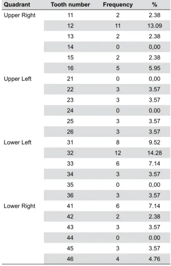
The quadrants and teeth in which the patients
perceived the highest pain during debonding and
their frequencies and percentages are presented
in Table 2 and 3, respectively. Approximately one
third of the patients (n=36) declared no pain
during debonding. According to the results in
Table 2, the chin area in which the frequency of
pain perception was maximum (26.7%) was the
lower left mandibular area.
The results of Table 3 showed the patients
perceived the highest pain in different teeth,
except in the upper right and left first premolar,
upper left central incisor, lower left second
premolar and lower right first premolar teeth.
The teeth having the most pain frequency were
the lower left (14.28%) and upper right (13.09%)
lateral incisors.
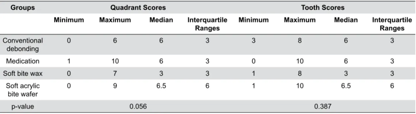
Table 4 shows the pain scores of the patients
in each group during debonding and the results
of between-groups comparisons. Although there was no statistically significant difference between
the groups, the patients in the soft bite wax group
declared lower pain scores in both quadrant
and tooth evaluations. The data in Table 4 were
classified according to gender, and pain scores of
the male and female patients in each group and
the results of between-groups comparisons of each gender are shown in Table 5.
As can be seen from Table 5, quadrant scores of
Group Gender No Yes p-value
(χ² between gender)
Frequency % Frequency %
Conventional debonding Female 12 10 8 6.6 1.000
Male 6 5 4 3.3
Medication Female 12 10 14 11.6 0.467
Male 4 3.3 _ _
Soft bite wax Female 16 13.3 4 3.3 1.000
Male 6 5 4 3.3
Soft acrylic bite wafer Female 14 11.6 4 3.3 0.792
Male 10 8.3 2 1.6
Total 80 67.7 40 33.3
Between-groups results (Kruskal-Wallis) : p=0.658 for males; p=0.292 for females
Table 1- Distribution of the anxiety scores and their between-groups and between genders comparisons
Quadrant Frequency %
Upper right 22 18.3
Lower right 18 15
Upper left 12 10
Lower left 32 26.7
No pain 36 30
Table 2- Frequencies and percentages of quadrants in which the highest pain or no pain was perceived
Quadrant Tooth number Frequency %
Upper Right 11 2 2.38 12 11 13.09
13 2 2.38
14 0 0,00
15 2 2.38
16 5 5.95
Upper Left 21 0 0,00
22 3 3.57
23 3 3.57
24 0 0.00
25 3 3.57
26 3 3.57
Lower Left 31 8 9.52
32 12 14.28
33 6 7.14
34 3 3.57
35 0 0,00
36 3 3.57
Lower Right 41 6 7.14
42 2 2.38
43 3 3.57
44 0 0.00
45 3 3.57
46 4 4.76
females showed statistically significant differences
between the groups. Soft bite wax and soft acrylic
bite wafer groups showed lower pain scores. These
two groups also showed lower pain scores in
males, although it was not statistically significant (p=0.097).
Discussion
Bond strength is important for maintaining
orthodontic treatment efficiency, but easy
debonding of the brackets is preferred at the
end of the treatment. Many kinds of debonding
methods have been suggested to lessen the
patient discomfort. These debonding methods
include ultrasonic instrumentation, laser irradiation
and electrothermal heating, using special
pliers. In addition to these methods, modified
adhesive resins containing thermoexpandable
microcapsules have been used to lessen the pain
and discomfort.3,11,17-19
Pain is an inherently subjective symptom,
and thus no objective method exists for its
assessment. Visual analog scale (VAS), numerical rating scale (NRS), and verbal rating scale (VRS)
are commonly used measurement instruments
to quantify pain intensity of the patients. The
comparative studies regarding these instruments
showed no statistically significant difference
among them.17,20 In this study, numerical rating
scale was used to assess the pain perceived during
debonding because of its easy application.
Debonding procedures should be harmless,
Groups Quadrant Scores Tooth Scores
Minimum Maximum Median Interquartile
Ranges
Minimum Maximum Median Interquartile
Ranges
Conventional
debonding 0 6 6 3 3 8 6 3
Medication 1 10 6 3 0 10 6 3
Soft bite wax 0 7 3 3 1 8 3 3
Soft acrylic
bite wafer 0 9 6.5 6 1 10 6.5 6
p-value 0.056 0.387
p>0.05 Kruskal-Wallis test
Table 4- Distribution of Numerical Rate Scale (NRS) scores regarding quadrant and teeth in which the patients perceived the highest pain and their between-groups comparisons
Gender Groups Quadrant Scores Tooth Scores
Median Minimum Maximum Median Minimum Maximum
Female Conventional
debonding 6ab 0 8 7 3 8
Medication 6b 1 10 6 4 10
Soft bite wax 0.5a 0 7 3 1 8
Soft acrylic bite
wafer 0ab 0 9 7 1 10
p-value 0.023* 0.673
Male Conventional
debonding 5 4 7 5 5 8
Medication 3.5 2 5 3 0 6
Soft bite wax 0 0 3 4 3 5
Soft acrylic bite
wafer 0 0 7 6.5 6 7
p-value 0.097 0.287
* P<0.05 Kruskal-Wallis and Mann-Whitney U tests
There is no statistical difference between the median values that marked with the same letters (a,b,ab)
painless and quick.21 Economically acceptable
and clinically easy and useful techniques are
preferred in clinical applications. A complex
debonding technique is not useful for the clinical
perspective. For this purpose, we aimed to
compare the conventional debonding technique
with the modified ones. Soft bite wax and soft
acrylic bite wafer were used to stabilize the
teeth during debonding. A prophylactic analgesic
was used to prevent pain in another group. As
opposed to the procedure used in this study,
Polat and Karaman22 (2005) used four different
analgesic agents to prevent pain after bonding
and archwire placement, and they compared the
effects of analgesics through placebo. As a result,
they concluded that acetaminophen lessened
orthodontic pain more effectively than placebo.
Pain perception has been reported in different
phases of the orthodontic treatment. For
debonding, not pain perception but different
effects of debonding procedure generally has been
investigated in literature.2,14,17,22,23 The factors
causing pain and discomfort were studied by
different authors, revealing that gender, tooth
type, jaw side and the tooth restorations had
weak relations with discomfort. Tooth type may
affect discomfort more than the other variables.
Two factors may affect patient discomfort at the
time of debonding: tooth mobility and direction of
force application. Intrusive forces can be tolerated
because the organization of periodontal structures
is established to resist the intrusive forces of
mastication. At debonding time, stabilization of
the teeth by advising the patient to bite on a
cotton roll may diminish discomfort of the patient.
Stabilizing the teeth with a finger can also be
helpful for minimizing discomfort.13 Similar forces
can result in different individual responses.24
Williams and Bishara12 (1992)reported that
sex, tooth type, tooth mobility, quick application
of debonding force, and force direction have
significant effects on the discomfort threshold
in debonding. They also stated that the type of
debonding instrument or bracket is not related
to the pain threshold. On the other hand, Pithon,
et al.21 (2015) investigated different debonding
instruments and found debonding with a lift-off debonding instrument caused significantly lower
pain levels than those carried out by the other
instruments. In this study, all the patients were
debonded with the same bracket removing plier
to standardize the procedure. In addition, molar
debondings and evaluations were included to the
study protocol as opposed to the study carried
out by Bavbek, et al.25 (2016).
It has been known that intrusive forces in
debonding are tolerable force types.12,25,26,
Mangnall, et al.26 (2013) evaluated patients’ pain experiences during debonding with a soft
acrylic wafer or conventional debonding and
reported that biting a soft acrylic bite wafer could
be useful to reduce pain. This study showed that
there was no significant difference in pain scores
of the investigated groups, although the soft bite
wax group had the lowest pain scores (Table 4).
The location of the tooth has an impact on the
degree of pain,25 being the debonding of incisors
more painful than that of posterior teeth.15,21
This phenomenon may be related with the tactile
sensory threshold, since this threshold is about
1 gram in the anterior portion of the dentition in
normal subjects and gradually increases toward
the posterior segment, ranging from 5 to 10
gram.21 According to Mangnall, et al.26 (2013), a greater debonding force is distributed to the per
unit area in the lower anterior region than in the
posterior, and thus greatest pain was perceived
in the lower anterior teeth (39%) followed by the upper right posterior teeth (18%). The authors
also stated that debonding was started from the
upper right side, and thus the first debonded
quadrant was remembered as more painful.26 In
our study, debonding was made beginning from
the upper left side toward the upper right posterior
region, followed by the lower left quadrant around
to the lower right quadrant. The highest pain level
was found in the lower left quadrant. As explained
by Mangnall et al.26 (2013), explanation of why the lower left quadrant was reported as the most
painful is difficult, It may have been resulted from
the torsional forces applied to the teeth during
debonding.
Pre-debonding anxiety may induce pain during debonding. Pre-debonding anxiety levels of the
male and female patients in each group were
both females (p=0.292) and males (p=0.658).
This finding is consistent with the study by
Williams and Bishara12 (1992), who noted that
gender difference has little influence on pain. Koyama, et al.27 (2005) noted that the subjective
pain experience is related to expectations of pain
and alters the brain mechanism, in other words,
positive expectations result in a reduced pain
experience.
A statistically significant difference was
observed in the quadrant scores of female
patients. Soft bite wax group showed lower
debonding pain levels than the other groups and
no significant differences among the other groups
was observed. The subjectivity of pain perception
shown in this study was similar to that in the
previous reports.25,28
It might be thought that this study had some
limitations. For example, there may have been
a bias in patient recruitment into the different
groups because this study is a controlled clinical
trial. Again, adding the patients with ceramic
brackets could enhance the scientific value of
the study.
Conclusions
The results of this study can be summarized
as follows:
1- Pre-anxiety scores of the patients showed
no difference between genders and groups.
2- The quadrants and teeth in which the
patients perceived the highest pain level was the
left side of mandible and lower left and the upper
right lateral incisors, respectively.
3- No significant difference among the four
different debonding techniques was found. The
pain level perceived in conventional debonding
technique was not statitically different from the
others.
Acknowledgements
We would like to thank Professor Dr. Hüsamettin Oktay for his kindly help in the final checking of
the article.
References
1- Bergius M, Broberg AG, Hakeberg M, Berggren U. Prediction of prolonged pain experiences during orthodontic treatment. Am J Orthod Dentofacial Orthop. 2008;133(3):339.e1-8.
2- Bergius M, Berggren U, Kiliaridis S. Experience of pain during an orthodontic procedure. Eur J Oral Sci. 2002;110(2):92-8.
3- Brown DF, Moerenhout RG. The pain experience and psychological adjustment to orthodontic treatment of preadolescents, adolescents, and adults. Am J Orthod Dentofacial Orthop. 1991;100(4):349-56. 4- Krishnan V. Orthodontic pain: from causes to management - a review. Eur J Orthod. 2007;29(2):170-9.
5- Ngan P, Kess B, Wilson S. Perception of discomfort by patients undergoing orthodontic treatment. Am J Orthod Dentofacial Orthop. 1989;96(1):47-53.
6- Oliver RG, Knapman YM. Attitudes to orthodontic treatment. Br J Orthod. 1985;12(4):179-88.
7- Berkley KJ. Sex differences in pain. Behav Brain Sci. 1997;20(3):371-80.
8- Unruh AM. Gender variations in clinical pain experience. Pain. 1996;65(2):123-67.
9- Erdinç AM, Dinçer B. Perception of pain during orthodontic treatment with fixed appliances. Eur J Orthod. 2004;26(1):79-85.
10- Jones M, Chan C. The pain and discomfort experienced during orthodontic treatment: a randomized controlled clinical trial of
two initial aligning arch wires. Am J Orthod Dentofacial Orthop. 1992;102(4):373-81.
11- Lee-Knight CT, Wylie SG, Major PW, Glover KE, Grace M. Mechanical and electrothermal debonding: effect on ceramic veneers and dental pulp. Am J Orthod Dentofacial Orthop. 1997;112(3):263-70. 12- Williams OL, Bishara SE. Patient discomfort levels at the time of debonding: a pilot study. Am J Orthod Dentofacial Orthop. 1992;101(4):313-7.
13- Todd KH, Funk KG, Funk JP, Bonacci R. Clinical significance of reported changes in pain severity. Ann Emerg Med. 1996;27(4):485-9. 14- Pringle AM, Petrie A, Cunningham SJ, McKnight M. Prospectice randomized clinical trial to compare pain levels associated with 2 orthodontic fixed bracket systems. Am J Orthod Dentofacial Orthop. 2009;136(2):160-7.
15- Normando TS, Calçada FS, Ursi WJ, Normando D. Patients’ report of discomfort and pain during debonding of orthodontic brackets: a comparative study of two methods. World J Orthod. 2010;11(4):e29-34.
16- Hjermstad MJ, Fayers PM, Haugen DF, Caraceni A, Hanks GW, Loge JH, et al. Studies comparing Numerical Rating Scales, Verbal Rating Scales, and Visual Analogue Scales for assessment of pain intensity in adults: a systematic literature review. J Pain Symptom Manage. 2011;41(6):1073-93.
17- Fleming PS, Dibiase AT, Sarri G, Lee RT. Pain experience during initial alignment with self-ligating and a conventional fixed orthodontic appliance system. A randomized controlled clinical trial. Angle Orthod. 2009;79(1):46-50.
18- Sheridan JJ, Brawley G, Hastings J. Electrothermal debracketing. Part II. An in vivo study. Am J Orthod. 1986;89(2):141-5.
19- Tsuruoka T, Namura Y, Shimizu N. Development of an easy-debonding orthodontic adhesive using thermal heating. Dent Mat J. 2007;26(1):78-83.
20- Jensen MP, Karoly P. Self-report scales and procedures for assessing pain in adults. In Turk DC, Melzack R, editors. Handbook of pain assessment. New York: Guilford Press; 2011. p. 19-44.
21- Pithon MM, Santos Fonseca Figueiredo D, Oliveira DD, Coqueiro RS. What is the best method for debonding metallic brackets from the patient’s perspective? Prog Orthod. 2015;16(1):17.
23- Tuncer Z, Ozsoy FS, Polat-Ozsoy O. Self-reported pain associated with the use of intermaxillary elastics compared to pain experienced after initial archwire placement. Angle Orthod. 2011;81(5):807-11. 24- Burstone CJ, Every TW, Pryputniewicz RJ. Holographic measurement of incisor extrusion. Am J Orthod. 1982;82(1):1-9.
25- Bavbek NC, Tuncer BB, Tortop T, Celik B. Efficacy of different methods to reduce pain during debonding of orthodontic brackets. Angle Orthod. 2016;86(6):917-24.
26- Mangnall LA, Dietrich T, Scholey JM. A randomized controlled trial to assess the pain associated with the debond of orthodontic fixed appliances. J Orthod. 2013;40(3):188-96.
27- Koyama T, McHaffie JG, Laurienti PJ, Coghill RC. The subjective experience of pain: where expectations become reality. Proc Natl Acad Sci U S A. 2005;102(36):12950-5.