Analgesic Efficacy of the Intra-Articular
Administration of High Doses of
Morphine in Patients Undergoing Total
Knee Arthroplasty
João Batista Santos Garcia, TSA, M.D., José Osvaldo Barbosa Neto, M.D., José Wanderley Vasconcelos, M.D., Letácio Santos Garcia Ferro, M.D., Rafaelle Carvalho e Silva, M.D.
INTRODUCTION
It is known that the postoperative period of total knee arthro-plasty is very painful, and patients often require analgesics and present elevated pain scores resulting in important morbidity1-3. Several approaches for adequate pain control in patients under-going knee surgeries have been investigated, from the systemic administration of non-hormonal anti-inflammatories (NSAIDs) to systemic and spinal opioids, and patient-controlled analgesia, exposing the patient to the inherent risks of invasive procedures and adverse effects of systemic analgesics2,4-6.
The intra-articular administration of opioids, which arose when experimental studies identified mobilization of opioid receptors in peripheral tissues induced by anti-inflammatory stimuli, whose effects are reversible by the administration of the specific opioid antagonist, is a therapeutic option. Anti-inflammatory effects on the synovial tissue, producing analge-sia similar to that of dexamethasone, as well as the reduction in the number of leukocytes in the chronically inflamed joint, were also observed7,8.
Due to the possibility of using the IA administration of morphine, several clinical assays have compared doses of 1 mg of morphi-ne with placebo with controversial results, especially regarding analgesia in the immediate postoperative (PO) period (0 to 2 hours). On the other hand, they are important by demonstrating a positive late effect (6 to 24 hours) of this therapy9-12. This dis-covery encouraged subsequent studies using higher doses of morphine, which observed progressive reduction in postoperati-ve pain scores and analgesic consumption with increasing doses of the opioid, characterizing a dose-dependent effect13.
In the case of total knee arthroplasty (TKA), few authors have evaluated the use of morphine doses higher than 5 mg combined or not with local anesthetics with controversial results1,14-17. Due to the lack of knowledge on the effects of elevated doses of IA morphine in the control of postoperative pain in TKA, this study was undertaken to assess the analgesic efficacy of intra-articular morphine 10 mg in patients undergoing this procedure.
METHODS
This protocol was approved by the Ethics on Research Com-mittee of the Hospital Universitário Presidente Dutra, and pa-tients signed an informed consent before the first evaluation. Fifty patients undergoing total knee arthroplasty (TKA) were
included in the study and randomly divided into two groups: Treatment Group and Control Group. Patients who refused to participate, classified as ASA IV or ASA V according to the American Society of Anesthesiologists, with psychiatric disor-ders, drug addiction, with known allergy to morphine, and who were discharged from the hospital before the first 24 postope-rative hours were excluded from the study.
All procedures were performed under spinal block with 15 mg of 0.5% hyperbaric bupivacaine without opioids. Benzodiaze-pines were allowed for sedation when the anesthesiologist deemed necessary.
A pneumatic tourniquet applied to the root of the thigh was used for the surgery, which consisted of a median incision for the approach to the knee, followed by luxation and lateral displacement of the patella. Cemented prosthesis was used, with or without patellar prosthesis, according to the orthopedic indication. The inclusion of patellar prosthesis was not consi-dered an exclusion criterion.
At the end of the surgery, local hemostasia was performed followed by placement of a suction drain through a different opening than the surgical wound, and synthesis of the wound planes. Before complete skin closure, the solution specified for the case was injected in the intra-articular space. In all patients, the drain was opened after 15 minutes.
Patients were divided in groups by random drawing without participation of the evaluator, surgeon, or patient. The solution was prepared by the Pharmacy, identified only by the case number, and transported to the operating room. One card for each patient, containing the group he/she belonged to, was prepared and placed on a sealed envelope identified only by the case number to be opened only at the end of the interven-tion. The treatment group received 10 mg (1 mL) of morphine diluted in 19 mL of NS (total of 20 mL), while the control group received 20 mL of NS.
All patients were granted access to rescue analgesia with the administration of 5 mg of morphine upon request, and a minimal four-hour period between doses was established. Additional 5-mg doses could be administered in case of pain. Patients were clearly instructed to request analgesics in case of pain, and the nursing staff was trained accordingly.
Data regarding age, gender, race, weight, and height, preope-rative pain scores, and duration of the surgery were recorded. Systematic pain evaluations, with the patient at rest, were per-formed in the following moments: 2 hours after the IA injection (M1), 6 hours after IA injection (M2), 12 hours after IA injection (M3), and 24 hours after IA injection (M4). The numeric scale (NS) was used for pain evaluation after properly explained to the patients. This scale has an axis numbered from 0 to 10, in which one extremity (zero) indicates the absence of pain and the other (ten) indicates the worse pain possible.
Results were tabulated on an electronic database program and exported to the BIO STAT 4.5 software18 for statistical analysis. To detect whether parameters had a normal distri-bution, the Shapiro-Wilks test, followed by parametric tests for parameters with normal distribution was used, and non-parametric test for the others.
As for anthropometric data, the Student t test was used to compare weight and height, and the Mann-Whitney test for age and duration of the surgery. Fisher’s exact test was used to compare both groups according to gender.
The Friedman test was used to compare intragroup NS scores among moments (M), and the Mann-Whitney test was used to compare intergroup NS scores. The latter was also used to compare analgesic consumption and the intergroup interval until the first request for analgesic.
Spearman test was used to determine whether preoperative pain correlated with the increase in Tr and reduction in anal-gesic consumption. To evaluate the significance of the side effects, the Chi-square test was used. A level of significance of 5% was adopted in all tests.
Calculation of the sample size was based on total analgesic consumption. It was determined that 25 patients per group would be enough to detect a difference of approximately 50% in mean analgesic consumption in each group with 98% po-wer and type one error of 0.01.
RESULTS
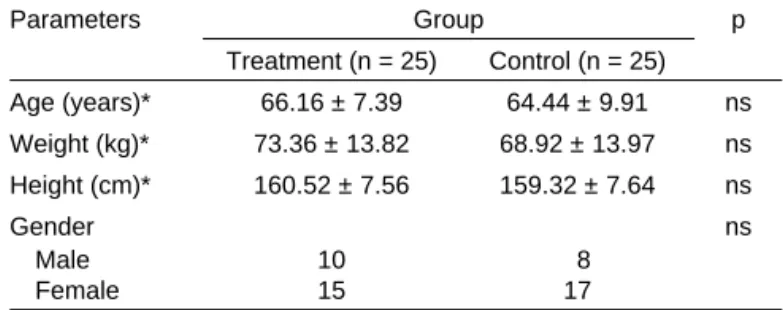
Both groups did not differ regarding age, gender, height, and weight. The surgery had a mean duration of 150 minutes that was similar in both groups. Table I shows the demographic data of the patients in the study.
Table II shows NS scores (median and variation) in the di-fferent moments (M). Figure 1 shows mean NS scores over 24 hours, comparing both groups. A statistically significant difference was observed only in M1 (p = 0.0215) and M2 (p = 0.0059), with lower scores in the treatment group. Comparing moments in each group over 24 hours, the treat-ment group showed statistically significant differences in pain severity in the following moments: preoperative and M3 (Pre-OP > M3; p = 0.0051) and preoperative and M4 (Pre(Pre-OP > M4; p = 0.0051). In the control group, statistically significant diffe-rences were observed between preoperative and M4 scores (PreOP > M4; p = 0.0093); M1 and M4 (M1 > M4; p = 0.0051); and M2 and M4 (M2 > M4; p < 0.0001).
When rescue medication was compared between both groups, consumption was significantly lower in the treatment group (p = 0.0001). Mean analgesic consumption in 24 hours was 12.2 mg of morphine in the treatment group, and 20.6 mg in the control group. Figure 2 shows total consumption during the study.
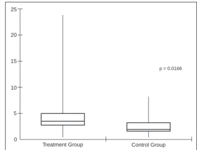
The time for the first request for rescue medication was sig-nificantly lower in the control group (p = 0.0166), 3.5 hours in the treatment group and 2 hours in the control group. Figure 3 shows medians, and maximal and minimal values in hours for the first request for rescue medication.
A correlation between preoperative pain and time until the first request for rescue medication was not observed in the treatment group (p = 0.8627) and control group (p = 0.8952). Similarly, a statistically significant correlation between the se-verity of preoperative pain and analgesic consumption was not observed in the treatment group (p = 0.8904) and control group (p = 0.4044).
Some adverse events were observed during the study, but wi-thout statistically significance between the groups: somnolence (treatment group: 4/25; control group: 4/25), nausea (treatment group: 10/25; control group: 13/25), and vomiting (treatment group: 7/25; control group: 7/25) were the most common. Table I – Demographic Data (Mean ± Standard Deviation)
Parameters Group p
Treatment (n = 25) Control (n = 25)
Age (years)* 66.16 ± 7.39 64.44 ± 9.91 ns
Weight (kg)* 73.36 ± 13.82 68.92 ± 13.97 ns
Height (cm)* 160.52 ± 7.56 159.32 ± 7.64 ns
Gender ns
Male 10 8
Female 15 17
**Results expressed as Mean ± Standard Deviation
ns = non-significant; Mann-Whitney test; Student t test; Fisher’s exact test.
Table II – Pain Severity According to the Numeric Scale (NS)
Parameters Group p
Treatment (n = 25) Control (n = 25)
Preoperative 5 (3-9) 7 (5-9) ns
M1 0 (0-8) 8 (5-10) 0.0215*
M2 5 (2-8) 8 (7-9) 0.0059*
M3 2 (0-6) 4 (0-7) ns
M4 2 (0-4) 2 (0-6) ns
Results expressed as median (minimal – maximal)
M1 = 2 h after IA morphine; M2 = 6 h after IA morphine; M3 = 12 h after IA morphine; M4 = 24 h after IA morphine;
ns = non-significant; (*) = significant (p < 0.05) – Mann-Whitney test.
Figure 1 – Comparison of Pain Scores in Both Groups in the First 24 Postoperative Hours.
Data presented as means. Friedman test.
Treatment Group Control Group *p < 0.05
8
7
7.3
6
6.1 6.5
5 5.48
4 4.6
4.4
3 3.64
2 2.9
2.3 2.8
1
0
PreOp m1 (2h)* m2 (6h)* m3 (12h) m4 (24h)
N S
DISCUSSION
The sample size of the present study was in conformity with the recommendations of some authors in systematic reviews, who indicate that this factor as an important cause of failure of the random distribution of patients with moderate to severe pain, leading to misinterpretation of the results by increasing the risk of false-positives. They also state that populations with more than 40 patients (20 per group) are recommended to minimize this problem19.
In the present study, a pneumatic tourniquet that was de-flated 15 minutes after the IA injection to allow greater time for the binding of morphine to its receptors was used in all procedures. A study suggested that tissue binding and, the-refore, the efficacy of the local anesthetic, could be increa-sed by maintaining longer the tourniquet in place after the IA injection. The author demonstrated, when evaluating the pharmacokinetics of this drug, an increase in plasma con-centration with a reduction in the time between the intra-articular injection and the release of the tourniquet, possibly by increasing local blood flow, leading to greater systemic absorption of the drug20.
Based on this evidence, Whitford investigated the contribution of the duration of the use of the pneumatic tourniquet for anal-gesia of patients undergoing knee arthroscopy. Patients recei-ved the intra-articular injection of 5 mg of morphine in 25 mL of NS and the tourniquet remained inflated for 10 minutes, in the first group, while in the second group it was removed immedia-tely after the administration of the drug. A significant reduction in pain and analgesic consumption, besides an increase in the time until the request of the first dose of rescue analgesic, was observed in the first group. The author attributed this pheno-menon to the removal of morphine from its receptors due to an increase in local blood flow secondary to post-ischemic reper-fusion with the early release of the tourniquet21.
The choice of the dose of morphine (10 mg) in the present study was based on the analysis of two known aspects from studies with patients undergoing arthroscopy. First, it has been demonstrated, and reaffirmed in a systematic review of the subject, a reduction in postoperative pain when doses hi-gher than 5 mg of the opioid are used, characterizing a dose-dependent analgesic effect13,22.
Second, total knee arthroplasty, which differs from arthroscopy, is associated with greater tissue trauma and pain; therefore, the doses recommended for arthroscopy could not be used in the present study. This problem has been indicated since the first studies on TKA23, when it was suggested that the addition of morphine to IA bupivacaine was not effective in reducing posto-perative pain due to the low dose of opioid used.
Regarding safety, contraindications to the IA administration of morphine for postoperative analgesia do not seem to exist. In an in vitro study, Jaureguito cultivated human cartilage removed during TKA of patients with osteoarthritis. He added different concentrations of morphine in NS (0.04, 0.2, and 0.4 mg.mL-1) and morphine associated with 0.25% bupivacaine to the culture, besides incorporating radionucleotides (10 mCi.mL-1 35SO
4) at the end of the incubation period to evaluate the synthesis of pro-teoglycans. Serial histologic slides stained with hematoxylin/eo-sin and electron microscopy were used to evaluate structural and cellular abnormalities, as well as histologic integrity. The author demonstrated a dose-dependent reduction in the incorporation of the radio sulfate in the samples after 12 hours. However, norma-lization was observed after 72 hours, even when higher doses of morphine were used. Those results indicated a transitory reduc-tion in the synthesis of proteoglycans, changes in metabolism, and cellular damage, which reverted after the third day. Besides, histologic or ultrastructural damages of the cartilage were not ob-served on microscopy when it was exposed to morphine24. The method of rescue analgesia chosen for the present study was the subcutaneous administration of 5 mg of morphine, Figure 2 – Comparison of Rescue Analgesic Consumption between
Both Groups.
Results presented as median and variation (maximal and minimal). Mann-Whitney test.
Figure 3 – Comparison of the Time Interval Until the First Request of Rescue Analgesic.
Data presented as median and variations (minimal and maximal). Mann-Whitney test.
p < 0.0001
30
25
20
15
10
5
0
Treatment Group Control Group
25
20
15
10
5
0
Treatment Group Control Group
which was enough to promote satisfactory postoperative anal-gesia in the control group in M3 and M4. In M1 and M2, in which this group showed higher NS scores, additional doses of subcutaneous morphine were administered until adequate pain control was achieved. The decision to use the same drug as co-intervention was aimed at trying to avoid masking the effect of the study treatment by the synergistic effects of ano-ther class of drug. The subcutaneous route, which is largely used in the control of pain exacerbation, was chosen since its safety and efficacy are similar to that of the intravenous route, with minimal side effects25,26.
In the present study, assessment of the analgesic efficacy was direct and indirect: the first was based on the compara-tive analysis of the intergroup and intragroup pain scores in the different moments (M); in the second, the time (Tr) until the first request of rescue medication and total analgesic con-sumption between both groups was evaluated. This type of analysis was used in a review22 article and follows the general tendency of most studies on the subject. It is believed that this is the best way to assess treatment efficacy since considering that the effects of the co-intervention with rescue analgesic shows a tendency to homogenize NS scores analysis of the indirect data can be a more reliable mean of characterizing the efficacy of IA morphine.
The possibility that pain reduction and the decreased need of analgesics after IA morphine was secondary to a systemic effect was investigated by several authors who demonstrated that the IA was superior than the intravenous route in pain reduction and, in some cases, the intragroup difference did not achieve statistical significance. However, the superiority of the intravenous over the IA route was not demonstrated when similar doses were used10,27,28.
It was also suggested that the effects of IA morphine were more prolonged that that of the intravenous administration. It has been postulated that this difference would be related with the intra-articular glucuronidation, which would produ-ce morphine-6-glucuronide, a metabolite with longer half-life that would be responsible for the longer time of action. In that study, the same author demonstrated plasma levels of mor-phine after the IA injection of 5 mg of mormor-phine lower than 10 mg.mL-1, which are not enough according to the author to produce systemic analgesia29.
In another study, the plasma levels of morphine after the ad-ministration of 5 mg of this drug reached a mean concentra-tion of 3.5 ng.mL-1 two hours after the IA administration, and 6.5 ng.mL-1 after the intravenous administration27. Despite the greater plasma concentration in the IV group, intergroup diffe-rences in numeric scale (NS) scores in the early (1, 2, and 4 postoperative hours) were not observed, but the IA group had lower NS scores at 6 and 24 hours27.
When 10 mg of morphine were administered IA and IV, the IA group showed a significant reduction in pain scores and anal-gesic consumption, but intragroup differences in the plasma concentration of morphine in the different moments (15 minu-tes, 1, 2, 4, and 24 hours) were not observed; however, the group that received the IA medication had lower NS scores at 6 and 24 h27.
When IA and intramuscular morphine 10 mg were compa-red the IA group showed significant compa-reduction in pain scores and consumption of analgesics, but serum levels of morphine (at 15 minutes, 1, 2, 4, and 24 hours) did not differ between both groups. Serum levels remained constantly low and ne-ver achieved the minimal effective concentration. The author suggested that the results were due mainly to the peripheral actions of the opioid30.
Postoperative assessment can be divided in three moments: early phase (0 to 2 hours), in which the residual effect of in-traoperative anesthesia/analgesia could lead to a bias; inter-mediate phase (2 to 6 hours), in which the effects of those medications would normally start to decrease; and late phase (6 to 24 hours), in which the analgesic effect would be predo-minantly local22. In the present study, patients were evaluated in the preoperative period, and at 2 (M1), 6 (M2), 12 (M3), and 24 (M4) hours after the IA injection of morphine.
To reduce the influence of the anesthesia on M1 evaluation, we decided to use spinal block with 15 mg of bupivacaine wi-thout the addition of opioids, and local anesthesia was not used during the procedure. Intra- and postoperative analgesic drugs were not used.
Direct assessment demonstrated a reduction in NS scores in the treatment group in all studies moments, but statistically significant differences were observed only in M1 and M2. Ano-ther author observed similar results using 5 mg of morphine, but with statistically significant differences only four hours af-ter the IA injection32.
The efficacy of the reduction in pain scores with IA morphine after arthroscopies remains controversial. So far, four syste-matic reviews on the subject were undertaken without con-clusions on its efficacy19,22,31,32. Several authors state the pre-sence of evidence that this route of administration would be effective in the reduction of the pain scores and reduction in the consumption of analgesics33,34.
However, those results were questioned recently based on the fact that few controlled studies with good methodological quality exist. The author also stated that clinical assays of bet-ter quality and larger study population demonstrated that IA morphine would not be an effective analgesic method, ques-tioning the evidence of assays favorable to the use of this rou-te of administration22.
bupivacaine used in the spinal block, which, along with IA mor-phine, would show more important reduction in pain scores in the first two hours, but it would not have such an important repercussion in the control group.
In the indirect assessment, the first parameter evaluated was the time until the first request for rescue analgesic (Tr), which was significantly different, longer in the treatment group with median of 3.5 hours vs. 2 hours in the control group. This result was similar to that of another study, in which the authors observed a longer time until the first dose of analgesic in the group that recei-ved 5 mg of morphine associated with bupivacaine, with means of 5.5 and 5 hours for the rheumatoid arthritis and osteoarthritis groups, respectively1. Those results were also similar to those of another author who compared IA morphine, tramadol, and place-bo and observed a significantly longer time in the opioid groups, with a mean of 34 and 33 minutes for morphine and tramadol, respectively. However, a significant difference was not observed between the treatment groups36.
This type of assessment seems to be the best way to analyze the efficacy of IA morphine, since it is based on the period the patient is not under the effects of the anesthetic and before the use of any other type of intervention, allowing the asses-sment of the effect of the local opioid. This method has been suggested as a mean to increase the sensitivity of the study, as well as the quantification of the analgesic used by the pa-tient in the postoperative period32.
The second parameter investigated was analgesic consump-tion in the first 24 hours, which was significantly higher in the control group, with comparable results to those of a similar study1. However, other authors did not observe a significant reduction in analgesic consumption, but those results can be attributed to the low doses of IA morphine used14,23.
Evaluating the hypothesis that low postoperative scores and reduced inflammation would be responsible for the inconclusi-ve results on the efficacy of the IA administration, the use of IA morphine in arthroscopic surgery was analyzed in a clinical as-say dividing patients in two groups: “surgery with little inflamma-tion” and “very inflammatory surgery”, followed by the random allocation of the patients in subgroups that received morphine, bupivacaine, or placebo. In the second group (very inflammatory surgery) the author observed statistically significant differences among the subgroups regarding the reduction in pain scores and analgesic consumption, especially in the morphine group. In the first group, bupivacaine was more effective with significant re-duction in pain scores despite the lack of difference in analgesic consumption among the subgroups. Based on those results, the author26 suggested that the lower expression of opioid receptors in the joint would be responsible for the reduced efficacy of mor-phine in the “little inflammatory” group37.
In the present study, a different approach was used to evaluate this hypothesis. Preoperative pain scores were correlated with postoperative analgesic consumption in 24 hours and with Tr, parameters that seem to have a better correlation with the local effects of morphine. However, a significant correlation was not observed, and this result contradicts the hypothesis that grea-ter level of preoperative pain and, possibly, more inflammation, would imply better control with the use of IA morphine.
Some side effects were observed during the study, and the most common included nausea (40% in the control group; 35% in the treatment group) and vomiting (28% in both groups). Those symptoms were self-limited and did not compromise the continuity of the study. Mild somnolence without further re-percussions was observed in only four patients (16%) in each group. None of the patients included in the study requested to be excluded due to the side effects. In previous studies, the development of side effects was not a limiting factor for the use of IA morphine, both in arthroscopies and TKA1,13,14,37,38. When the side effects of this analgesic technique are compa-red to that of other techniques, similar percentage of episodes of nausea and vomiting are observed. In a study comparing epidural patient-controlled analgesia (PCA) with sufentanil for postoperative pain after TKA, the author observed a percen-tage ranging from 38% to 40% in the study groups. In another study in which the author investigated the use of 250 µg of spi-nal morphine, isolated or in association with clonidine, he ob-served a 20% incidence of nausea and vomiting in the different groups3,4,39.
The predominance of female patients in all groups is a limitation of the present study, since this was indicated in clinical assays as a possible confounding factor4. It was observed that the in-cidence of complaints of postoperative pain is higher in female patients undergoing knee arthroscopies, considering that the re-lative risk of postoperative pain in those procedures is 1.47, for mild to moderate pain, although a difference in the incidence of severe pain in males and females does not exist40.
It was possible to conclude that 10 mg of intra-articular mor-phine increased the time until the first request for rescue analgesic and reduced analgesic consumption in the first 24 postoperative hours, and it also decreased postoperative pain scores at 2 and 6 hours.
REFERÊNCIAS – REFERENCES
1. Tanaka N, Sakahashi H, Sato E et al. The efficacy of intra-articular anal-gesia after total knee arthroplasty in patients with rheumatoid arthritis and in patients with osteoarthritis. J Arthroplasty 2001;16:306-311.
2. Pitimana-Aree S, Visalyaputra S, Komoltri C et al. An economic evaluation of bupivacaine plus fentanyl versus ropivacaine alone for patient-controlled epidural analgesia after total-knee replacement pro-cedure: a double-blinded randomized study. Reg Anesth Pain Med 2005;30:446-451.
3. Klasen JA, Opitz SA, Melzer C et al. – Intraarticular, epidural, and intravenous analgesia after total knee arthroplasty. Acta Anaesthesiol Scand 1999;43:1021-1026.
4. Sitsen E, van Poorten F, van Alphen W et al. Postoperative epidu-ral analgesia after total knee arthroplasty with sufentanil 1 microg/ml combined with ropivacaine 0.2%, ropivacaine 0.125%, or levobupiva-caine 0.125%: a randomized, double-blind comparison. Reg Anesth Pain Med 2007;32:475-480.
5. Sites BD, Beach M, Biggs R et al. Intrathecal clonidine added to a bupivacaine-morphine spinal anesthetic improves postoperative anal-gesia for total knee arthroplasty. Anesth Analg 2003;96:1083-1088. 6. Good RP, Snedden MH, Schieber FC et al. Effects of a preoperative
femoral nerve block on pain management and rehabilitation after total knee arthroplasty. Am J Orthop 2007;36:554-557.
8. Stein C – The control of pain in peripheral tissue by opioids. N Engl J Med, 1995;332:1685-1690
9. Dalsgaard J, Felsby S, Juelsgaard P et al. Analgesi af lavdosis in-traartikulaer morfin efter ambulant knaeartroskopi. Ugeskr Laeger 1993;155:4166-4169.
10. Björnsson A, Gupta A, Vegfors M et al. Intraarticular morphine for postoperative analgesia following knee arthroscopy. Reg Anesth 1994;19:104-108.
11. Chan ST. Intra-articular morphine and bupivacaine for pain relief af-ter therapeutic arthroscopic knee surgery. Singapore Med J 1995; 36:35-37.
12. De Andres J, Valia JC, Barrera L et al. Intra-articular analgesia after arthroscopic knee surgery: comparison of three different regimens. Eur J Anaesthesiol 1998;15:10-15.
13. Likar R, Kapral S, Steinkellner H et al. Dose-dependency of intra-artic-ular morphine analgesia. Br J Anaesth 1999;83:241-244.
14. Mauerhan DR, Campbell M, Miller JS et al. Intra-articular morphine and/or bupivacaine in the management of pain after total knee arthro-plasty. J Arthroplasty 1997;12:546-552.
15. Ritter MA, Koehler M, Keating EM et al. Intra-articular morphine and/ or bupivacaine after total knee replacement. J Bone Joint Surg Br 1999;81:301-303.
16. Solheim N, Rosseland LA, Stubhaug A. Intra-articular morphine 5 mg after knee arthroscopy does not produce significant pain relief when administered to patients with moderate to severe pain via an intra-articular catheter. Reg Anesth Pain Med 2006;31:506-513.
17. Jensen MP, Karoly P, Braver S. The measurement of clinical pain intensity: a comparison of six methods. Pain 1986;27:117-126. 18. Ayres M, Ayres Jr M, Ayres DL et al. Bioestat versão 4.0: Aplicações
Estatísticas nas Áreas das Ciências Biológicas e Médicas. Brasília, Ministério da Ciência e Tecnologia, 2005.
19. Rosseland LA. No evidence for analgesic effect of intra-articular mor-phine after knee arthroscopy: a qualitative systematic review. Reg Anesth Pain Med 2005;30:83-98.
20. Katz JA, Kaeding CS, Hill JR et al. The pharmacokinetics of bupiva-caine when injected intra-articularly after knee arthroscopy. Anesth Analg 1988;67:872-875.
21. Whitford A, Healy M, Joshi GP et al. The effect of tourniquet release time on the analgesic efficacy of intraarticular morphine after ar-throscopic knee surgery. Anesth Analg 1997;84:791-793.
22. Gupta A, Bodin L, Holmström B et al. A systematic review of the pe-ripheral analgesic effects of intraarticular morphine. Anesth Analg 2001;93:761-770.
23. Badner NH, Bourne RB, Rorabeck CH et al. Addition of morphine to intra-articular bupivacaine does not improve analgesia following knee joint replacement. Reg Anesth 1997;22:347-350.
24. Jaureguito JW, Wilcox JF, Thisted RA et al. The effects of morphine on human articular cartilage of the knee: an in vitro study. Arthroscopy 2002;18:631-636.
25. Elsner F, Radbruch L, Loick G et al. Intravenous versus subcutaneous morphine titration in patients with persisting exacerbation of cancer pain. J Palliat Med 2005;8:743-750.
26. Koshy RC, Kuriakose R, Sebastian P et al. Continuous morphine infu-sions for cancer pain in resource-scarce environments: comparison of the subcutaneous and intravenous routes of administration. J Pain Pall Care Pharmacother 2005;19:27-33.
27. Richardson MD, Bjorksten AR, Hart JA et al.The efficacy of intra-artic-ular morphine for postoperative knee arthroscopy analgesia. Arthros-copy 1997;13:584-589.
28. Dierking GW, Ostergaard HT, Dissing CK et al. Analgesic effect of intra-articular morphine after arthroscopic meniscectomy. Anaesthe-sia 1994;49:627-629.
29. Joshi GP, McCarroll SM, Cooney CM et al. Intra-articular morphine for pain relief after knee arthroscopy. J Bone Joint Surg Br 1992;74:749-751. 30. Raj N, Sehgal A, Hall JE et al. Comparison of the analgesic efficacy and
plasma concentrations of high-dose intra-articular and intramuscular morphine for knee arthroscopy. Eur J Anaesthesiol 2004;21:932-937. 31. Kalso E, Tramer MR, Carroll D et al. Pain relief from intra-articular
morphine after knee surgery: a qualitative systemic review. Pain 1997;71:127-134.
32. Kalso E, Smith L, McQuay HJ et al. No pain, no gain: clinical excel-lence and scientific rigour-lessons learned from IA morphine. Pain 2002;98:269-275.
33. Karlsson J, Rydgren B, Eriksson B et al. Postoperative analgesic ef-fects of intra-articular bupivacaine and morphine after arthroscopic cruciate ligament surgery. Knee Surg Sports Traumatol Arthroscopy 1995;3:55-59.
34. Cepeda MS, Uribe C, Betancourt J et al. Pain relief after knee arthros-copy: intra-articular morphine, intra-articular bupivacaine, or subcuta-neous morphine? Reg Anesth 1997;22:233-238.
35. Brandsson S, Karlsson J, Morberg P et al. Intraarticular morphine af-ter arthroscopic ACL reconstruction: a double-blind placebo-controlled study of 40 patients. Acta Orthop Scand 2000;71:280-285.
36. Akinci SB, Sarıcaoglu F, Atay OA et al. Analgesic effect of intra-articu-lar tramadol compared with morphine after arthroscopic knee surgery. Arthroscopy 2005;21:1060-1065.
37. Marchal JM, Delgado-Martinez AD, Poncela M et al. Does the type of arthroscopic surgery modify the analgesic effect of intraarticular morphine and bupivacaine? A preliminary study. Clin J Pain 2003; 19:240-246.
38. Alvarez-Cabo JM, Lopez-Rouco M, Gonzalez-Paleo JR. Analgesic ef-fect of intra-articular morphine after arthroscopic knee surgery. Ambul Surg 1998;6:179-182.
39. Bourke M, Hayes A, Doyle M et al. A comparison of regularly admin-istered sustained release oral morphine with intramuscular morphine for control of postoperative pain. Anesth Analg 2000; 90:427-430. 40. Rosseland LA, Stubhaug A. Gender is a confounding factor in pain
trials: women report more pain than men after arthroscopic surgery. Pain 2004;112:248-253.
RESUMEN
Garcia JBS, Barbosa Neto JO, Vasconcelos JW, Ferro LSG, Silva RC – Eficacia Analgésica del Uso de Dosis Alta de Morfina Intra-articular en Pacientes Sometidos a la Artroplastia Total de Rodilla.
JUSTIFICATIVA Y OBJETIVOS: A pesar de que la eficacia de la morfina intra-articular (IA), permanece como algo controvertido, ha quedado demostrado que las dosis mayores generan mejores re-sultados y consecuentemente, un menor consumo postoperatorio de analgésico, caracterizando así, el efecto dosis-dependiente en la acción periférica. Fue realizado un estudio controlado, aleatorio y doble ciego para evaluar la eficacia de 10 mg de morfina por vía intra-articular en pacientes sometidos a la artroplastia total de rodilla.
MÉTODO: Se evaluaron 50 pacientes sometidos a la artroplastia total de rodilla, distribuidos aleatoriamente en dos grupos: el grupo tratamiento recibió 10 mg (1 mL) de morfina por vía intra-articular diluido en 19 mL de solución fisiológica al 0,9% (SF), mientras que el grupo control recibió una inyección intra-articular con 20 mL de SF, ambos después del cierre de la cápsula articular, al final de la operación. La morfina subcutánea bajo demanda, estuvo disponible para el dolor residual. Se evaluaron las siguientes variables: intensidad del dolor graduada en la Escala Nu-mérica (EN) a las 2h (M1), 6h (M2), 12h (M3) y 24h (M4), después de la inyección IA; tiempo para la primera solicitación de analgésico; y consu-mo de analgésicos y efectos adversos.