This study determined the reduction threshold in thickness of the dentin shade composite necessary to result in perceptible and acceptable color changes on simulated restorations. Three composite systems (Charisma Diamond, IPS Empress Direct, and Filtek Z350 XT) were evaluated using cylinder-shaped specimens built-up with dentin and enamel shades. The opacity of the composites was assessed using 1.0 mm thick specimens over black and white backgrounds. A baseline color was established for each system by combining 1.0 mm thick enamel shade with 3.0 mm of dentin shade cylinders over a dark background (n = 9). Then, the color changes (∆E00) caused by sequential 0.1 mm reductions on
dentin shade cylinders were calculated. Opacity changes on dentin shade cylinders and combined enamel-dentin pair cylinders were also assessed after each thickness reduction. Polynomial regression was performed with averages of ∆E00 as a function of thickness
of dentin shade cylinders; and acceptability (∆E = 1.77) and perceptibility (∆E = 0.81) thresholds were calculated. Linear regressions were also performed for ∆E00 as function of
opacity of dentin shade cylinders and combined enamel-dentin pair of cylinders. Except for Charisma, enamel shades presented the lowest opacity than dentin one. Perceptible and acceptable color changes were observed for dentin shade cylinders thinner than 2.0-2.4 mm and 1.1-1.4 mm, respectively, were used. No difference among the composite systems was observed. In conclusion, reductions on dentin shade composite lower than 0.6-mm did not yield perceptible color changes, and clinically significant color changes only were observed within reductions higher than 1.6-mm.
Color Changes Caused by Reduction on
the Dentin Shade Composite Thickness
Sheila Mara Morais Santos1, Paula Damasceno Silva1, André Luis Faria-e-Silva2
1Graduate Programa in Dentistry, UFS - Universidade Federal de Sergipe, Aracaju, SE, Brazil 2Department of Dentistry, UFS - Universidade Federal de Sergipe, Aracaju, SE, Brazil
Correspondence: Prof. Dr. André Luis Faria-e-Silva, Rua Cláudio Batista, s/n, Sanatório, CEP 49060-100 Aracaju, SE, Brasil. Tel.: +55-79-3194-7220. e-mail: [email protected]
Key Words: Color, composite dental resin, esthetics, dental, optical phenomena.
Introduction
Direct composite restorations in anterior teeth has been demonstrated be an excellent option to solve esthetic concerns involving alteration in color and/or shape of teeth, or to close diastemata (1-3). Further to easy handling characteristics, the composite systems available nowadays present several shades allowing to mimic both enamel and dentinal tissues (4-5). When compared to ceramic veneers, composite resin presents reduced cost that allows its indication for patients with reduced willingness to pay (6). However, the ability and knowledge of clinicians regarding esthetic aspects of smile, and optics characteristics of hard tooth tissue and composites are essential to achieve esthetical direct restorations.
Enamel is a crystalline tissue characterized by high mineral content, which results in a chromatic translucent structure that allows to visualize the underlying dentin (5,7,8). Furthermore, the chromaticity of enamel modify the dentin color and affect the ultimate tooth color (5). On the other hand, the higher organic content of dentin increases its opacity and the chromaticity of this tissue strongly affects the tooth color (8). Similarly, composites shades are available in different degrees of translucency seeking out to simulate the optical characteristics of enamel and dentin (3,5). Further to translucency degree, the thickness of composite increment also affects the light
transmission through the material bulk (9-11). Therefore, using the same thickness of hard tooth tissue only would result in similar optical aspects if the translucency/ opacity of the composite was like those observed from dentin and enamel. However, composites with different degrees of translucency/ opacity are available in the market. An important matter hindering the stratification of composite restoration is that the enamel shade composites of some manufacturer are more translucent than the tooth enamel, while some dentin shade composites are more opaque than tooth dentin (12,13).
S. M. M. Santos et al.
hypothesis of the study was that similar reductions are necessary to cause perceptible and clinically relevant color changes despite the differences in the opacity of composite systems.
Material and Methods
Three systems of composite resins were evaluated in the present study: Charisma Diamond (Heraeus Kulzer, Hanau, Germany), IPS Empress Direct (Ivoclar Vivadent, Schaan, Liechtenstein), and Filtek Z350 XT (3M ESPE, St. Paul, MN, USA). For the IPS Empress Direct and Filtek Z350 XT, two shades with opacities corresponding to enamel (EA1 and A1E, respectively) and dentin (DA1 and A1B, respectively) were selected. Since the rationale for shade choice was to obtain similar color for composite systems, the lighter dentin shade (OL) of Charisma Diamond system was used. To characterize the opacity of dentin and enamel shades from each system, cylinder-shaped specimens (10 mm diameter x 1.0 mm thickness) were built-up by insertion of composites into a metallic mold. The composites were light-cured with a LED-based unit Radii Cal (irradiance ≈ 1,000 mW/cm2; SDI, Victoria, Australia) for 40 s, followed by the specimens polishing with 600 grit Silicon Carbide (SiC) abrasive papers (Norton Saint-Gobain, Guarulhos, SP, Brazil), and the final diameter was measured with a digital caliper.
The opacity of specimens (n = 3) was assessed with a sphere spectrophotometer (SP60, X-Rite, Grand Rapids, MI, USA), in reflectance mode, using CIE L*a*b* system (L*: white/black; a*: red/green; b*: yellow/blue). Specimens were positioned in focus on a clear acrylic stand, and the measurements were performed with a D65 illuminant, in the wavelength ranging from 400 to 700 nm, and with the specular light included (SPIN mode). Due to sphere geometry of spectrophotometer, the object was illuminated diffusely and the detector received the reflected light at an 8° angle from the surface of the composite cylinder. The color parameters were measured over white (L* = 95.2, a*= -1.2, b*= 0.3) and black (L* = 0.2, a* = 0.3, b* = 0.2) backgrounds, while the opacity was automatically calculated by spectrophotometer by difference between the colors measured using these backgrounds. The average of opacity and the confidence interval at 95% were calculated for each composite.
Afterwards, three other cylinder-shaped specimens (10 mm diameter x 3.0 mm thickness) were confectioned for each dentin shade composite. Each dentin shade cylinder was combined with the three 1-mm thick enamel shade cylinders confectioned previously, resulting in nine pairs (n = 9) of enamel-dentin cylinders per composite system (3 dentin shade cylinders x 3 enamel shade cylinder). Then, the enamel shade cylinder was positioned over the dentin
shade one with a thin layer of glycerol between them to avoid significant changes of refractive index caused by air; and the combined enamel-dentin shades pairs of cylinders were placed over a dark background (L* = 24.7, a* = 0.1, b* = 0.1). The color of cylinders pairs was measured and the values of L*, a* and b* recorded. Three measurements were performed for each pair and the averages of these values were used to calculate the mean parameters of color for each composite system. This first color assessment using 3-mm thick dentin shade cylinders was defined as the color standard. The opacity of dentin shade cylinders alone and combined with enamel shade cylinders were also assessed using the same protocol described previously.
Following, the dentin shade cylinders were manually abraded until reach 2.9 mm of thickness with 600-grit SiC paper. The color and opacity of the combined cylinder (1.0-mm of thickness enamel shade and 2.9-(1.0-mm of thickness dentin shade) were measured according prior description. Based on changes on color parameters, the pooled color change (∆E00) was calculated according with the following formula (15):
The opacities of dentin shade alone and combined with corresponding enamel shade cylinder were also measured. The opacity and color measurements were repeated after each 0.1-mm reduction on dentin shade composite cylinder until using 1.0-mm of thickness dentin shade composite cylinders. Color changes (∆E00) were calculated after each thickness reduction based on color measured with 3.0-mm thick dentin shade cylinder.
Data of ∆E00 (mean and standard error) were plotted as function of dentin shade thickness from each composite system, followed by calculation of polynomial regressions at 3rd order. The formulas of these polynomial regressions were used to estimate the minimum reduction on thickness of dentin shade composite cylinder required to obtain perceptible and clinically relevant color changes. The values of ∆E00 reported by a prior study (16) as the threshold values indicating perceptible (0.81) and clinically acceptable (1.77) color changes were used. Further, the confidence intervals at 95% for each minimum thickness of dentin shade composite were calculated based on standard errors of ∆E00 obtained in the present study. Graphics of ∆E00 as function of opacity changes of dentin shade composite and enamel-dentin pairs of cylinders were also plotted. Linear regressions for each data set were calculated. All data analyses and graphics plotting were performed using the SigmaPlot 12.0 statistical software package (Systat Software Inc., Chicago, IL, USA) and significance level was set at α = 0.05.
Results
Color changes by thin dentin shade composite
thick specimens. No significant differences were observed among the dentin shades composites. Regarding the enamel shades, IPS Empress Direct and Filtek Z350 XT presented similar opacities and lower than that measured for Charisma Diamond. For this last composite system, similar opacities were observed between enamel and dentin shades for Charisma Diamond. Enamel shades were more translucent than dentin shade for the other composite systems.
Curves of ∆E00 as function of thickness of dentin shade composite are displayed in Figure 1. All regressions presented p-values lower than 0.001; and the determination coefficient ranged from 0.893 (IPS Empress Direct) to 0.955 (Filtek Z350 XT). Table 2 presents the values of minimum thickness reduction of dentin shade cylinders to results in perceptible and clinically acceptable color changes. To obtain
perceptible changes from color obtained with 3.0 mm thick dentin cylinders, it was necessary to reduce the dentin shade cylinder until obtain thickness ranging from 2.41 mm (Filtek Z350 XT) to 2.01 mm (IPS Empress Direct). However, no significant difference was observed among the composite systems. Regarding to maintain the color changes clinically acceptable, the dentin shades cylinders couldn’t be thinner than 1.44-mm, 1.30-mm and 1.15-mm for Filtek Z350-XT, IPS Empress Direct and Charisma Diamond, respectively. No significant statistical difference among the composites systems was also observed regarding the thickness of dentin shade cylinders required to maintain the color change clinically acceptable.
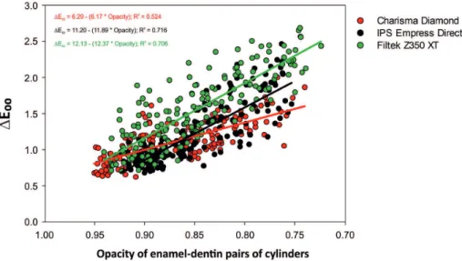
Linear regressions of ∆E00 as function of opacity of dentin shade cylinders and enamel-dentin shades pairs cylinders are illustrated in Figure 2 and 3, respectively. Determinant coefficients were higher using data of the dentin shade cylinders (ranged from 0.707 to 0.932) than correlating with opacity of the enamel-dentin shades pairs cylinders (ranged from 0.524 to 0.716). All linear regressions presented p-value lower than 0.001.
Discussion
Despite the reduced cost and adequate clinical behavior, obtaining aesthetic direct restorations with composites remains a challenge for the clinicians since to reproduce the optical features of hard dental tissues can be a difficult task (3-5, 9, 17-20). Moreover, resin-based materials with different optical features are now available in the market
Table 1. Description of composites systems and their shades used in the present study and the means of opacity (95% confidence interval) measured using 1.0-mm thick disks
Composite
system Manufacturer Shades Opacity Charisma
Diamond
Heraeus Kulzer, Hanau, Germany
A1 (enamel) 62.1 (60.2/ 64.0) OL (dentin) 64.4 (62.9/ 65.8) IPS
Empress Direct
Ivoclar Vivadent, Schaan, Liechtenstein
EA1 (enamel) 53.7 (51.7/ 55.8) DA1 (dentin) 64.8 (63.4/ 66.2) Filtek
Z350 XT
3M ESPE, St. Paul, MN, USA
A1E (enamel) 56.4 (54.9/ 57.8) A1B (dentin) 61.1 (58.7/ 63.6)
Figure 1. Behavior of data (average; standard error) of ∆E00 as function of thickness of dentin shade
composite measured for different composite systems. The lines represent the results of polynomial regression (3rd order) for each composite. The formulas polynomial regression (3rd order) and determination
coefficients (R2) are described in the figure. Red line and formula – Charisma diamond; Black line and
S. M. M. Santos et al.
requiring changes in the restorative approaches according to the material chosen (12-14). Further to color of the composites, the opacity of the material also strongly affects the ultimate color of restoration, mainly when dental substrates with significant color alteration are underlying the restoration or translucent incisal areas are restored (9). In this last case, any visualization of the dark oral cavity through the restorative material presenting improper level of translucency results in grayer (reduced lightness) restoration and can compromise the final aesthetic. In the present study, a dark background was used to simulate this clinical situation and only the thickness of the dentin shade was modified.
It has been reported that the enamel thickness significantly affects the ultimate color of tooth structure (8), and so is the enamel shade composite on restoration color (5). In the present study, we choose to maintain the thickness of enamel shade in 1.0 mm for all experimental set-ups since this is the average of human enamel thickness, which varies depends on the type of tooth and anatomical location (21). Using this standard thickness, the opacity of enamel shade composites ranged from 53.7 % for IPS Empress Direct (more translucent) to 62.1 % for Charisma Diamond (more opaque). These differences on opacities among the composite systems evaluated are related to the composition of materials. Composites containing smaller and well-distributed fillers, and similar refractive indexes between those observed for the resin matrix and fillers allows increased light transmission (22,23). Therefore, more translucent materials require thicker increments to hide the color of the underlying substrate. Regarding the
initial dentin shade thickness, we choose to start with 3.0 mm seeking out to completely hire the dark background. However, combining 1.0 mm thick enamel shade and 3.0 mm thick dentin shade allowed some light transmission and slight visualization of the dark background (the opacity ranged from 91.7 % for IPS Empress Direct to 94.8% for Charism Diamond). Indeed, no significant difference between the composite brands in the opacity of dentin shades were observed either for cylinders with 1.0 mm (61.1 to 64.8 %) or 3.0 mm (88.2 to 88.5 %) of thickness; and the opacities of enamel-dentin shade combination were strongly affected by the enamel shade opacity. An explanation can be that the 1.0-mm enamel shade cylinders blocked higher than 50% of the light-transmission.
Therefore, based only in the enamel shades opacities, it could be expected that reducing the dentin shade
Table 2. Estimated values (95% confidence intervals*) of composite thickness corresponding to dentin shade required to not affect the perceptibility and acceptability of ultimate color measured with dentin shade (3.0 mm of thick at baseline) under 1-mm thick enamel shade over black background.
Composite system
Parameter**
Perceptibility Acceptability Charisma Diamond 2.20 (1.87 – 2.51) 1.15 (0.97 – 1.67) IPS Empress Direct 2.01 (1.81 – 2.27) 1.30 (1.09 – 1.49) Filtek Z350 XT 2.41 (2.16 – 2.71) 1.44 (1.16 – 1.73) *Estimated using the variation coefficient of ∆E00 values. **Values of
50:50% color changes threshold determined by Paravina et al. 2015 (∆E00 for perceptibility = 0.81; ∆E00 for acceptability = 1.77).
Figure 2. Behavior of data of ∆E00 as function of opacity of dentin shade composite measured for
different composite systems. The lines represent the results of linear regression for each composite. The formulas linear regression and determination coefficients (R2) are described in the figure. Red
Color changes by thin dentin shade composite
thickness would result in more pronounced changes in ∆E00 for the restorative systems presenting more translucent enamel shades. In fact, the correlation tests showed the lowest slope of lines (demonstrated by linear regression) for Charisma Diamond (more opaque enamel shade) for either correlation between ∆E00 and the opacity of the dentin shade cylinders (3.41) or the enamel-dentin pairs of cylinders (6.17). IPS Empress direct and Filtek Z350 XT present more translucent enamel shade composites (53.7 and 56.4, respectively), resulting in increased slope in lines of correlations between ∆E00 and the opacity of the dentin shade cylinders (7.08 and 6.02, respectively) or the enamel-dentin pairs of cylinders (11.59 and 12.37, respectively). Interestingly, the determination coefficients were higher for linear regressions with data of dentin shade cylinders opacity than those of enamel-dentin pairs; suggesting that the changes in dentin shade opacity affected more the ultimate color than the changes in overall opacity of specimens. However, these findings are explained due to maintaining the thickness of enamel shade cylinders constant during the measurements and any change in the overall opacity was due to reductions in opacity of the dentin cylinders.
Changes on dentin shade cylinders opacity were performed by abrasion of the cylinders to obtain sequential 0.1 mm reductions on their thickness until to evaluate the 1.0 mm thick dentin shade specimens. Thinner cylinders were not used in the experiment due to difficulty to control the abrasion procedures of thin specimens. Therefore, polynomial regressions were performed to estimate the ∆E00 for using dentin shade cylinders with intermediate
thicknesses or thinner than 1.0 mm. These regression analyses presented high determination coefficients (ranging from 0.894 to 0.955) demonstrating the formulas calculated strongly explain the relation between the thickness of dentin shade composites and changes on the ultimate color. Furthermore, the changes on curve slopes demonstrate the complexity between the reduction on the composite thickness and changes in the ultimate color. Based on these formulas, the thicknesses of dentin shade cylinders required to yield perceptible and clinically relevant color changes were calculated. Data of ∆E00 from a prior study that calculated the color change thresholds required to 50% of observers consider the difference of color between two objects as visually perceptible or clinically acceptable were used (16). The results of present study did not demonstrate any significant difference among the composite brands analyzed. Reductions of 0.59 to 0.99 mm in dentin shade cylinders were necessary to yield perceptible color changes; while only reductions higher than 1.56-1.85 mm in dentin shade cylinders resulted in color changes clinically unacceptable. Therefore, for the composites evaluated in the present study, the placement of dentin shade composites thicker than 1.15-1.44 mm seems does not modify significantly the ultimate color of restorations when a 1.0 mm thick enamel shade composite is also used.
In the present study, the composite A1B was used as dentin shade for the system Filtek Z350 XT because this shade presents opacity closer than that observed for human dentin than the more opaque composite A1D from the same manufacturer (12) Regarding the Charisma Diamond, this composite system presents four shades indicated to be used
Figure 3. Behavior of data of ∆E00 as function of opacity of specimens (dentin shade disks over 1.0-mm
thick enamel shade disks) measured for different composite systems. The lines represent the results of linear regression for each composite. The formulas linear regression and determination coefficients (R2)
S. M. M. Santos et al.
as dentin shades: OL (lighter), OM (medium), OD (darker), and OB, which is indicated for bleached tooth. Therefore, we choose to evaluate the shade OL that is closer shade to A1 used for the other composites. In conclusion, the results of present study demonstrated that, despite differences on the opacity among the composite systems evaluated, similar thickness reductions cause similar perceptible and clinically relevant color changes. Therefore, the hypothesis of study was accepted. However, it is important to emphasize that only lighter composites were evaluated in the present study and the findings cannot be extrapolated for other shades since the opacity also depends on the composite shade (21).
Resumo
Este estudo objetivou determinar o limiar de redução na espessura do compósito de cor da dentina necessário para resultar em mudanças de cor perceptíveis e aceitáveis em restaurações simuladas. Três sistemas de compósitos (Charisma Diamond, IPS Empress Direct e Filtek Z350 XT) foram avaliados utilizando corpos-de-prova cilíndricos construídos com cores de dentina e esmalte. A opacidade dos compósitos foi avaliada usando cilindros de 1,0 mm de espessura sobre fundo preto e branco. Uma cor inicial padrão foi estabelecida para cada sistema pela combinação de esmalte de 1,0 mm de espessura com 3,0 mm de cilindros de resina na cor de dentina sobre um fundo escuro (n = 9). Em seguida, foram calculadas as alterações de cor (∆E00) causadas por reduções sequenciais de 0,1 mm nos cilindros de resina na cor de dentina. Mudanças de opacidade em cilindros na cor de dentina e cilindros combinados de esmalte e dentina foram também avaliadas após cada redução de espessura. Regressão polinomial foi realizada com médias de ∆E00 em função da espessura dos cilindros na cor de dentina; e os limiares de aceitabilidade (∆E00 = 1,77) e perceptibilidade (∆E00 = 0,81) foram calculados. Regressões lineares também foram realizadas para ∆E00 em função da opacidade dos cilindros na cor de dentina e do par combinado de cilindros de esmalte-dentina. Com exceção da Charisma, os compósitos de esmalte apresentaram menor opacidade do que os de dentina. Alterações de cor perceptíveis e aceitáveis foram observadas quando os cilindros na cor de dentina foram mais finos que 2,0-2,4 mm e 1,1-1,4 mm, respectivamente. Nenhuma diferença entre os sistemas compostos foi observada. Em conclusão, reduções no compósito de dentina menor que 0,6 mm não produziu mudanças de cor perceptíveis, e mudanças de cor clinicamente significantes apenas foram observadas com reduções maiores que 1,6 mm.
References
1. Gresnigt MM, Kalk W, Ozcan M. Randomized clinical trial of indirect resin composite and ceramic veneers: up to 3-year follow-up. J Adhes Dent 2013;15:181-190.
2. Lempel E, Lovász BV, Meszarics R, Jeges S, Tóth Á, Szalma J. Direct resin composite restorations for fractured maxillary teeth and diastema closure: A 7 years retrospective evaluation of survival and influencing factors. Dent Mater 2017;33:467-476.
3. Katsarou T, Antoniadou M, Papazoglou E. Effectiveness of optical illusions applied on a single composite resin veneer for the diastema closure of maxillary central incisors. Int J Esthet Dent 2017;12:42-59. 4. Baratieri LN, Araujo E, Monteiro S Jr. Color in natural teeth and direct
resin composite restorations: essential aspects. Eur J Esthet Dent 2007;2:172-186.
5. Villarroel M, Fahl N, De Sousa AM, De Oliveira OB Jr. Direct esthetic restorations based on translucency and opacity of composite resins. J Esthet Restor Dent 2011;23:73-87.
6. Waning A. Direct or indirect restorative dentistry-a mere choice about cost in relation to longevity? Dent Update 2011;38:5-10.
7. Hariri I, Sadr A, Shimada Y, Tagami J, Sumi Y. Effects of structural orientation of enamel and dentine on light attenuation and local refractive index: an optical coherence tomography study. J Dent 2012;40:387-396.
8. Oguro R, Nakajima M, Seki N, Sadr A, Tagami J, Sumi Y. The role of enamel thickness and refractive index on human tooth colour. J Dent 2016;51:36-44.
9. An JS, Son HH, Qadeer S, Ju SW, Ahn JS. The influence of a continuous increase in thickness of opaque-shade composite resin on masking ability and translucency. Acta Odontol Scand 2013;71:120-129. 10. Khashayar G, Dozic A, Kleverlaan CJ, Feilzer AJ, Roeters J. The influence
of varying layer thicknesses on the color predictability of two different composite layering concepts. Dent Mater 2014;30:493-508. 11. Miotti LL, Santos IS, Nicoloso GF, Pozzobon RT, Susin AH, Durand LB.
The use of resin composite layering technique to mask discolored background: A CIELAB/CIEDE2000 Analysis. Oper Dent 2017;42:165-174.
12. Ryan EA, Tam LE, McComb D. Comparative translucency of esthetic composite resin restorative materials. J Can Dent Assoc 2010;76:a84. 13. Mikhail SS, Schricker SR, Azer SS, Brantley WA, Johnston WM. Optical
characteristics of contemporary dental composite resin materials. J Dent 2013;41:771-778.
14. Pecho OE, Ghinea R, do Amaral EA, Cardona JC, Della Bona A, Pérez MM. Relevant optical properties for direct restorative materials. Dent Mater 2016;32:105-112.
15. Sharma G, Wu W, Dalal EN. The CIEDE2000 color-difference formula: implementation notes, supplementary test data, and mathematical observations. Color Res Appl 2005; 30: 21-30.
16. Paravina RD, Ghinea R, Herrera LJ, Bona AD, Igiel C, Linninger M, Sakai M, Takahashi H, Tashkandi E, Perez Mdel M. Color difference thresholds in dentistry. J Esthet Restor Dent 2015; 27:1-9.
17. Mourouzis P, Koulaouzidou EA, Palaghias G, Helvatjoglu-Antoniades M. Color match of resin composites to intact tooth structure. J Appl Biomater Funct Mater. 2015;13:e259-265.
18. Dietschi D, Fahl N Jr. Shading concepts and layering techniques to master direct anterior composite restorations: an update. Br Dent J. 2016;221:765-771.
19. Marjanovic J, Veljovic DN, Stasic JN, Savic-Stankovic T, Trifkovic B, Miletic V. Optical properties of composite restorations influenced by dissimilar dentin restoratives. Dent Mater. 2018;34: 737-745 20. Kedici PS, Atsü S, Gökdemir K, Sarikaya Y, Gürbüz F. Micrometric
measurements by scanning electron microscope (SEM) for dental age estimation in adults. J Forensic Odontostomatol. 2000;18:22-26. 21. Hyun HK, Christoferson CK, Pfeifer CS, Felix C, Ferracane JL. Effect
of shade, opacity and layer thickness on light transmission through a nano-hybrid dental composite during curing. J Esthet Restor Dent. 2017;29:362-367.
22. Faria-E-Silva AL, Pfeifer CS. Impact of thio-urethane additive and filler type on light-transmission and depth of polymerization of dental composites. Dent Mater. 2017;33:1274-1285.
23. Maia RR, Oliveira D, D’Antonio T, Qian F, Skiff F. Comparison of light-transmittance in dental tissues and dental composite restorations using incremental layering build-up with varying enamel resin layer thickness. Restor Dent Endod. 2018;43:e22.