https://doi.org/10.1590/0004-282X20180004
ARTICLE
Migraine improvement correlates with posterior
cingulate cortical thickness reduction
Variação da espessura cortical no cíngulo posterior em pacientes com enxaqueca.
Correlação com a melhora pós-tratamento profilático
Vanise C. G. Amaral1, Gustavo Tukamoto2, Tadeu Kubo2, Ronir Raggio Luiz3, Emerson Gasparetto4,
Maurice B. Vincent5,6
Migraine, the most prevalent neurological disease, affects
mostly females at the peak of productivity in life1. It is
charac-terized by paroxysmal intense unilateral, throbbing head pain attacks accompanied by varying combinations of hypersensitiv-ity to light, sound and smell, nausea, vomiting, as well as a vari-ety of autonomic, cognitive, emotional and motor disturbances2.
Variations in cortical thickness have been found in many physiological conditions and neurological disorders, includ-ing migraine3, but the relevance, reversibility, and nature of
these changes remain controversial4,5. Among other find
-ings, voxel-based morphometry and diffusion tensor imag -ing have revealed thicken-ing of the somatosensory cortex3,4,6,
1Universidade Estadual do Amazonas, Departamento de Neurologia e Neurocirurgia, Manaus AM, Brasil; 2Clínica de Diagnóstico por Imagem (CDPI), Rio de Janeiro RJ, Brasil;
3Universidade Federal do Rio de Janeiro, Departamento de Saúde Pública, Rio de Janeiro RJ, Brasil;
4Universidade Federal do Rio de Janeiro, Hospital Universitário Clementino Fraga Filho, Departamento de Radiologia, Rio de Janeiro RJ, Brasil; 5Universidade Federal do Rio de Janeiro, Hospital Universitário Clementino Fraga Filho, Departamento de Neurologia, Rio de Janeiro RJ, Brasil; 6Neuroscience. Eli Lilly and Company, Indianapolis, USA.
Correspondence: Vanise C. G. Amaral; Universidade do Estado do Amazonas, Escola Superior de Ciências da Saúde; Av Carvalho Leal, 1777; 69065-130 Manaus AM, Brasil; E-mail: [email protected]
Conflict of interest: Maurice Vincent is a research fellow at Eli Lilly and Company. The other authors did not declare any conflict of interest.
Received 21 September 2017; Received in final form 29 November 2017; Accepted 09 December 2017.
ABSTRACT
Objective: The main goal of this study was to correlate migraine improvement, after prophylactic therapy, with cortical thickness changes.
Methods: Cortical thickness maps were obtained with magnetic resonance imaging (MRI) from 19 patients with migraine before (first scan) and after (second scan) prophylactic treatment, and these were compared with controls using the FreeSurfer MRI tool. Cortical changes were correlated with the headache index (HI). Results: Anincrease incortical thickness was found in the right cuneus and precuneus, somatosensory and superior parietal cortices in both patient scans, compared with the controls. No changes were observed in the left hemisphere. Following correction for multiple comparisons, no areas changed from the first to the second scan. Regression analysis showed a significant negative correlation between the HI improvement and cortical thickness changes in the left posterior cingulate, a region involved with nociception and, possibly, the development of chronic pain. Conclusion: There were changes in cortical thickness in patients with migraine relative to controls in areas involved with vision and pain processing. Left posterior cingulate cortical changes correlated with headache frequency and intensity.
Keywords: migraine disorders; magnetic resonance imaging - FreeSurfer; prophylactic treatment.
RESUMO
Objetivos: Correlacionar a melhora de pacientes enxaquecosos após tratamento preventivo com alterações na espessura do córtex cerebral.
Métodos: Espessura cortical foi determinada a partir de imagens de ressonância magnética (RM)em 19 pacientes com enxaqueca, antes (1ª RM) e após (2ª RM) o tratamento profilático, e comparada com controles, usando o programa FreeSurfer. Mudanças corticais foram correlacionadas com o índice de cefaleia (HI). Resultados: O hemisfério direito apresentou aumento da espessura no córtex do cúneus e pré-cúneus, parietal superior e somatossensitivo na primeira RM e na segunda RM, em comparação aos controles. Após correção para comparações múltiplas, nenhuma região cortical se mostrou estatisticamente diferente entre a primeira e a segunda RM. A regressão mostrou correlação (negativa) significativa entre melhora do HI e mudanças na espessura cortical do cíngulo posterior esquerdo. Conclusão: Existem alterações de espessura cortical em pacientes com enxaqueca em relação a controles em áreas envolvidas com processamento visual e com a dor. As alterações corticais no cíngulo posterior esquerdo variaram de acordo com a frequência e intensidade das crises.
increased gray matter density in the caudate nucleus7 and
gray matter volume loss in the superior temporal gyrus, infe-rior frontal gyrus, precentral gyrus, anteinfe-rior cingulate cortex, amygdala, parietal operculum, middle and inferior frontal gyrus and bilateral insula6. Changes in cortical or subcortical
structures depend on the frequency of migraine attacks for a number of cortical regions3,7. Functional imaging techniques
used to measure relative activations in different brain areas
point to a migraine generator in the brainstem, probably in the dorsal rostral pons8.
Available data suggest that some cerebral changes are associated with longer disease duration and increased migraine frequency. Migraine has a benign course and improves with age9. This contradicts possible cumulative
migraine-related effects on brain structure.
It remains unclear whether the migraine-related struc-tural changes are a consequence of repetitive attacks or a sub-stratum that contributes to the migraine susceptibility. Few studies have focused on the reversibility of migraine-related
cerebral changes. The main goal of this study was to correlate
migraine improvement, after prophylactic therapy, with cor-tical thickness changes.
METHODS
Participants
This study was performed at the Headache Unit, Hospital Universitário Clementino Fraga Filho, Rio de Janeiro, Brazil, and CDPI Neuroimaging facility, Rio de Janeiro (Local ethics
committee approval #123/11 according to the Declaration
of Helsinki).
Sixty-eight prophylaxis-naïve patients with episodic migraine with aura, migraine without aura, and chronic
migraine (IHS-II10 and, retrospectively, IHS-III beta11
diagnos-tic criteria), successively selected either from the University Hospital headache outpatient unit or private practice (MV),
were eligible to participate in this study. Patients with claustro-phobia and/or any other known contraindication did not qual-ify. Seventeen patients were excluded due to technical errors in
magnetic resonance imaging (MRI) data acquisition, 29 refused to be scanned or did not fulfill the inclusion criteria, and three were removed because of MRI post-processing technical con -cerns. From the original 25 historical headache-free partici-pants selected at the CDPI for comparison, six were removed for optimal age and gender matching. A comprehensive neu-rological examination was performed to exclude concomitant systemic or neurological disorders.
Before any prophylactic treatment, subjects provided
information about the average number of attacks per month for at least the last three months, attack duration, and sever-ity as mild, moderate, intense or very intense according to
their impact on daily activity. A headache index (HI) was
calculated as the attack duration (hours) multiplied by the frequency of attacks in one month. Subjects underwent a first scan and, after image acquisition, routine prophylaxis (usual care according to our individualized routine proce -dures as for every new migraine patient without any drug
preference) was prescribed. After a variable period of time (1,893.8 ± 555.3 days, min = 339; max = 2,247) patients were rescanned under identical protocols. The clinical variables and the HI were recorded again.
MRI acquisition and analysis
The MRI data were acquired on a 3 Tesla (T) scanner (Magnetom Trio Tim, Siemens, Erlangen, Germany), using a 12-channel head coil. The MRI protocol included: sagittal T1 3D magnetization prepared rapid gradient echo (MPRAGE) - weighted image (TR, 2530 ms; TE, 3.45 ms; inversion time, 1100 ms; FOV, 256 mm; matrix, 256 x 256; 1.0 mm section thickness, flip angle, 7; voxel size, 1.3 x 1.0 x 1.3 mm), The par -ticipants’ heads were stabilized with tape across the forehead
and padding around the sides. All MRIs were reviewed by
experienced neuroradiologists and were of good quality for post-processing.
Cortical thickness and statistics
Cortical reconstruction and volumetric segmentation
was performed using Free Surfer version 5.0.0 (http://surfer. nmr.mgh.harvard.edu) normalization of intensity; tessella
-tion of the gray matter/white matter boundary; automated topology correction; surface deformation following inten -sity gradients to optimally place the gray matter/white mat-ter and gray matmat-ter/CSF borders at the location where the
greatest shift in intensity defines these transitions; and infla -tion of the brain.
This method used both intensity and continuity infor
-mation from the entire three-dimensional MR volume in
segmentation and deformation procedures to produce rep-resentations of cortical thickness, calculated as the closest distance from the gray matter/white matter boundary to the gray matter/CSF boundary at each vertex on the tessellated
surface. Once an accurate white matter/gray matter surface
had been created, the pial surface was generated by expand-ing the white matter surface, so that it closely followed the gray/CSF intensity gradient without crossing the white mat-ter surface boundary. Afmat-ter the pial surface was generated, it was visually checked for defects that may have been created
during automatic topology fixing. If the surface appeared to
deviate from the gray matter/CSF boundary, manual editing was performed.
Cortical thickness maps were calculated for each partici-pant. Mean cortical thickness was measured and statistically
compared using the Query Design Estimate Contrast (QDEC)
tool, a single-binary application in FreeSurfer software, which
of 10. Free Surfer is hypothesis-free and can localize group
differences in cortical thickness. All cortical regions were
considered. Corrections for multiple comparisons in
corti-cal thickness data were performed by the QDEC tool using Monte-Carlo simulation (significance set at p < 0.05), avail -able in Free Surfer.
Normality of thickness data distributions were tested using the Kolmogorov-Smirnov test.
The data obtained from neuroimaging and clinical assess -ments were correlated and analyzed by Spearman’s
correla-tion coefficient, p < 0.05. Differences were tested using the Student’s t-tests for paired samples. The Wilcoxon test was used for non-parametric statistics. Effect sizes (Cohen’s) were included to demonstrate the magnitude of the differ
-ence between the migraine patients and controls. Effect sizes (Cohen’s d) of approximately 0.2, 0.4 and 0.8 were
con-sidered to be small, medium and large effects, respectively. The Statistical Package for the Social Sciences 16.0 was used
for this analysis.
RESULTS
Seven patients with migraine with aura and 12 patients
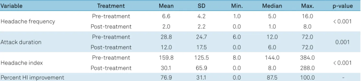
with migraine without aura (33.9 ± 8.6 years old, one male. According to IHS-III criteria: two with chronic migraine) were scanned pre- and post-treatment; and 19 matched healthy controls (33.1 ± 9 years old, one male) were scanned once. The HI in the patients improved by 76.9% on average between the first and second scan (initial value: 159.8 ± 125.5; final value: 30.1 ± 65.9; p < 0.001) (Table 1).
First scan vs. second scan comparisons in the patients
The cortical thickness decreased by more than 1% in the second scan in nine (left hemisphere) and eight (right hemi
-sphere) regions, and increased more than 1% in four (in each hemisphere) regions (Table 2).The areas in the left hemi
-sphere with a significant cortical thickness decrease were:
fusiform, lingual, posterior cingulate, and rostral middle
fron-tal. Only in the left transverse temporal area did the thickness increase significantly. The areas in the right hemisphere with
a significant thickness decrease were: caudal middle frontal,
isthmus of cingulate, pars triangularis, posterior cingulate, rostral middle frontal and superior frontal. No cortical area
showed a significant thickness increase on the right side. No thickness variation remained significant after the Monte
Carlo corrections for multiple comparisons.
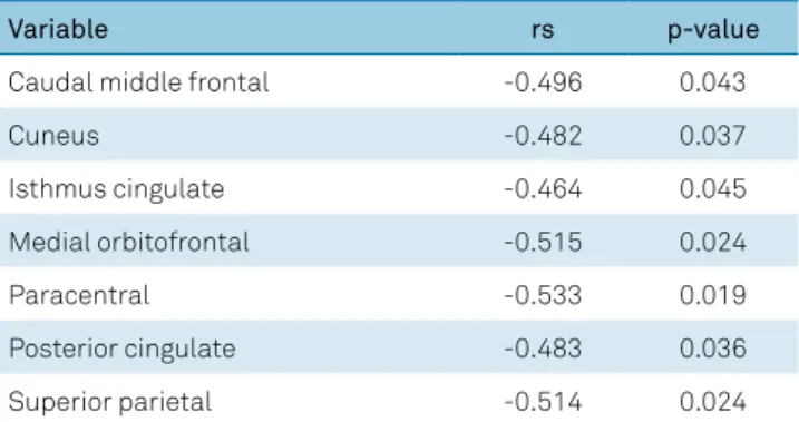
The HI variation correlated negatively with cortical thickness changes in different areas of the left hemisphere (Table 3). The left posterior cingulate thickness was the only region with a significant reduction in thickness after treat
-ment and correlation (negative) with the HI.
First scan vs. controls comparisons
In the left hemisphere, the cortex was significantly thicker, in patients, only in the pars opercularis. The effect
sizes were greater than 0.4 in the isthmus cingulate, paracen-tral, pericalcarine, post-cenparacen-tral, posterior cingulate, precu-neus, rostral anterior cingulate and superior parietal
corti-ces. The thickness was significantly reduced in patients in the entorhinal cortex and temporal pole. The effect sizes were
greater than 0.4 in the orbitofrontal, superior temporal and temporal pole cortices.
In the right hemisphere, the cortex was significantly
thicker in patients in the paracentral, post-central,
precu-neus and superior parietal areas. The effect sizes were greater
than 0.4 in the inferior parietal, isthmus cingulate, paracen-tral, pericalcarine, post-cenparacen-tral, posterior cingulate, precen-tral, precuneus, superior parietal and supramarginal areas.
The thickness was significantly reduced in patients in the entorhinal cortex. The effect sizes were greater than 0.4 in the
pars orbitalis, temporal pole and insulacortices.
Second scan vs. controls comparisons
In the left hemisphere, the cortex was significantly thicker in patients in the paracentral and pars opercularis. The effect
size was greater than 0.4 in the isthmus cingulate, paracen-tral, pars opercularis, pericalcarine, post-cenparacen-tral, precuneus, rostral anterior cingulate, posterior cingulate, and superior
parietal cortices. The thickness was significantly reduced in patients only in the entorhinal cortex. The effect sizes were
greater than 0.4 in the entorhinal, parahippocampal, superior temporal and temporal pole cortices.
Table 1. Monthly headache characteristics before and after treatment.
Variable Treatment Mean SD Min. Median Max. p-value
Headache frequency Pre-treatment 6.6 4.2 1.0 5.0 16.0 < 0.001
Post-treatment 2.0 2.2 0.0 1.0 8.0
Attack duration Pre-treatment 28.8 24.7 6.0 12.0 72.0 0.001
Post-treatment 12.0 17.5 0.0 6.0 72.0
Headache index Pre-treatment 159.8 125.5 8.0 144.0 384.0 < 0.001
Post-treatment 30.1 65.9 0.0 8.0 288.0
Percent HI improvement 76.9 31.1 0.0 87.5 100.0
In the right hemisphere, the cortex was significantly
thicker in patients in the paracentral, post-central,
precu-neus and superior parietal areas. The effect size was greater
than 0.4 in the isthmus cingulate, lingual, paracentral, peri-calcarine, post-central, precentral, precuneus supramarginal and superior parietal areas.
Monte-Carlo simulation
In the patient group, there was no significant thickness variation comparing the second scan with the first scan. The
Figure shows the cortical areas in the right hemisphere where
the cortex is significantly thicker comparing the patients’ first and second scans with controls.
DISCUSSION
This was one of the first attempts to correlate improve -ment of cortical thickness pre- and post-treat-ment in migraine. After multiple comparisons correction, only the
right hemisphere showed thickness changes (increase),
in patients vs. controls, located in the somatosensory and
superior parietal cortices, both in the first and second scans.
In addition, the right cortex was thicker in the second scan in the precentral, supramarginal, cuneus and precuneus when
compared to controls. The effect size analysis showed small changes in various areas, specifically in the lingual, fusiform and caudal anterior cingulate cortices on the left (thickness reduction); and caudal middle frontal, pars triangularis, and rostral middle frontal (thickness reduction), and transverse temporal (thickness increase) cortices on the right. Patients improved significantly following treatment as shown by the HI reduction, but there were no corresponding changes in
cortical thickness after correction for multiple comparison.
Regression analysis showed significant negative correlations between the HI improvement and cortical thickness changes
only in the left hemisphere.
The only area with significant thickness change (reduc
-tion, before Monte-Carlo correction) after treatment and simultaneous significant correlation (negative) with HI
improvement in the second scan was the left posterior
cin-gulate cortex. This region has been shown to be involved
with nociception12. Pain has been shown experimentally to Table 3. Correlations between % headache index (HI)
improvement and cortical thickness (second scan).
Variable rs p-value
Caudal middle frontal -0.496 0.043
Cuneus -0.482 0.037
Isthmus cingulate -0.464 0.045
Medial orbitofrontal -0.515 0.024
Paracentral -0.533 0.019
Posterior cingulate -0.483 0.036
Superior parietal -0.514 0.024
All values correspond to the left hemisphere. No significant correlation was found at the right hemisphere. rs: Spearman’s correlation coefficient. Table 2. Cortical thickness variation in the second scan.
Variable Left hemisphere Right hemisphere
% Variation Effect size p-value % Variation Effect size p-value
Caudal anterior cingulate -2.31 -0.33 0.067 -1.38 -0.18 0.096
Caudal middle frontal -1.11 -0.28 0.058 -1.55 -0.38 0.013
Inferior temporal -1.48 -0.39 0.053 - -
-Isthmus of cingulate - - - -1.63 -0.17 0.008
Lateral orbitofrontal 2.24 0.21 0.253 - -
-Lingual -1.45 -0.30 0.003 - -
-Fusiform -1,52 -0,33 0,029 - -
-Medial orbitofrontal 1.96 0.20 0.344 - -
-Parahippocampal -1.04 -0.11 0.310 - -
-Pars triangularis - - - -2.36 -0.39 0.002
Posterior cingulate -1.01 -0.18 0.031 -1.45 -0.22 0.001
Rostral anterior cingulate - - - -1.56 -0.17 0.363
Rostral middle frontal -1.13 -0.28 0.012 -1.82 -0.45 0.005
Superior frontal - - - -1.12 -0.26 0.039
Frontal pole -2.25 -0.27 0.069 1.38 0.14 0.564
Temporal pole 2.87 0.27 0.184 3.23 0.21 0.339
Transverse temporal 1.72 0.22 0.012 1.75 0.43 0.091
Insula - - - 1.33 0.19 0.24
deactivate the posterior cingulate cortex13. This deactiva
-tion was reduced in patients with chronic pain14. The pos
-terior cingulate cortex is part of the default mode network, a relevant area in pain physiology15. Reduced brain volumes
in posterior cingulate cortex, a region reported to have anti-nociceptive functions, have been reported in various chronic pain conditions, such as phantom limb pain16, fibromyal
-gia17, headache18 and trigeminal neuropathic pain19. In
con-trast, we did not find differences in posterior cingulate cortex thickness between the controls and patients. However, with our finding of decreased left posterior cingulate cortex thick
-ness with HI improvement, we speculate that, in migraine,
less antinociceptive activity in the left posterior cingulate cortex is required as the pain decreases.
In our study, the migraineurs showed a significant
increase in cortical precuneus thickness in the right hemi-sphere when compared with controls. Maleki et al. reported
that female migraine patients have thicker posterior insula and precuneus cortices compared with male migraineurs and healthy controls of both sexes20. The precuneus was
shown to be more activated in females20, who are particularly
susceptible to migraine. The anterior, central and posterior
precuneus are involved with sensorimotor processing, cog-nition/associative processing, and visual processing,
respec-tively. The three areas are functionally connected to the supe
-rior parietal cortex, paracentral lobule and motor cortex; the
dorsolateral prefrontal, dorsomedial prefrontal and
multi-modal lateral inferior parietal cortex; and adjacent visual
cortical regions21. Schwedt et al. also found negative
correla-tions between pain thresholds and cortical thickness in the left posterior cingulate/precuneus in healthy individuals22.
In contrast, migraineurs without aura exhibited positive cor-relations between pain thresholds and cortical thickness in the right precuneus22. Previous task-based fMRI studies have Figure. The figure shows cortical areas in the right hemisphere where the cortex is significantly thicker comparing the patients’ first and second scans with controls. Regions of increased cortical thickness or surface area are shown in red (color-coded according to t
value), and regions of decreased cortical thickness or surface area are shown in blue (color-coded according to t value). The color bar scale is logarithmic and represents –log 10(p): 2.5 corresponds to a p value of 0.05 and 5 corresponds to a p value of 0.00001. Only clusters surviving multiple comparisons using the Monte Carlo simulation (10,000 permutations) are displayed. Cortical areas: A: Right precentral. B: Right post-central. C: Right superior parietal. D: Right precuneus. E: Right supramarginal. F: Right cuneus.
-5.00 -2.50 0.00 2.50 5.00
A
B
C
D
E
shown greater visual stimuli-induced precuneus activation in migraineurs without aura than in controls23. The changes
found in the present and previous studies, associated with
the fact that migraine patients may suffer visual, cognitive and sensorimotor deficits during attacks24, suggest that the
cuneus is particularly dysfunctional in migraine.
In a previous report, migraineurs exhibited enhanced brain activation in the cerebellum anterior lobe/culmen,
lingual gyrus, precuneus (all bilaterally), and the left cuneus
while viewing negative pictures compared with neutral pic-tures25. There are extensive connections between the pre
-cuneus and the dorsal premotor area, the supplementary motor area and the anterior cingulate cortex26. Compared
with controls, migraineurs without aura showed a significant
decrease in functional resting-state connectivity between the left precuneus and the left inferior and superior occipital gyrus, bilateral middle occipital gyrus, bilateral cuneus, bilat-eral superior parietal lobules, bilatbilat-eral somatosensory cortex, bilateral dorsolateral prefrontal cortices, right premotor cor-tex, pons, bilateral cerebellar posterior lobes, right paracen-tral lobule, right middle cingulate gyrus and bilateral supple-mentary motor areas27. All these brain regions are involved
with pain processing23.
The somatosensory cortex has been shown to be thicker in migraine patients, which is in line with the present findings.
Activation of the somatosensory cortices has been reported in
approximately 75% of human imaging studies of pain28. This
area is strongly implicated in the ascending trigemino-cortical
nociceptive pathway. Thickening of the somatosensory cor -tex has been demonstrated in migraine6,29. Migraine patients
with a higher headache frequency (8-14 days/month) showed
increased post-central gyrus thickness in comparison with
low frequency (less than two days/months) patients3. Kim et
al. compared migraine patients with controls and found that migraine patients had cortical thickening in bilateral post-central gyrus30. Whether these abnormalities contribute to
migraine susceptibility or, conversely, are the consequence of repeated attacks, is still a matter of debate.
Our results showed that migraineurs presented with cor
-tical thickening in two areas of the parietal lobe (superior parietal lobe and supramarginal gyrus). The inferior pari -etal lobe is mainly involved in top-down control of execu-tive functions and in cogniexecu-tive aspects of processing sensory stimuli, including pain31. Migraine with aura patients showed
bilateral thickening of regions in the inferior parietal lobe32,
in contrast with another study33.
Although several studies, including ours, confirm cortical
thickness changes in migraine, a larger patient cohort failed
to replicate cortical thickness findings in both migraine
patients with, and without, aura relative to controls5.
Methodological issues are probably the best explanation for those discrepancies.
Our study confirms that neuroimaging may demonstrate
changes in cortical areas previously shown to be involved
with the lateral pain system. That system consists of neurons of the spinothalamic tract that project to the somatosensory
nuclei of thalamus, which in turn transfer nociceptive infor-mation to the primary and secondary somatosensory areas,
and posterior insula; these discriminatory areas determine
the localization, the intensity, and the quality of pain and are connected to the parietal cortex34.
Most of our findings were lateralized. Since our patients presented with bilateral ( five individuals), alternating (nine), predominantly left ( four), or right (one) pain, the laterality of
our data cannot be explained by headache side preference, even though predominantly left-sided headache was more
frequent. Bilateral reduction in regional cerebral blood flow
has been documented in the cingulofrontal transitional cor-tex and posterior cingulate corcor-tex during noxious stimula-tion of the left hand35. Similar to our results, in patients with
HIV-associated distal neuropathic pain, changes (increase)
were restricted to the left posterior cingulate cortex36.
A negative correlation between pain thresholds and cortical thickness was also reported on the left22. Further data with
larger samples are required for better understanding of the
laterality findings.
The potential reversibility of cortical changes following
treatment suggested in this study raises multiple questions. Firstly, it remains to be determined what the pathological bases of the thickness changes are, and how migraine trig-gers the tissue adaptations4. Secondly, what are the
mecha-nisms responsible for the return to previous thickness, an
effect probably related to headache frequency and duration? Thirdly, what are the implications of these changes in corti
-cal spreading depression? Fourthly, how could these changes,
either isolated or in combination with other neuroimaging
findings, eventually serve as biomarkers for diagnosis, sever
-ity, or drug efficacy?
Cortical thickness changes occur because of hypo- or hyper-function secondary to local, near or distant connec-tions37. Although treatment can theoretically change
corti-cal thickness in affected regions, the present results do not allow causation conclusions. Our study has strong aspects. All patients were treatment naïve at the first scan; they were
examined by the same physicians under a unique
proto-col and usual care, reproducing real life treatment; and the observation was over a long period of time, on average. There
are many weak points to be considered. Firstly, the time span
between the first and second scan in our patients varied con -siderably. Secondly, due to the study length, patients did not record a precise headache diary, leading to potential recall
biases. Thirdly, the sample size was relatively small. Fourthly,
controls were not scanned twice, at the same time as the patients. Finally, although we are aware that cortical
reduction in regions known to be affected in migraineurs,
point to the hypothesis that anatomical variations induced by the disease are not irreversible and allow the hypothesis
that treatment may reverse cortical thickness modifications
in migraine.
Taken together, the data show that there are differences
in cortical thickness between migraine patients and controls.
Among the regions with significant change in cortical thickness
following preventive treatment, the degree of improvement cor-related with reduction in thickness in the left posterior cingu-late. Previous studies have shown that this area is involved with
pain processing and is affected in headache disorders. To inves -tigate the reasons for intriguing right-to-left changes and to
clar-ify controversies still remaining in this field, young migraine-free subjects with migraine parents should be scanned at base-line
and re-examined later in the event of migraine development.
References
1. Woldeamanuel YW, Cowan RP. Migraine affects 1 in 10 people worldwide featuring recent rise: A systematic review and meta-analysis of community-based studies involving 6 million participants. J Neurol Sci. 2017 Jan;372:307-15. https://doi.org/10.1016/j.jns.2016.11.071
2. Burstein R, Noseda R, Borsook D. Migraine: multiple processes, complex pathophysiology. J Neurosci. 2015 Apr;35(17):6619-29. https://doi.org/10.1523/JNEUROSCI.0373-15.2015
3. Maleki N, Becerra L, Brawn J, Bigal M, Burstein R, Borsook D. Concurrent functional and structural cortical alterations in migraine. Cephalalgia. 2012 Jun;32(8):607-20. https://doi.org/10.1177/0333102412445622
4. Hadjikhani N. Relevance of cortical thickness in migraine sufferers. Expert Rev Neurother. 2008 Mar;8(3):327-9. https://doi.org/10.1586/14737175.8.3.327
5. Datta R, Detre JA, Aguirre GK, Cucchiara B. Absence of changes in cortical thickness in patients with migraine. Cephalalgia. 2011 Oct;31(14):1452-8. https://doi.org/10.1177/0333102411421025
6. DaSilva AF, Granziera C, Snyder J, Hadjikhani N. Thickening in the somatosensory cortex of patients with migraine. Neurology. 2007 Nov;69(21):1990-5. https://doi.org/10.1212/01.wnl.0000291618.32247.2d
7. Maleki N, Becerra L, Nutile L, Pendse G, Brawn J, Bigal M et al. Migraine attacks the Basal Ganglia. Mol Pain. 2011 Sep;7:71. https://doi.org/10.1186/1744-8069-7-71
8. Schwedt TJ, Dodick DW. Advanced neuroimaging of migraine. Lancet Neurol. 2009 Jun;8(6):560-8. https://doi.org/10.1016/S1474-4422(09)70107-3
9. Lipton RB, Bigal ME, Scher AI, Stewart WF. The global burden of migraine. J Headache Pain. 2003 Mar;4 Suppl 1:s3-11. https://doi.org/10.1007/s101940300001
10. Headache Classification Committee of the International Headache S. The International Classification of Headache Disorders. 2nd edition. Cephalalgia. 2004;24(1 Suppl):9-160.
11. Headache Classification Committee of the International Headache S. The International Classification of Headache Disorders. 3rd edition (beta version). Cephalalgia. 2013;33(9):629-808. https://doi.org/10.1177/0333102413485658
12. Emerson NM, Zeidan F, Lobanov OV, Hadsel MS, Martucci KT, Quevedo AS et al. Pain sensitivity is inversely related to regional grey matter density in the brain. Pain. 2014;155(3):566-73). https://doi.org/10.1016/j.pain.2013.12.004
13. Kong J, Loggia ML, Zyloney C, Tu P, Laviolette P, Gollub RL. Exploring the brain in pain: activations, deactivations and their relation. Pain. 2010;148(2):257-67). https://doi.org/10.1016/j.pain.2009.11.008
14. Baliki MN, Geha PY, Apkarian AV, Chialvo DR. Beyond feeling: chronic pain hurts the brain, disrupting the default-mode network dynamics. J Neurosci. 2008;28(6):1398-403). https://doi.org/10.1523/JNEUROSCI.4123-07.2008
15. Raichle ME, MacLeod AM, Snyder AZ, Powers WJ, Gusnard DA, Shulman GL. A default mode of brain function. Proc Natl Acad Sci U S A. 2001;98(2):676-82). https://doi.org/10.1073/pnas.98.2.676
16. Draganski B, Moser T, Lummel N, Gänssbauer S, Bogdahn U, Haas F et al. Decrease of thalamic gray matter following
limb amputation. Neuroimage. 2006;31(3):951-7). https://doi.org/10.1016/j.neuroimage.2006.01.018
17. Robinson ME, Craggs JG, Price DD, Perlstein WM, Staud R. Gray matter volumes of pain-related brain areas are decreased in fibromyalgia syndrome. J Pain. 2011;12(4):436-43). https://doi.org/10.1016/j.jpain.2010.10.003
18. Schmidt-Wilcke T, Leinisch E, Straube A, Kämpfe N, Draganski B, Diener HC et al. Gray matter decrease in patients with chronic tension type headache. Neurology. 2005;65(9):1483-6). https://doi.org/10.1212/01.wnl.0000183067.94400.80
19. DaSilva AF, Becerra L, Pendse G, Chizh B, Tully S, Borsook D. Colocalized structural and functional changes in the cortex of patients with trigeminal neuropathic pain. PLoS One. 2008;3(10):e3396. https://doi.org/10.1371/journal.pone.0003396
20. Maleki N, Linnman C, Brawn J, Burstein R, Becerra L, Borsook D. Her versus his migraine: multiple sex differences in brain function and structure. Brain. 2012 Aug;135(Pt 8):2546-59.
https://doi.org/10.1093/brain/aws175
21. Margulies DS, Vincent JL, Kelly C, Lohmann G, Uddin LQ, Biswal BB et al. Precuneus shares intrinsic functional architecture in humans and monkeys. Proc Natl Acad Sci USA. 2009 Nov;106(47):20069-74. https://doi.org/10.1073/pnas.0905314106
22. Schwedt TJ, Chong CD. Correlations between brain cortical thickness and cutaneous pain thresholds are atypical in adults with migraine. PLoS One. 2014 Jun;9(6):e99791. https://doi.org/10.1371/journal.pone.0099791
23. Schwedt TJ, Chiang CC, Chong CD, Dodick DW. Functional MRI of migraine. Lancet Neurol. 2015 Jan;14(1):81-91. https://doi.org/10.1016/S1474-4422(14)70193-0
24. Vincent MB, Hadjikhani N. Migraine aura and related phenomena: beyond scotomata and scintillations. Cephalalgia. 2007 Dec;27(12):1368-77. https://doi.org/10.1111/j.1468-2982.2007.01388.x
25. Wang M, Su J, Zhang J, Zhao Y, Yao Q, Zhang Q et al. Visual cortex and cerebellum hyperactivation during negative emotion picture stimuli in migraine patients. Sci Rep. 2017 Feb;7:41919. https://doi.org/10.1038/srep41919
26. Petrides M, Pandya DN. Projections to the frontal cortex from the posterior parietal region in the rhesus monkey. J Comp Neurol. 1984 Sep;228(1):105-16. https://doi.org/10.1002/cne.902280110
27. Zhang J, Su J, Wang M, Zhao Y, Yao Q, Zhang Q et al. Increased default mode network connectivity and increased regional homogeneity in migraineurs without aura. J Headache Pain. 2016 Dec;17(1):98. https://doi.org/10.1186/s10194-016-0692-z
28. Apkarian AV, Bushnell MC, Treede RD, Zubieta JK. Human brain mechanisms of pain perception and regulation in health and disease. Eur J Pain. 2005 Aug;9(4):463-84. https://doi.org/10.1016/j.ejpain.2004.11.001
30. Kim JH, Kim JB, Suh SI, Seo WK, Oh K, Koh SB. Thickening of the somatosensory cortex in migraine without aura. Cephalalgia: an international journal of headache. 2014; 34(14):1125-33. https://doi.org/10.1177/0333102414531155
31. Weidner R, Krummenacher J, Reimann B, Müller HJ, Fink GR. Sources of top-down control in visual search. J Cogn Neurosci. 2009 Nov;21(11):2100-13. https://doi.org/10.1162/jocn.2008.21173
32. Messina R, Rocca MA, Colombo B, Valsasina P, Horsfield MA, Copetti M et al. Cortical abnormalities in patients with migraine: a surface-based analysis. Radiology. 2013 Jul;268(1):170-80. https://doi.org/10.1148/radiol.13122004
33. Schmitz N, Arkink EB, Mulder M, Rubia K, Admiraal-Behloul F, Schoonman GG et al. Frontal lobe structure and executive function in migraine patients. Neurosci Lett. 2008 Aug 1;440(2):92-96. https://doi.org/10.1016/j.neulet.2008.05.033
34. Grazzi L, Chiapparini L, Ferraro S, Usai S, Andrasik F, Mandelli ML et al. Chronic migraine with medication overuse pre-post withdrawal of symptomatic medication: clinical results and fMRI correlations. Headache. 2010 Jun;50(6):998-1004.
https://doi.org/10.1111/j.1526-4610.2010.01695.x
35. Vogt BA, Derbyshire S, Jones AK. Pain processing in four regions of human cingulate cortex localized with co-registered PET and MR imaging. Eur J Neurosci. 1996;8(7):1461-73). https://doi.org/10.1111/j.1460-9568.1996.tb01608.x
36. Keltner JR, Connolly CG, Vaida F, Jenkinson M, Fennema-Notestine C, Archibald S et al HIV Distal Neuropathic Pain Is Associated with smaller ventral posterior cingulate cortex. Pain Med. 2017 Mar;18(3):428-40. https://doi.org/10.1093/pm/pnw180