Protective effects of acerola juice on genotoxicity induced by iron
in vivo
Roberta Nunes Horta
1,2,*, Vivian Francilia Silva Kahl
2,*, Merielen da Silva Sarmento
2,
Marisa Fernanda Silva Nunes
1,2, Carem Rejane Maglione Porto
2, Vanessa Moraes de Andrade
4,
Alexandre de Barros Falcão Ferraz
3and Juliana Da Silva
21
Centro de Ciências da Saúde, Universidade da Região da Campanha (URCAMP), Bagé, RS, Brazil.
2Laboratório de Genética Toxicológica, Universidade Luterana do Brasil, Canoas, RS, Brazil.
3
Laboratório do Farmacognosia e Fitoquímica, Universidade Luterana do Brasil, Canoas, RS, Brazil.
4Laboratório de Biologia Celular e Molecular, Programa de Pós-Graduação em Ciências de Saúde,
Unidade de Ciências de Saúde, Universidade do Estado de Santa Catarina, Criciúma, SC, Brazil.
Abstract
Metal ions such as iron can induce DNA damage by inducing reactive oxygen species (ROS) and oxidative stress. Vitamin C is one of the most widely consumed antioxidants worldwide, present in many fruits and vegetables, espe-cially inMalpighia glabra L., popularly known as acerola, native to Brazil. Acerola is considered a functional fruit due to its high antioxidant properties and phenolic contents, and therefore is consumed to prevent diseases or as adjuvant in treatment strategies. Here, the influence of ripe and unripe acerola juices on iron genotoxicity was ana-lyzedin vivo using the comet assay and micronucleus test. The comet assay results showed that acerola juice ex-erted no genotoxic or antigenotoxic activity. Neither ripe nor unripe acerola juices were mutagenic to animals treated with juices, in micronucleus test. However, when compared to iron group, the pre-treatment with acerola juices ex-erted antimutagenic activity, decreasing significantly micronucleus mean values in bone marrow. Stage of ripeness did not influence the interaction of acerola compounds with DNA, and both ripe and unripe acerola juices exerted pro-tective effect over DNA damage generated by iron.
Keywords: genotoxicity; comet assay; micronucleus; acerola juice; iron.
Received: June 17, 2015; Accepted: October 07, 2015.
Introduction
Fruits play a prominent role in the prevention of vari-ous diseases, such as cancer, cardiovascular and neuro-degenerative conditions. People who eat fruits in childhood have 38% less probability to develop cancers (Maynardet al., 2003) and balanced diets can contribute for the genomic stability (Fenech, 2014). More attention has been paid to these foods, since epidemiological evidence has shown that regular consumption of vegetables is associated with re-duced mortality and morbidity from some chronic diseases (Strandhagenet al., 2000), and its protective effect has been attributed to the presence of constituents with antioxidant properties such as pholyphenols, carotenoids, and vitamins (Nunes et al., 2011; Kahl et al., 2012; Liu, 2013; Kozlowska and Szotask-Wegierek, 2014).
Malpighia glabraL., popularly called “acerola” in Brazil or “Barbados cherry”, is a native species from tropi-cal America. Acerola compounds, such as vitamin C, carot-enoids, precursors of vitamin A, lycopene, among others (Chaveset al., 2004), depends on the cultivars, environ-mental conditions and the stage of fruit ripeness (Chaveset al., 2004; Nuneset al., 2011).
Despite scientific data reporting benefits from juice consumption (Frankeet al., 2006; Leffaet al., 2013), some compounds present in juices have also been identified as being mutagenic or carcinogenic (Spadaet al., 2008). Al-though the genotoxicity of several components of acerola juice have already been evaluated individually, it is impor-tant to test the effect of whole juice, a complex mixture, in different biological systems. Genotoxic effects, for exam-ple, may be mediated by the interaction of juice compounds with transition metals or by-products of juice auto-oxi-dation; Vitamin C can act as a pro-oxidant, because its re-ducing ability, through Fenton and Fenton-like reactions (De Freitas and Meneghini, 2001).
One feature of the normal human diet is the simulta-neous presence of both essential and toxic metals
(Beyers-Send correspondence to Juliana da Silva. Programa de Pós-Gra-duação em Genética e Toxicologia Aplicada, Universidade Lute-rana do Brasil, Av. Farroupilha 8001, Prédio 22, Sala 22, 92425-900, Canoas, RS, Brazil. E-mail: juliana.silva@ulbra.br
*
Shared authorship; both authors participated equally in all steps of the work.
mann and Hartwig, 2008). Metal ions generate DNA dam-age directly or indirectly by formation of reactive oxygen species (ROS) (De Freitas and Meneghini, 2001; Phaniendraet al., 2015). In recent years, many studies have been conducted on the role of ROS in the etiology of vari-ous diseases since some free radicals, in a condition of oxi-dative stress, are not neutralized by antioxidant cell protec-tive mechanisms or antioxidant compounds (Halliwell and Guterridge, 2000; Phaniendra et al., 2015). Vitamin C, present in acerola juice is an example of an antioxidant compound that can chelate metals, thus preventing the gen-eration of ROS (De Freitas and Meneghini, 2001; Pha-niendraet al., 2015), apart from playing a role in the regula-tion of DNA repair enzymes (Jomova and Valko, 2011). Considering the importance of consumption of fruits and the intense growth of the culture of acerola in Brazil and its widespread use, the aim of this study was to test the geno-toxic effects of acerola juice at two ripeness stages associ-ated with metallic agentsin vivoin mice, using the comet assay and the micronucleus test, in order to improve our un-derstanding of the role of dietary antimutagens and anticarcinogens in humans.
Material and Methods
Preparation of acerola juice
Acerola samples were collected from an organic cul-ture in May 2008, in the district of Ubajara, CE, Brazil. The fruits were collected randomly across the whole plant, and were sorted according to ripeness using a skin color gradi-ent, as follows: (1) green fruits (unripe), fruits showing a green hue on more than 75% of its skin; and (2) red fruits (ripe), those showing a red or burgundy hue on 100% of the skin (Campeloet al., 1998). After collection, fruits were kept frozen and protected from light in order to preserve their chemical and physical characteristics upon juice prep-aration (Nunes et al., 2011). Approximately 20 units of acerola fruit were used to produce 150 g of pulp extract,
af-ter fruits were peeled and seeds removed. The pulp extract from ripe and unripe fruits were diluted with water (150 g of pulp extract per liter of water), forming the juice used in treatments. A glass of juice consumed by humans usually has 200 mL, and therefore, is composed by around 30 g of pulp extract. The acerola cultivar used in this study was AC-69.
Animals and treatment
Animals used were CF1 miceMus musculus, aged 5-7 weeks old, weighing 20-40 g, provided by the Fun-dação Estadual de Produção e Pesquisa em Saúde (FEEPS), Porto Alegre, RS, Brazil. The animals were kept in the cen-tral animal house of Lutheran University of Brazil (ULBRA), Canoas, RS, Brazil. The temperature in the ex-perimental room was about 24 °C, and relative humidity was roughly 60%. The light cycle was 12 h light/12 h dark. All animals received commercial standard mouse cube diet (Labina-PURINE) and waterad libitum. All experimental procedures were approved by CONEP 2008-008-A, Brazil (National Commission of Ethics in Research).
Ten mice per group (five females and five males) were used for the treatment (0.1 mL/10 g body weight) di-vided in six groups: (a) Negative control, water; (b) Posi-tive Control, FeSO4(35 mg/kg), (c) Unripe acerola juice;
(d) Ripe acerola juice; (e) Unripe acerola juice plus FeSO4;
(f) Ripe acerola juice plus FeSO4.The dose, exposure time
and the administration route of FeSO4were based on a
pre-vious study (Nuneset al., 2011). Table 1 summarizes the treatment period. Acerola, FeSO4and water were
adminis-trated by gavage. The test substance was handled as poten-tially mutagenic, according to the safety procedures required.
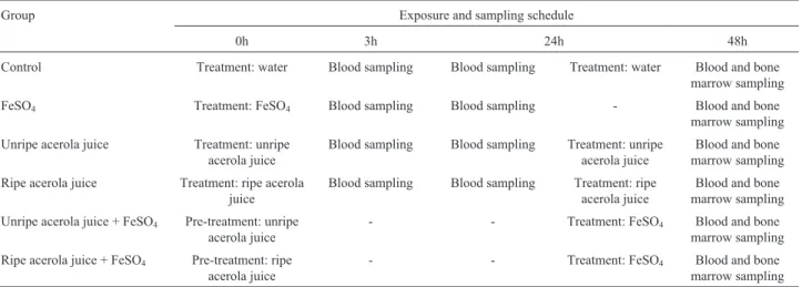
Table 1- Experimental procedures: treatment protocols and blood sampling schedules.
Group Exposure and sampling schedule
0h 3h 24h 48h
Control Treatment: water Blood sampling Blood sampling Treatment: water Blood and bone
marrow sampling
FeSO4 Treatment: FeSO4 Blood sampling Blood sampling - Blood and bone
marrow sampling
Unripe acerola juice Treatment: unripe acerola juice
Blood sampling Blood sampling Treatment: unripe acerola juice
Blood and bone marrow sampling
Ripe acerola juice Treatment: ripe acerola juice
Blood sampling Blood sampling Treatment: ripe acerola juice
Blood and bone marrow sampling
Unripe acerola juice + FeSO4 Pre-treatment: unripe
acerola juice
- - Treatment: FeSO4 Blood and bone
marrow sampling
Ripe acerola juice + FeSO4 Pre-treatment: ripe
acerola juice
- - Treatment: FeSO4 Blood and bone
Genotoxicity assays
Comet assay
The analysis was conducted in accordance with the protocol described by Singhet al.(1998) with some modifi-cations (Nuneset al., 2011). Blood cells (10mL of whole blood with heparin) were embedded in 90mL of 0.75% (w/v) low melting point agarose and the mixture added to a microscope slide pre-coated with 1.5% (w/v) of normal melting point agarose and topped with a coverslip. The slides were briefly placed on ice for agarose to solidify and then the coverslips were carefully removed. Next, the slides were immersed in lysis solution (2.5 M NaCl, 100 mM EDTA and 10 mM Tris, pH 10.0–10.5) containing freshly added 1% Triton X-100 and 10% dimethyl sulfoxide (DMSO) for at least 1 h at 4 °C. Subsequently, the slides were incubated in freshly made alkaline buffer (300 mM NaOH and 1 mM EDTA, pH>13) for 20 min for DNA un-winding, and electrophoresis was performed in the same buffer. The electrophoresis conditions were 15 min at 300 mA and 25 V (0.7 V/cm). All these steps were carried out under dim indirect light. Following electrophoresis, slides were neutralized in 400 mM Tris buffer (pH 7.5) and fixed (15% w/v trichloroacetic acid, 5% w/v zinc sulfate, 5% glycerol), washed in distilled water and dried overnight. The gels were re-hydrated for 5 min in distilled water, and then stained for 15 min (37 °C) with a solution containing the following sequence: 34 mL of Solution B (0.2% w/v ammonium nitrate, 0.2% w/v silver nitrate, 0.5% w/v tungstosilicic acid, 0.15% v/v formaldehyde, 5% w/v so-dium carbonate) and 66 mL of Solution A (5% soso-dium car-bonate). The staining was stopped with 1% acetic acid and the gels were air-dried. To calculate a damage index (DI), cells were visually allocated into 5 classes according to tail size (0 = no tails, to 4 = maximum-length tails) which re-sulted in a single DNA damage score for each sample and consequently for each group studied. Thus, the damage in-dex (DI) of the group could range from 0 (completely un-damaged = 100 cells X 0) to 400 (maximum damage = 100 cells X 4). The damage frequency (DF in %) was calculated for each sample based on the number of cells with tailvs.
those without. All slides were coded for blinded analysis. The results of the potential of acerola juice to modu-late the effect of FeSO4treatment were expressed as
de-scribed in the literature (Kapiszewska et al., 2005), as percentage inhibition of damage index (DI) according to the expression: (I%): percentage of inhibition of DI = [FeSO4DI - DI of the extract with FeSO4] / [FeSO4DI - DI
negative control] x 100
Micronucleus test
Each complete test was made according to a report by Mavourninet al.(1990) and OECD (2013). Bone marrow smears were prepared for the 48-h exposure sample, when animals were killed by decapitation. The bone marrow was
extracted from the two femurs and the smears were pre-pared directly on slides, two per animal, with a drop of fetal calf serum. The slides were stained with 10% Giemsa for 5 min, air-dried and coded for blinded analysis. To avoid false negative results and as a measure of toxicity in bone marrow, the polychromatic erythrocytes: normochromatic erythrocytes (PCE/NCE) ratio was scored in 1,000 cells. The incidence of micronuclei was observed in 2,000 PCE for each animal (i.e.1,000 from each of the two slides pre-pared from the duplicate), using bright-field optical micros-copy at a magnification of 200–1000. The test groups were compared to the respective negative controls by gender, separately and in combination.
Statistical analysis
The normality of variables was evaluated using the Kolmogorov–Smirnov test. Statistical differences between the groups were analyzed using the non-parametric two-tailed Kruskal–Wallis Test with the Dunn correction for multiple comparisons for comet assay and micronucleus test results. Student’st-test was used to compare damage between genders. The critical level for rejection of the null hypothesis was considered to be aPvalue of 5%.
Results
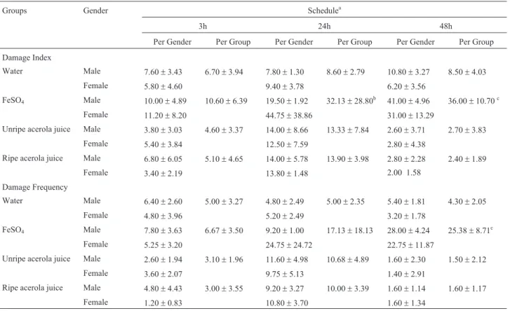
Table 2 summarizes the comet assay data expressed as damage index (DI) and damage frequency (DF) for blood cells of mice exposed for 3 h, 24 h and 48 h to water, FeSO4and acerola juices, at different maturation stages.
The FeSO4group showed a significant increase in DI, as
compared to the negative control, at 24 h (P < 0.01) and 48 h (P < 0.001). After treatment with FeSO4at 48 h, the mean
DF values were also significantly elevated (P < 0.001). Concerning the acerola juice groups, no genotoxic activity was observed, when compared with the negative control group.
Figure 1 shows that groups pre-treated with acerola presented lower DI than the FeSO4group. Nevertheless, no
statistically significant difference in antigenotoxic activity of acerola juice was observed in relation to FeSO4. The
pre-treatment with unripe acerola juice presented modula-tion of DNA damage of 21.33%, while the modulamodula-tion by ripe acerola juice was 56.65%. Our study also found no dif-ferences between juice maturation stage and animal gender in the comet assay results.
Table 3 shows the results of the micronucleus test for bone marrow samples. Bone marrow cells of mice treated with unripe and ripe acerola juice showed no significant in-crease in micronucleus mean, when compared with nega-tive control, therefore not presenting mutagenicity. FeSO4
Bone marrow cells of mice pre-treated with juices from unripe and ripe acerola fruits showed a significant de-crease in micronucleus (P < 0.001 and P < 0.01, respec-tively), when compared with the FeSO4group, revealing
antimutagenic activity (Figure 2).
Discussion
A recent study considers acerola as a functional food since its pulp shows great antioxidant activity and phenolic content and, therefore is consumed to prevent diseases or as
Table 2- Comet assay parameters (damage index and damage frequency index; mean±standard deviation) for blood samples of mice treated with unripe and ripe acerola juice. For each group, n = 10 (five males and five females), with 100 cells per animal.
Groups Gender Schedulea
3h 24h 48h
Per Gender Per Group Per Gender Per Group Per Gender Per Group
Damage Index
Water Male 7.60±3.43 6.70±3.94 7.80±1.30 8.60±2.79 10.80±3.27 8.50±4.03
Female 5.80±4.60 9.40±3.78 6.20±3.56
FeSO4 Male 10.00±4.89 10.60±6.39 19.50±1.92 32.13±28.80b 41.00±4.96 36.00±10.70c
Female 11.20±8.20 44.75±38.86 31.00±13.29
Unripe acerola juice Male 3.80±3.03 4.60±3.37 14.00±8.66 13.33±7.84 2.60±3.71 2.70±3.83
Female 5.40±3.84 12.50±7.59 2.80±4.38
Ripe acerola juice Male 6.80±6.05 5.10±4.65 14.00±5.78 13.90±3.98 2.80±2.28 2.40±1.89
Female 3.40±2.19 13.80±1.48 2.00 1.58
Damage Frequency
Water Male 6.40±2.60 5.00±3.27 4.80±2.49 5.00±2.35 5.40±1.81 4.30±2.05
Female 4.80±3.96 5.20±2.49 3.20±1.78
FeSO4 Male 7.80±3.63 6.67±3.50 9.20±1.00 17.13±18.13 28.00±4.24 25.38±8.71c
Female 5.25±3.20 24.75±24.72 22.75±11.87
Unripe acerola juice Male 2.60±1.94 3.10±1.96 11.60±4.98 10.68±4.89 1.60±2.30 1.50±2.12
Female 3.60±2.07 9.75±5.13 1.40±2.91
Ripe acerola juice Male 4.80±4.43 3.00±3.55 9.20±3.27 10.00±3.39 1.60±1.14 1.60±1.17
Female 1.20±0.83 10.80±3.70 1.60±1.34
aFor more details see Table 1. b
Significant difference in relation to water (negative control) at P < 0.01, Kruskall-Wallis test.
c
Significant difference in relation to water (negative control) at P < 0.001, Kruskall-Wallis test.
Table 3- Detection of micronucleus mean (±standard deviation) in bone marrow polychromatic erythrocytes (MNPCE) of mice individuals exposed to acerola juice. Each group, n = 10 (five males and five females), with 2000 cells/animal.
Groups Gender Bone marrow (MNPCE) Ratio (PCE:NCE)
Per gender Per group Per gender Per group
Water Male 1.80±0.83 2.50±1.35 1.09±0.28 1.14±0.29
Female 3.20±1.48 1.19±0.32
FeSO4 Male 7.33±1.52 7.28±1.49a, b, c 1.18±0.27 1.26±0.23
Female 7.25±1.70 1.33±0.21
Unripe acerola juice Male 2.00±1.00 1.90±0.87 0.86±0.11 0.91±0.13
Female 1.80±0.83 0.95±0.14
Ripe acerola juice Male 4.60±1.14 4.00±1.70 0.94±0.23 0.93±0.16
Female 3.40±2.07 0.92±0.09
aP < 0.001 in relation to unripe acerola juice, Kruskall-Wallis test; b
P < 0.01 in relation to ripe acerola juice, Kruskall-Wallis test.
adjuvant in treatment strategies (Pazet al., 2015). Acerola shows complex traits, interacting with biological organ-isms in many ways. In this study, genotoxicity and anti-genotoxicity of acerola juices in different stages of ripeness in relation to FeSO4DNA damages were evaluated in blood
cells of mice using the comet assay and in bone marrow cells using the micronucleus test. The choice of these two types of fruit stage is due to the fact that the physico-chemical characteristics of acerola undergo changes as the fruit ripens (Chaveset al., 2004; Nuneset al., 2011; Pazet al., 2015).
Acerola juice from unripe or ripe juices did not show genotoxic and mutagenic potential in mice afterin vivo
treatment. Our previous studies have shown that acerola samples of lyophilized fruit pulp collected in two Brazilian states (São Paulo and Ceará) were not genotoxic in mice blood cellsex vivo(Nuneset al., 2012). A similar result was observed for genotoxicityex vivoalso when two ripeness stages of acerola were used (Nunes et al., 2011). The genotoxicity of juices of different fruits and vegetables, such as orange, guava,Pterodon emarginatus and carrot has been studied, also showing no genotoxicity (Frankeet al., 2006; Fernandéz-Bedmaret al., 2011; Leeet al., 2011).
Iron plays an essential role in metabolism, since it participates in the transport of oxygen and xenobiotic
me-tabolizing enzymes (De Freitas and Meneghini, 2001; Coo-per and Groves, 2011). However, exposure to excessive amounts of iron can damage cells and organs, since the metal takes part in reactions which generate reactive oxy-gen species (ROS) (Frankeet al., 2006; Beyersmann and Hartwig, 2008; Jomova and Valko, 2011). The World Health Organization (WHO, 2002) has established as the recommended dietary allowance (RDA) between 8 and 18 mg/day iron, depending on the gender. The tested dose in this study corresponds to about 12% of the LD50for FeSO4
and was genotoxic 24 h after treatment, inducing DNA damage in the comet assay (24 h and 48 h) and micro-nucleus test (48 h). The evolution of systemic effects and the peak of body iron intoxication occur 4-6 h after admin-istration of large quantities of the compound (SACN, 2010), which can justify the non-genotoxic effect 3-h after exposure. It is likely that the DNA damage was generated early after exposure, as a consequence of the iron peak in the blood and inside the cells. Despite the DNA damage ob-served after 24 h and 48 h, it is not clear whether iron was free or bound in blood. Although free iron is not likely to exist in biological systems, it is well known that the pres-ence of trace amounts of “free” iron (i.e., weakly bound) is involved in the generation of oxidative stress (Kosken-korva-Franket al., 2013). Similarly to the present work, a Figure 2- Antimutagenic activity of acerola unripe and ripe juices evaluated by Micronucleus test.aSignificant compared to control group at P < 0.001; b
significant compared to FeSO4group at P < 0.01, andcP < 0.001; Kruskall-Wallis (Dunn).
Figure 1- Modulation of DNA damage induced by FeSO4by pre-treatment with unripe (21.33%) and ripe acerola (56.65%) juices evaluated by Comet
previous study demonstrated analogous results in mice ex-posed to FeSO4in the comet assay (Frankeet al., 2006).
When pre-treatment was evaluated, acerola juice in both maturation stages did not significantly reduce DNA damage evaluated by the comet assay, though unripe and ripe acerola juices showed an interesting modulation of 21.33% and 56.65%, respectively, in relation to FeSO4
treatment only, apart from antimutagenic activity in the micronucleus test. Thus, pre-treatment with acerola juice protected the blood cells against DNA damage generated by FeSO4. Although the main positive aspect of acerola
fruit is its high vitamin C content, it also presents large quantities of antioxidants, such as carotenoids, flavonoids, and anthocyanins (Chaves et al., 2004; Nóbrega et al., 2014; Silvaet al., 2014). Some studies stated that the anti-oxidant potential of acerola juice depended on its content of phenolic compounds and vitamin C (Leffa et al., 2013; Dias et al., 2014; Paz et al., 2015). Vitamin C has been studied for its protective action against different diseases (Folchettiet al., 2014; Iveset al., 2014; Koikeet al., 2014). The mechanisms by which vitamin C acts include antioxi-dant activity as well as bio-antimutagenic and/or desmu-tagenic actions (De Freitas and Meneghini, 2001; Frankeet al., 2006). Although many other factors are involved in the differences observed, considerable evidence demonstrated that acerola juice has beneficial potential to DNA, protect-ing it against damages caused by FeSO4.
In a previous study from our laboratory (Nuneset al., 2011) we observed that ripe acerola showed antigeno-toxicity activity against damage caused by hydrogen perox-ideex vivo. In the present study, we observed modulation of damage caused by FeSO4to both unripe and ripe acerola
juice. Furthermore, pre-treatment showed significant anti-mutagenic activity. Despite the differences in antigeno-toxic activities between unripe and ripe fruits (Nuneset al., 2011), in the present study we did not observe this effect, probably due to the biological response of metabolismin vivoand the complex mixture of nutrients of acerola juice. The chemical composition of fruits evaluated by HPLC (the same samples as in the previous study) demonstrated that unripe acerola showed higher levels of vitamin C (8,104 mg/100 g of acerola sample) than ripe acerola (4,447.6 mg/100 g of acerola sample). Quantification anal-ysis shows that the ripe acerola has a higher content of flavonoids (15.3±0.35%) in comparison to unripe acerola (8.7±0.21%). For rutin and quercetin no significant differ-ences were observed (150 mg of rutin/100 g of acerola sam-ple and 57 mg of quercetin/100 g of acerola samsam-ple). It was also observed in this previous study (Nuneset al., 2011) that the amount of extract needed to capture the DPPH free radical is lower in unripe than in ripe acerola, and that the antioxidant capacity of unripe fruit at IC50was two times
higher than that of ripe sample. In conclusion, both unripe and ripe acerola juice exerted protective effect on DNA damage generated by FeSO4. Stage of ripeness did not
in-fluence the interaction of acerola compounds with DNA, showing that consumption of acerola juice, combined with a healthy and balanced diet, can lead to a protective effect.
Acknowledgments
We thank Nutrilite Farm, Ceará, Brazil, for help and assistance. This work was supported by Conselho Nacional de Desenvolvimento Científico e Tecnológico (CNPq), Fundação de Amparo à Pesquisa do Estado do Rio Grande do Sul (FAPERGS) and Universidade Luterana do Brasil (ULBRA).
References
Beyersmann D and Hartwig A (2008) Carcinogenic metal com-pounds: recent insight into molecular and cellular mecha-nisms. Arch Toxicol 82:493-512.
Campelo E, Martins M, Carvalho I and Pedrosa E (1998) Teores de vitamina C em polpas de acerola (Malpighia glabra L.) congeladas. Bol CEPPA 6:107-113 (in Portuguese). Chaves MCV, de Gouveia JPG, Almeida FAC, Leite JCA and da
Silva FLH (2004) Caracterização físico-química do suco da acerola. Rev Biol Ciênc Terra 4:1-10 (in Portuguese). Cooper HL and Groves JT (2011) Molecular probes of the
mecha-nism of cytochrome P450. Oxygen traps a substrate radical intermediate Arch Biochem Biophys 507:111-118. De Freitas JM and Meneghini R (2001) Iron and its sensitive
bal-ance in the cell. Mutat Res 475:153-159.
Dias FM, Leffa DD, Daumann F, Marques SO, Luciano TF, Possato JC, Santana AA, Neves, RX,, Rosa JC, Oyama LM,
et al.(2014) Acerola (Malpighia emarginataDC) juice take protects against alterations to proteins involved in in-flammatory and lipolysis pathways in the adipose tissue of obese mice fed a cafeteria diet. Lipids Health Dis 13:e24. Fenech M (2014) Nutriomes and personalised nutrition for DNA
damage prevention, telomere integrity maintenance and cancer growth control. Cancer Treat Res159:427-441. Fernandéz-Bedmar Z, Anter J, de La Cruz-Ares S,
Muñoz-Serrano A, Alonso-Moraga A and Pérez-Guisado J (2011) Role of citrus juices and distinctive components in the mod-ulation of degenerative processes: genotoxicity, antigeno-toxicity, cytoxicity and longevity inDrosophila. J Toxicol Environ Health A 74:1052-1066.
Folchetti LD, Monfort-Pires M, Barros C, Martini L and Ferreira S (2014) Associations of fruits and vegetables consumption and related-vitamins with inflammatory and oxidative stress markers in prediabetic individuals. Diabetol Metab Syndr 6:e22.
Franke SIR, Prá S, Dias JF, Giulian R, Yoneama,ML, Da Silva J, Erdtmann B, and Henriques JAP (2006) Influence of orange juice in the levels and in the genotoxicity of iron and cooper. Food Chem Toxicol 44:425-435.
Halliwell B and Guterridge JMC (2000) Free radicals in biology and medicine, 4rd edition. Oxford University Press, New York, 704 p.
Ives SJ, Harris RA, Witman MAH, Fjeldstad AS, Garten RS, McDaniel J, Wray WD and Richardson RS (2014) The role of redox balance. Hypertension 63:459-467.
Kahl VFS, Reyes JM, Sarmento MS and Da Silva J (2012) Mitiga-tion by vitamin C of the genotoxic effects of nicotine in mice, assessed by comet assay and micronucleus induction. Mutat Res 744:140-144.
Kapiszewska M, Soltys E, Visioli F, Cierniak A and Zajac G (2005) The protective ability of the Mediterranean plant ex-tract against the oxidative DNA damage. The role of the rad-ical oxygen species and the polyphenol content. J Physiol Pharmacol 56:183-197.
Koike K, Ishigami A, Sato Y, Hirai T, Yuan Y, Kobayashi E, Tobino K, Sato T, Sekiya M, Takahashi K,et al.(2014) Vi-tamin C prevents cigarette smoke-induced pulmonary em-physema in mice and provides pulmonary restoration. Am J Respir Cell Mol Biol 50:347-357.
Koskenkorva-Frank TS, Weiss G, Koppenol WH and Burckhardt S (2013) The complex interplay of iron metabolism, reactive oxygen species, and reactive nitrogen species: insights into the potential of various iron therapies to induce oxidative and nitrosative stress. Free Radical Biol Med 65:1174-1194. Kozlowska A and Szotask-Wegierek D (2014) Flavonoids - food
source and health benefits. Rocz Panstw Zakl Hig 65:79-85.
Lee HJ, Park YK and Kang MH (2011) The effect of carrot juice,
b-carotene supplementation on lymphocyte DNA damage, erythrocyte antioxidante enzymes and plasma lipid profiles in Korean smoker. Nutr Res Pract 5:540-547.
Leffa DD, da Silva J, Daumann F, Dajori ALF, Longaretti LM, Damiani AP, de Lira F, Campos, F, Ferraz ABF, Côrrea DS and Andrade VM (2013) Corrective effects of acerola (Malpighia emarginataDC) juice intake on biochemical and genotoxical parameters in mice fed on a high-fat diet. Mutat Res 770:144-152.
Liu RH (2013) Health-promoting components of fruits and vege-tables in the diet. Adv Nutr 4:348S-392S.
Mavournin KH, Blakey DH, Cimino MC, Salamone SF and Heddle JA (1990) The in vivo micronucleus assay in mam-malian bone marrow and peripheral blood. A report of the U.S. Enviromental Protection Agency Gene-Tox Program. Mutat Res 239:29-80.
Maynard M, Gunnel D and Emmett P (2003) Fruit, vegetable and antioxidant in childhood and risk of adult cancer: the Boyd Orr cohort. J Epidemiol Comm Health 57:218-225.
Nóbrega EM, Oliveira EL, Genovese MI and Correia RTP (2014) The impact of hot air drying on the physical-chemical char-acteristics, bioactive compounds and antioxidant activity (Malpighia emarginata) residue. J Food Process Preserv 39:131-141.
Nunes RS, Kahl VFS, Sarmento MS, Richter MF, Costa-Lotufo LV, Rodrigues FAR, Abin-Carriquiry JA, Martinez MM, Ferronatto S, Ferraz ABF and Da Silva J (2011 Antioxidant and antigenotoxicity activity of acerola fruit (Malpighia glabraL.) at two stages of ripeness. Plant Foods Hum Nutr 66:129-135.
Nunes RS, Kahl VFS, Sarmento MS, Richter MF, Abin-Carri-quiry JA, Martinez MM, Ferraz ABF and Da Silva J (2012) Genotoxic and antigenotoxic activity of acerola (Malpighia glabra L.) extract in relation to the geographic origin. Phytother Res 27:1495-1501.
OECD (2013) Organization for Economic Cooperation and De-velopment. Guidelines for the testing of chemicals: in vitro mammalian chromosome aberration test, revised and new guidelines. Paris, 20 p.
Paz M, Gúllon,P, Barroso MF, Carvalho AP, Domingues VF, Gomes AM, Becker H, Longhinotti E and Delerue-Matos C (2015) Brazilian fruit pulps as functional foods and addi-tives: evaluation of bioactive compounds. Food Chem 172:462-468.
Phaniendra A, Jestadi DB and Perivasamv L (2015) Free radicals: properties, sources, targets and their implication in various diseases. Indian J Clin Biochem 30:11-26.
SACN (2010) Iron and Health, Scientific Advisory Committee on Nutrition. London, 374 p.
Silva LMR, Figueiredo EAT, Ricardo NMPS, Vieira I, Figueiredo R, Brasi I and Gomes C (2014) Quantification of bioactive compounds in pulps and by-products of tropical fruits from Brazil. Food Chem 143:398-404.
Singh NP, McCoy MT, Tice RR and Schneider EL (1998) A sim-ple technique for quantitation of low levels of DNA damage in individual cells. Exp Cell Res 175:184-191.
Spada PDS, de Souza GGN, Bortolini GV, Henriques JAP and Salvador M (2008) Antioxidant, mutagenic, and anti-mutagenic activity of frozen fruits. J Med Food 11:144-151. Strandhagen E, Hansson PO, Bosaeus I, Isaksson B and Eriksson
H (2000) High fruit intake may reduce mortality among middle-aged and elderly men. The Study of Men Born in 1913. Eur J Clin Nutr 54:337-341.
WHO (2002) World Health Organization, Food and Agriculture Organization of the United Nations. 2002. Human Vitamin and Mineral Requirements. Food and Nutrition Division. FAO Rome. Report of a joint FAO/WHO expert consulta-tion Bangkok, Thailand. Rome, 303 p.
Associate Editor: Catarina S. Takahashi