Associated with Fibrous Adhesion Formation after
Murine Flexor Tendon Injury
Alayna E. Loiselle1, Benjamin J. Frisch2, Matthew Wolenski1, Justin A. Jacobson1, Laura M. Calvi2, Edward M. Schwarz1, Hani A. Awad1,3, Regis J. O’Keefe1*
1Center for Musculoskeletal Research, University of Rochester, Rochester, New York, United States of America,2Endocrine Division, Department of Medicine, University of Rochester School of Medicine and Dentistry, Rochester, New York, United States of America,3Department of Biomedical Engineering, University of Rochester, Rochester, New York, United States of America
Abstract
The pathogenesis of adhesions following primary tendon repair is poorly understood, but is thought to involve dysregulation of matrix metalloproteinases (Mmps). We have previously demonstrated that Mmp9 gene expression is increased during the inflammatory phase following murine flexor digitorum (FDL) tendon repair in association with increased adhesions. To further investigate the role ofMmp9, the cellular, molecular, and biomechanical features of healing were examined in WT and Mmp92/2mice using the FDL tendon repair model. Adhesions persisted in WT, but were reduced in Mmp92/2mice by 21 days without any decrease in strength. Deletion ofMmp9resulted in accelerated expression of neo-tendon associated genes,Gdf5andSmad8,and delayed expression ofcollagen Iand collagenIII. Furthermore, WT bone marrow cells (GFP+) migrated specifically to the tendon repair site. Transplanting myeloablated Mmp92/2mice with WT marrow cells resulted in greater adhesions than observed in Mmp92/2mice and similar to those seen in WT mice. These studies show thatMmp9is primarily derived from bone marrow cells that migrate to the repair site, and mediates adhesion formation in injured tendons.Mmp9is a potential target to limit adhesion formation in tendon healing.
Citation:Loiselle AE, Frisch BJ, Wolenski M, Jacobson JA, Calvi LM, et al. (2012) Bone Marrow-Derived Matrix Metalloproteinase-9 Is Associated with Fibrous Adhesion Formation after Murine Flexor Tendon Injury. PLoS ONE 7(7): e40602. doi:10.1371/journal.pone.0040602
Editor:Elizabeth G. Laird, University of Liverpool, United Kingdom
ReceivedFebruary 7, 2012;AcceptedJune 11, 2012;PublishedJuly 11, 2012
Copyright:ß2012 Loiselle et al. This is an open-access article distributed under the terms of the Creative Commons Attribution License, which permits unrestricted use, distribution, and reproduction in any medium, provided the original author and source are credited.
Funding:This work was partially supported by National Institutes of Health grant number AR056696 from NIAMS. The funders had no role in study design, data collection and analysis, decision to publish, or preparation of the manuscript. No additional external funding received for this study.
Competing Interests:The authors have declared that no competing interests exist.
* E-mail: [email protected]
Introduction
While flexor tendon repair following traumatic transection restores the continuity of the tendon tissue, the healing process is frequently complicated by scarring and adhesion formation [1,2]. The scar tissue formed in association with the injury and repair process impedes the ability of low friction gliding of the tendon through the tendon synovial sheath. Clinical interventions such as controlled passive motion (CPM) [3,4] therapy protocols have reduced but not eliminated adhesion formation. The lack of motion of one of the fingers interferes substantially with the function of the entire hand. Although flexor tendon injuries are common and simple to surgically repair, the injury is associated with significant disability and physical impairment.
Tendon healing is a complex process that involves the coordinated interaction of numerous cell types. Following injury, an initial inflammatory reaction including hematoma formation occurs within 24 hours. Inflammatory cells such as neutrophils and macrophages migrate to the repair site, and engulf cellular debris [5], while tenocytes initiate synthesis of tendon extracellular matrix. The proliferative phase of healing is marked by the rapid deposition of an abundant but disorganized collagen matrix that is composed primarily of type III collagen. The newly synthesized collagen matrix re-establishes continuity of the tendon and
provides some mechanical strength. During this phase of healing the tendon ends adjacent to the injury site undergo catabolism [5] and are replaced with granulation tissue. This expands the initial zone of injury and increases the total volume of tissue involved in the injury and repair process. While the exuberant injury response ensures healing and reconstitutes tendon strength, it increases scarring. The tendon becomes adherent to adjacent structures and the impaired tendon gliding ultimately decreases the overall finger arc of motion. The proliferative phase begins about four days after repair, and lasts for four weeks in human tendon healing [6]. In this murine model, the proliferative phases occurs until about 14 days post-repair [7]. Over time the repair tissue undergoes remodeling, synthesis of type I collagen is increased, and scar tissue is replaced with normal tendon. Thus, minimizing the formation of scar tissue while maximizing tendon strength is a goal of successful flexor tendon repair [1].
degenerative patellar tendinopathy, rotator cuff tendinitis, and Achilles tendon rupture, Mmp9 up-regulation is observed [14,15,16]. Mmp9 is implicated in scar formation in the spinal cord [17] and the lung [18]. In an experimental model of intrasynovial murine flexor tendon repair, we recently demon-strated elevated Mmp9expression during the early-inflammatory period of tendon healing [7]. This is the period of maximal tissue catabolism and extension of the zone of injury.
Mmp9 has been implicated in scarring following myocardial injury, central nervous system injury, lung injury, liver injury, and in kidney disease [19,20,21,22,23,24,25,26]. Furthermore,Mmp9
gene deletion results in reduced interstitial fibrotic lesions in mice with obstructive nephropathy, inhibits allergen induced lung fibrosis, and attenuates fibrosis in the liver following injury [20,25,27]. A polymorphism that results in decreased expression of theMmp9gene is associated with decreased scar formation and blindness in patients with trachoma eye infection [28]. Collective-ly, these observations suggest thatMmp9is directly involved in scar formation during adult tissue repair.
The source ofMmp9in injury and repair processes can either be local stromal cells, bone marrow cells, or systemically derived macrophages or mesenchymal precursors recruited to the injury site [29,30,31,32]. Inflammatory cytokines induced by injury or other conditions stimulate Mmp9 expression in tissue fibroblasts [32]. For example, neural stem cells in a hypoxic environment have a five-fold up-regulation of Mmp9 [33]. Astrocytes, kidney mesangial cells, and cardiac, synovial and dermal fibroblasts also have been shown to expressMmp9 [22,31,34,35,36,37]. Macro-phages recruited to the injured tissues have been shown to be a major source ofMmp9expression in central nervous system injury, liver fibrosis, and pulmonary disease [19,27,38]. Bone marrow-derived hematopoietic cells expressingMmp9are recruited to skin cancers where they modulate tumor invasiveness in a mouse model [12].
This study defines the role ofMmp9during tendon healing and in the process of adhesion formation using a murine intrasynovial flexor digitorum longus tendon injury and repair model [7,39]. Tendon injuries heal in Mmp92/2mice with decreased adhesions, more robust tissue remodeling and with accelerated expression of molecular markers of tendon differentiation. Transplantation of bone marrow from Mmp92/2 donor mice into irradiated WT (wild type) recipients and vice versa, demonstrates that Mmp9
expression during tendon repair is derived from cells that originate in the bone marrow and migrate to the injury site. This work identifies a novel molecular and cellular target for therapeutic intervention to preempt formation of fibrous adhesions after flexor tendon injury.
Results
Loss of Mmp9 Alters Gene Expression during Flexor Tendon Healing
An intrasynovial FDL tendon repair model [7,39] was used in Mmp92/2and WT mice to examine the role ofMmp9in tendon healing. Prior experiments in this model have shown peaks of
Mmp9andCol3expression during the early-inflammatory phase of healing (seven-10 days), followed by a shift to Col1 expression (14 days), and induction of Mmp2 at the initiation of the remodeling phase of tendon healing when adhesion formation is highest (21 days). The expression of the tendon-associated transcription factorSmad8[40,41] and tendon associated growth factor Gdf5 are both markedly up-regulated between 28 and 35 days. This pattern of gene expression is associated with a reduction in the gliding coefficient (a biomechanical measure of adhesions),
and a transition to tissue morphology that more closely resembles normal tendon tissue rather than scar tissue [7].
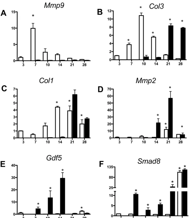
The expression of genes associated with the inflammatory, proliferative and remodeling phases of healing in FDL injured tendons of WT and Mmp92/2mice were analyzed by real-time RT-PCR and normalized to their expression level in day three repairs in WT mice (Figure 1). Peak expression of Cox-2, an important mediator of inflammation, occurred at three days post-repair in WT and Mmp92/2 tendons, however, expression was not significantly different between groups at this time (p.0.05).
Cox-2was not differentially expressed between WT and Mmp92/2 at any time during healing, with slight, but in-significant decreases after day three (Figure S1).Mmp9was not expressed in reparative tissues in Mmp92/2 mice, but its expression in WT mice was similar to that observed in prior studies [7], with peak expression at seven days and a return to control levels by 14 days (Figure 1A). Type III Collagen (Col3), which is associated with granulation tissue, was increased at seven days post-repair in WT mice (3.2-fold), and had peak expression at 10 days post-repair (10.8-fold increase).Col3expression remained significantly elevated through 14 days (5.5-fold increase), after which time expression returned to baseline levels in WT mice (Figure 1B). In WT mice, peak expression of Col1was observed on day 14 post-repair (4.4-fold increase) and persisted through day 28 (2-fold increase; Figure 1C). In contrast, both Col3 and Col1 expressions were delayed in Mmp92/2mice.Col3expression was not significantly increased in Mmp92/2 mice until day 21 (8.3-fold increase), but remained significantly elevated through day 28 (7.7-fold increase). The pattern ofCol1expression in Mmp92/2mice was similar to that observed withCol3. Peak expression ofCol1occurred on day 21 (6.2-fold increase), and remained elevated through day 28 (2.7-fold increase; Figure 1B&C).
Mmp2 (Gelatinase A) expression is associated with the tissue remodeling phase of tendon healing when adhesions decrease [7].
Mmp2 was expressed earlier and at increased levels in tendon repairs in Mmp92/2 mice compared to WT mice (Figure 1D).
Mmp2expression was increased beginning at day 14 post-repair (21-fold increase; Figure 1D) in Mmp92/2 mice, and peak expression occurred on day 21 (56-fold increase). At day 28Mmp2
expression remained elevated (4.7-fold increase). In contrast, elevation ofMmp2expression was not observed until day 21 in WT mice (12-fold increase), although this also represented the point of maximal expression in these mice. At day 28, expression ofMmp2
decreased relative to day 21, but remained elevated (5.1-fold increase) compared to day three WT controls.
The earlier and more robust expression ofMmp2in the tendon repairs of Mmp92/2 mice suggests more vigorous and rapid tendon regeneration and remodeling. To further examine this possibility, the expression of genes associated with neo-tendon formation,Gdf5andSmad8were examined in tendon repair tissues in Mmp92/2and WT mice [41,42,43]. In healing tendons in WT mice,Gdf5andSmad8expression became elevated only late in the repair process (day 28) and following the peak expression ofMmp2.
Gdf5was increased 2.5-fold andSmad8was increased 106-fold at 28 days following injury in WT mice. In contrast, the expression of these neo-tendon related genes was markedly accelerated in Mmp92/2mice.Gdf5was elevated by seven days post-repair
Figure 1. Early expression of neo-tendon associated genes during flexor tendon healing in Mmp92/2mice.Gene expression of (A) Mmp9(B)Col3a1, (C)Col1a1, (D)Mmp2, (E)Gdf5, and (F)Smad8in FDL tendon repair tissue over time up to 28 days post-op. Total RNA was extracted and pooled from five tendon repairs per time-point and processed for real-time RT-PCR. Gene expression was standardized with the internalb-actin control and then normalized by the level of expression in day three WT FDL tendon repairs. Data presented as the mean fold induction (over WT day three repairs)6SEM. * p,0.05 vs. WT day three tendon repair. White bars represent WT mice. Black bars represent Mmp92/2mice.
Mmp92/2Tendons have a Reduced Zone of Injury
Histology was used to assess changes in the structure and morphology of healing FDL tendons. There were no observable differences in the structure of sham control tendons between WT and Mmp92/2mice (Figure 2 A&B).
In WT mice at seven days post repair, a reactive, cellular granulation tissue developed adjacent to the tendon repair site and at the repair site (Figure 2C). At day 10 post-repair, granulation tissue was more cellular and the native collagen tissue adjacent to the repair site underwent catabolism and invasion by the reactive granulation tissue (Figure 2E). At 14 days the ends of the tendon were bridged at the repair site by granulation tissue and an external callus of thickened granulation tissue encased the repair site and adjacent tendon tissue (Figure 2G). At day 21, the granulation tissue reached maximal thickness. The tissue was composed of disorganized collagen fascicles, which replaced the tendon ends adjacent to the repair (Figure 2I). At day 28 remodeling of the tendon occurred with thinning of the granulation tissues that encased the tendon. At this time point extensive remodeling and replacement of the original tendon tissue was observed, with more organized tissue that was oriented parallel to the long axis of the tendon. The area of tendon replacement by the healing scar tissues comprised the entire zone of injury and is show with the green box (Figure 2K).
The tendon repair process in Mmp92/2mice had similarities and some unique features compared to WT repairs. While WT and Mmp92/2tendons healed with increased granulation tissue at
the repair site by day seven (Figure 2C&D), the width of the granulation tissue was reduced at subsequent time points in Mmp92/2repairs (Days 10, 14, 21, and 28; Figures 2F, 2H, 2J, and 2L). The difference in the width of the reparative tissue is apparent in the photomicrographs following repair in the WT and Mmp92/2mice. The black bar (Figure 2I and 2J) shows the width of the granulation tissue in WT mice compared to the width of the granulation tissue in the Mmp92/2mice (yellow bar; Figure 2J). Another distinguishing feature is that the degree of catabolism of native tendon tissue adjacent to the repair site is reduced. This results in a reduction of the extent of the zone of injury. This is readily apparent on photomicrographs of WT and Mmp92/2 repair tissues at day 28. The green boxes show the extent of the zone of injury and area of native tendon replacement and remodeling in WT mice (Figure 2K) and Mmp92/2 mice (Figure 2L). Altogether, the molecular and histological data show that Mmp92/2mice undergo tendon healing with less catabolism and extension of the zone of injury and have a concomitant acceleration of the reduction and remodeling of granulation tissues.
Adhesions are Remodeled Earlier in Mmp92/2 Flexor Tendons
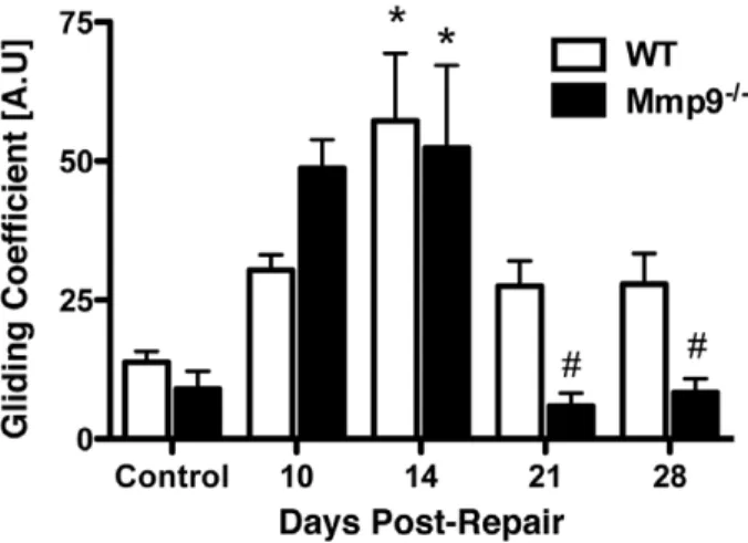
The gliding coefficient is derived from a biomechanical measurement of metatarsal-phalangeal (MTP) joint range of motion following a series of applied loads to the tendon [7,39]. An elevated gliding coefficient is associated with a higher force required for a given degree of flexion, and therefore is an index of scar and adhesion formation [7,39]. The baseline WT gliding
coefficient was not statistically different than the controls from Mmp92/2 mice. The gliding coefficient was increased at day 10 and day 14 in the repairs from both WT and Mmp92/2 mice. However, there was no significant difference in the gliding coefficients at these time points, and in both WT and Mmp92/
2 mice maximal adhesions occurred at 14 days (WT, 4.1-fold;
Mmp92/2 mice, 5.8-fold; Figure 3). A significant difference was observed beginning at 21 days following repair. In Mmp92/2 mice the gliding coefficient had returned to baseline levels, consistent with tendon remodeling. In contrast, the gliding coefficient remained elevated in the repairs of the WT mice at 21 days (2-fold). Similar differences were observed at 28 days. Thus the biomechanical measure of the tissue adhesions further confirms that Mmp92/2 mice undergo tendon repair with reduced scar formation.
Strength and Stiffness are Maintained in the Repairs of Mmp92/2Mice
Biomechanical measurement of the maximum tensile load at failure of repaired and control tendons were determined in both the WT and the Mmp92/2mice (Table 1). Control tendons in the WT and Mmp92/2mice had a similar load at failure. Following repair, the maximum load at failure was markedly decreased after 14 days of healing in both WT mice (8.8% of control) and Mmp92/2mice (16.4% of control) (Table 1). The maximal load at failure progressively increased as the repair progressed. However, by 28 days, repaired tendons in WT mice had reached only 25% of the maximum load at failure of un-operated WT tendon, while repaired tendons in Mmp92/2mice had regained only 31% of the maximum load at failure of un-operated Mmp92/2tendon.
The stiffness (the slope of the linear region of the force displacement curve) of the control and repaired tendons in WT
Figure 2. Decreased area of remodeling during tendon healing in Mmp92/2mice.Representative histological sections of sham control WT
(A) and Mmp92/2(B) FDL Tendons. Repaired WT and Mmp92/2FDL Tendons at days 7 (C, D), 10 (E, F), 14 (G, H) 21 (I, K) and 28 (K, L). Sections were
stained with Alcian Blue/Hematoxylin and Orange G. Of note is the fibroblastic granulation tissue that fills in the repair site between tendon ends (marked as T) and is progressively remodeled (green box) with collagen becoming oriented in a parallel fashion with the long axis of the tendon. Black bar represents width of WT tendon; yellow bar represents width of Mmp92/2tendon at Day 21. Scale bars represent 200 microns. 5X
magnification.
doi:10.1371/journal.pone.0040602.g002
Figure 3. Earlier resolution of fibrous adhesions in Mmp92/2 tendon repairs.Gliding coefficient of WT and Mmp92/2mouse FDL
tendon repairs over time up to 28 days post-repair. Repair data is compared to sham control tendons (mean 6 SEM). (*) Indicates significant difference versus respective control, while (#) indicates significant difference versus Day 14 Mmp92/2repair.
mice and Mmp92/2mice were also measured (Table 2). Similar stiffness was observed in the control (un-operated) tendons from WT mice and Mmp92/2mice (Table 2). As with measurements of strength, the stiffness of the repaired tendons also increased with the time of healing. The stiffness of the tendon repairs of the WT and Mmp92/2mice were similar at each time point. At 14 days, Mmp92/2 repairs were 36% stiffer than WT, although this difference was not significant (p = 0.13). At 28 days repaired tendons in the WT mice and Mmp92/2mice regained 44.9% and 45.9% of their stiffness, respectively (Table 2).
Bone Marrow Derived Cells Migrate to the Flexor Tendon Repair Site
To determine the ability of bone marrow-derived cells to migrate to the flexor tendon repair site, bone marrow mononu-clear cells from C57B6/J GFP+transgenic mice were transplanted
into WT C57B6/J mice following conditioning whole body irradiation. After allowing the transplanted cells to engraft, the mice underwent flexor tendon transection and repair.
The presence of bone marrow-derived cells at the repair site was determined by examining histological sections of healing tendon with fluorescent imaging for the expression of GFP. GFP+cells
were not observed in un-injured contralateral sham control tendons (Figure 4A). However, GFP+cells were observed in the
healing tendon tissues where they localized to the reparative granulation tissues. GFP+cells were present three days after repair
(Figure 4B) and increased numbers were observed by seven days (Figure 4C). GFP+bone marrow-derived cells remained abundant
at 14 days following repair and they were localized to the healing granulation tissue (Figure 4D). By day 21, GFP+bone marrow
derived cells were diffusely located throughout the granulation tissue and in areas of remodeled tendon tissue (Figure 4E). At 28
days post-repair the remaining GFP+cells were present in the area of tendon remodeling (Figure 4F).
Mmp9 is Expressed by Bone Marrow-derived Cells during Flexor Tendon Healing
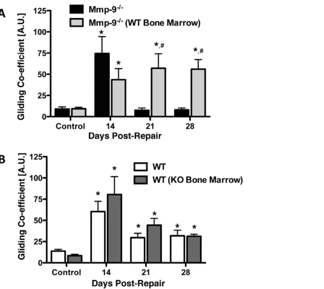
In order to determine if bone marrow-derived cells migrating to the injury site provide local Mmp9expression in tendon repair, reciprocal bone marrow transplants were conducted in which WT mice received Mmp92/2 bone marrow transplantation, and Mmp92/2mice received WT bone marrow transplantation.
The expression ofMmp9was examined by real time RT-PCR in tissue extracted from the healing tendons of WT mice; WT mice following transplantation with Mmp92/2 bone marrow cells; Mmp92/2 mice; and Mmp92/2 mice following transplantation with WT bone marrow cells (Figure 5A). Since we demonstrated earlier (Figure1A) that during tendon repair Mmp9 expression peaks at day seven post injury, tissues from the repaired tendons were harvested at three and seven days post-repair and analyzed by real-time RT-PCR for expression of Mmp9. WT mice had
Mmp9expression measured at three days following repair and this level of expression was used as a baseline level of normalized expression (1.060.54).Mmp9expression increased 4.1-fold at day seven in the WT mice. In contrast, WT mice with transplantation of Mmp92/2bone marrow cells did not have measurable levels of expression ofMmp9at day three and had minimal expression at day seven following repair (0.05960.03; p,0.05, compared to the expression level in day three post-repair WT tendons). As expected, repairs from Mmp92/2 mice lacked Mmp9 gene expression. However, in Mmp92/2mice transplanted with bone marrow cells from WT mice, no detectableMmp9expression was observed three days following repair, whereas a 2.3-fold increase (p,0.05; compared to WT day three expression) was observed at seven days after tendon repair (Figure 5). Collectively, these data show that Mmp9 expression during tendon repair is dependent upon migration of bone marrow-derived cells to the injury site.
To determine the influence of host cells on bone marrow transplantation, the level of Mmp9 expression in bone marrow from WT mice receiving Mmp92/2bone marrow, and Mmp92/
2 mice receiving WT bone marrow were analyzed. Mmp92/2
mice with WT bone marrow had a significant 18.5-fold induction (p = 0.001) ofMmp9expression relative to WT mice with Mmp92/
2bone marrow at seven days post-repair (Figure S2). There was a
2.84-fold increase inMmp9expression in bone marrow from WT mice with Mmp92/2bone marrow, relative to Mmp9expression in tendon tissue in these mice, suggesting the presence of some host WT cells in the bone marrow of WT mice with Mmp92/2bone marrow.
Mmp9Deficient Bone Marrow Cells are Associated with Decreased Tendon Adhesions
Quantification of adhesions and biomechanical testing was done to determine the functional consequences of both bone marrow cell-specific deletion ofMmp9, and expression of Mmp9solely in bone marrow cells during flexor tendon healing.
To further define the role of bone marrow cell derivedMmp9in adhesion formation, MTP joint flexion tests were performed in Mmp92/2mice and in Mmp92/2mice following transplantation with WT bone marrow cells (Figure 6A). As previously shown, Mmp92/2 mice had a reduced gliding coefficient during tendon healing, compared to WT controls. At 14 days following repair, the gliding coefficient was increased 8.3-fold, but was reduced to basal levels at day 21 and 28. In contrast, the transplantation of WT bone marrow cells into Mmp92/2 mice resulted in an
Table 1.Maximum load at failure [N] of WT and Mmp92/2 mouse FDL tendon repairs over time up to 28 days.
Days Post-Repair WT Mmp92/2
Control 10.160.39 9.261.01
10 0.8960.31* 1.5160.27*
14 0.8960.24* 2.2960.22*
21 2.1560.30* 2.3460.21*
28 2.5260.34* 2.8860.31*
Repair data is compared to sham control tendons (mean6SEM), (*) indicates p,0.05 compared to respective control.
doi:10.1371/journal.pone.0040602.t001
Table 2.Stiffness [N/mm] of WT and Mmp92/2mouse FDL tendon repairs over time up to 28 days.
Days Post-Repair WT Mmp92/2
Control 5.6560.33 4.8860.17
10 1.6360.15* 1.1960.02*
14 1.4660.17* 2.0060.21*
21 1.8960.26* 2.2360.18*
28 2.5460.26* 2.5960.19*
Repair data is compared to sham control tendons (mean6SEM), (*) indicates p,0.05 compared to respective control.
increased in gliding coefficient that mimicked the pattern of adhesions observed in WT mice undergoing tendon repair. Mmp92/2 mice with WT bone marrow transplantation had a maximal gliding coefficient at 21 days (9-fold), but had sustained elevation of the gliding coefficient at 28 days (6-fold). The gliding coefficient was significantly elevated in the tendon repairs of the Mmp92/2mice with WT bone marrow transplantation compared
to the tendon repairs of the Mmp92/2mice at 21 and 28 days. Therefore, rescue of Mmp9 expression in bone marrow-derived cells is sufficient to reestablish tendon scarring after injury in
Mmp9-deficient mice.
Adhesions were also measured in WT mice and in WT mice following transplantation with Mmp92/2 bone marrow cells (Figure 6B). As previously shown, WT mice had a maximal increase in the gliding coefficient at 14 days after repair (approximately 4-fold) followed by a decrease at 21 (2.1-fold) and 28 (2.3-fold) days following repair. WT mice with transplanted Mmp92/2 bone marrow cells similarly had maximal gliding coefficients at 14 days followed by a decrease at 21 and 28 days after repair. The gliding coefficients in the WT mice and in WT mice with Mmp92/2 bone marrow cell transplants were not significantly different, suggesting that even very low levels ofMmp9
produced locally in the healing tissues might be produced by cells other than migrating marrow cells. This could include the low levels ofMmp9produced by tenocytes, or alternatively, some other cell types. Another explanation is that other factors or non-marrow cell populations are also involved in the adhesion formation that occurs in this model. Finally, based upon low, but detectable expression of Mmp9 in the bone marrow of WT mice with Mmp92/2 bone marrow, there appear to be a small number residual WT bone marrow cells after conditioning radiation, which could provide sufficientMmp9to result in scarring.
As a control for any possible effects of whole body irradiation and bone marrow transplantation, tendon repairs were performed and the gliding coefficient measured in transplant control mice (WT bone marrow cells transplanted in WT mice; and Mmp92/2 bone marrow cells transplanted into Mmp92/2mice). There was no significant difference between the gliding coefficient in
Figure 4. Bone marrow cells migrate specifically to the FDL repair site in vivo.Representative sections of repaired flexor tendons from C57Bl6/J mice that were myeloablated, and reconstituted with bone marrow from GFP transgenic mice. Tendons were repaired after bone marrow cells had engrafted, and tissues were harvested between three and 28 days post-repair. Sections were counterstained with the nuclear dye DAPI (blue) and bone marrow derived cells were identified based on the expression of GFP. Contralateral sham controls (A) did not have any GFP expressing cells indicating a lack of bone marrow cells in un-injured tendon, while bone marrow derived cells are present at the FDL repair site at (B) three, (C) seven, (D) 14, (E) 21, and (F) 28 days post-repair. Tendon tissue is outlined in orange and marked as ‘T’. All images are 10x magnification. Scale bars represent 200 microns.
doi:10.1371/journal.pone.0040602.g004
Figure 5.Mmp9is not expressed in healing tendons from WT mice with Mmp92/2 bone marrow. Real-time RT-PCR for Mmp9 expression in WT, Mmp92/2 (KO), WT mice transplanted with bone
marrow from an Mmp92/2donor (WT [KO Bone Marrow]), Mmp92/2
mice transplanted with bone marrow from a WT donor (KO [WT Bone Marrow]) in repaired flexor tendons at three and seven days post-repair. Expression is normalized tob-actin, and compared day three expression in WT mice. Data are presented as mean 6 SEM. (*) Indicates a significant difference compared to WT day three expression.
transplant controls compared to WT and Mmp92/2 mice respectively at any time post-repair (data not shown).
Bone Marrow-specific Loss of Mmp9 does Not Decrease the Maximum Load at Failure
Following adhesion testing, the tensile properties of repaired tendons were measured. There was no difference in the basal maximum load at failure of tendons from mice that received bone marrow transplants or un-transplanted WT mice and Mmp92/2 mice (Table 3). After repair, the maximum load at failure was significantly lower than controls in all groups, but the maximum load at failure of repairs from Mmp92/2 mice with WT bone marrow cells was not significantly different than those from WT mice with Mmp92/2 bone marrow at any time. On day 28, tendons from WT mice with Mmp92/2 bone marrow regained 22% of the maximum load at failure of control tendons, while Mmp92/2mice with WT bone marrow transplantation regained 19% of load to failure in control tendons (Table 3).
The stiffness of repaired tendons from these groups of mice was also analyzed. There was no significant difference between control specimens for any of the groups. At 14 days post-repair, the
stiffness of all groups was significantly decreased compared to all controls, and stiffness of the repaired tendons remained signifi-cantly decreased compared to controls in all groups through 28
Figure 6. WT bone marrow in Mmp92/2mice results in prolonged presence of adhesions during tendon healing.Gliding coefficient
based on range of applied load of FDL tendon repairs from [A] Mmp92/2mice, and Mmp92/2mice with WT bone marrow; [B] WT, and WT with
Mmp92/2bone marrow between 14 and 28 days post-repair. (*) Indicates p
,0.05 compared to respective control,#indicates p,0.05 compared to Mmp92/2repair at the same time-point. Data are presented as mean
6SEM. doi:10.1371/journal.pone.0040602.g006
Table 3.Maximum load at failure of flexor tendon repairs following reciprocal bone marrow transplants between WT and Mmp92/2mice to result in WT mice with Mmp92/2bone marrow, and Mmp92/2mice with WT bone marrow.
Days Post-Repair
WT Mice/
Mmp92/2bone marrow
Mmp92/2Mice/ WT bone marrow
Control 9.7361.64 9.9362.04 14 2.0660.17* 1.4760.19*
21 2.0160.30* 1.9060.22*
28 2.1560.22* 2.5160.42*
days post-repair. At day 28 post-repair, the stiffness of tendons from Mmp92/2mice with WT bone marrow transplantation was significantly decreased (p,0.05) compared to WT mice with Mmp92/2bone marrow transplantation (Table 4).
Discussion
We show that complete loss of Mmp9 during flexor tendon healing results in earlier remodeling of adhesions compared to WT mice, without loss of repaired tendon strength. Accelerated, and increased expression of Mmp2, Gdf5 and Smad8 suggest the remodeling phase of flexor tendon healing occurs earlier in Mmp92/2 mice, resulting in the subsequent decrease in the gliding coefficient. Additionally, the contribution of bone marrow-derived cells to the flexor tendon healing process was demonstrat-ed by presence of GFP-expressing cells in WT mice following bone marrow transplant of GFP+ cells. The functional role ofMmp9
expression in bone marrow-derived cells was demonstrated by the increased adhesions that occurred in myeloablated Mmp92/2 mice transplanted with WT bone marrow cells. The gliding coefficient was similar to that observed in tendon repairs of WT mice. Notably, the strength and stiffness of repairs in Mmp92/2 mice were similar to those observed in WT mice. Thus, lack of
Mmp9reduces tendon adhesions but does not result in a weaker repair tissue.
Based on the causal association between inflammation and adhesion formation [44,45], previous work has altered the healing course with anti-inflammatory treatment [46,47]. These studies have demonstrated that an attenuated inflammatory response can decrease adhesions, however, decreased adhesions are often accompanied by a decrease in the strength of the repair during the early phases of healing, rendering these tendons unsuitable for aggressive physical therapy, and prone to re-rupture [46,47]. Virchenko et al have shown that an early decrease in force at failure resulted from Cox-2 inhibition [47], suggesting that altering the normal cascade of inflammatory events leads to a decrease in the force at failure. Furthermore, a greater proportion of untreated specimens had a higher histology score, corresponding a greater amount of mature tissue, suggesting a larger zone of injury in mice treated with a Cox-2 inhibitor. In contrast, our work shows that deletion ofMmp9results in reduced catabolism of native tendon and a development of a smaller zone of injury compared to WT repairs. However, there is no reduction in the maximum load at failure compared to tendon repairs in WT mice. This is an important distinction because a decrease in the maximum load at failure increases the likelihood of rupture. Our data indicate that
targeted inhibition of earlyMmp9expression may improve flexor tendon healing.
The robust remodeling of adhesions and the decreased gliding coefficient by day 21 in Mmp92/2mice suggests an accelerated remodeling phase. Prior work in our laboratory has shown that tendon remodeling is associated with elevated expression ofMmp2
[7]. Elevated expression ofMmp2occurs earlier in Mmp92/2mice (14 days) compared to WT mice (21 days). Mmp2expression is increased immediately before a significant decrease in the gliding coefficient, suggesting an important role for Mmp2 in scar remodeling. The reduced area of initial catabolism of tendon adjacent to the repair site in tendons from Mmp92/2 mice suggests that while degradation of some native tissue is beneficial, too much catabolism results in a more abundant response with a prolonged period of increased adhesions. While lack of Mmp9
results in a change in the kinetics of tendon remodeling, the overall mechanical strength of the tendon repair, a key component of tissue regeneration, is not reduced in the Mmp92/2 mice. The return of tendon strength requires a balance between degradation, collagen formation, and tissue remodeling. Alteration in the expression of Mmp9 changes the anabolic phase of healing, suggesting thatMmp9expression is a key regulator of subsequent tissue responses.
We show that bone marrow cells migrate to the flexor tendon repair site consistent with a prior report showing migration to the patella tendon repair site [29]. By day seven post-repair there is a large influx of cells that declines, but persists through day 28. The important role of inflammatory mediators or chemotactic signaling in this ‘‘homing’’ process is demonstrated by the complete absence of GFP+ cells in contralateral control tendons, indicating that BMSCs migrate specifically to the site of injury. Our findings differ somewhat from other models of bone marrow cell migration to the tendon repair site. In a murine partial patella tendon defect model there is a massive influx of GFP+cells to the repair site within 24 hours [29], while we found very few bone marrow cells at three days post-repair. The rate of healing and the disruption in vascularization in a partial defect model may be different than that of a complete transection. The presence of intact tendon at the repair site may increase the early intrinsic phase of healing, or increase the release of chemotactic signals from the tendon tissue that recruits bone marrow cells to the repair site. Additionally, the synovial sheath surrounding the flexor tendon may be an additional barrier that bone marrow cells must cross before arriving at the intrasynovial repair site.
Expression ofMmp9specifically in cells derived from the bone marrow was achieved by bone marrow transplantation, while the functional consequences of bone marrow-specificMmp9deletion were also shown. Loss ofMmp9in the bone marrow during flexor tendon healing resulted in earlier remodeling of adhesions, compared to those mice that express normal levels ofMmp9 in the bone marrow, as well as in mice with bone marrow cell-specific
Mmp9 expression. Resolution of fibrous adhesions after flexor tendon injury did not occur to the same degree in mice with bone marrow cell-specific loss of Mmp9 compared to healing in Mmp92/2 mice. Based on the low-level expression of Mmp9
transcripts in tendons from WT mice with Mmp92/2 bone marrow, it appears that marrow cells are a major source ofMmp9
during flexor tendon healing. It appears that some WT host cells persisted in the bone marrow and provided a slight contribution of
Mmp9during flexor tendon healing. This would explain why there was not complete remodeling of adhesions to the extent of complete loss ofMmp9, and also why there was low, but detectable expression ofMmp9 in tendons from WT mice with Mmp92/2 bone marrow by real-time PCR.
Table 4.Stiffness of flexor tendon repairs following reciprocal bone marrow transplants between WT and Mmp92/2mice to result in WT mice with Mmp92/2bone marrow, and Mmp92/ 2mice with WT bone marrow.
Days Post-Repair
WT Mice/
Mmp92/2bone marrow
Mmp92/2Mice/ WT bone marrow
Control 5.3860.46 5.1660.44 14 1.3060.31* 1.7260.27*
21 1.4260.32* 1.3760.19*
28 2.1060.17*,#
1.4360.22*
Data are6SEM, (*) indicates p,0.05 compared to respective control, (#) indicates p,0.05 compared to Mmp92/2mice with WT bone marrow at the same time-point.
Bone marrow-specific loss ofMmp9leads to earlier remodeling of adhesions, without a decrease in the strength of the repair. This study presents a novel mechanism of improved flexor tendon healing, by inhibition of Mmp9, which in the setting of tendon repair seems to be primarily produced by bone marrow-derived cells. Small moleculeMmp9inhibitors are currently in clinical trials for Multiple Sclerosis, Chronic Obstructive Pulmonary Disease and Prostate Cancer [38,48], and may aid the pursuit ofMmp9
inhibition during tendon healing. This work demonstrates that it is possible to regain gliding function of the flexor tendon sooner, without compromising the strength of the repair. One limitation of this study is that we have not directly assessed the in vivo gelatinolytic activity of Mmp9 during flexor tendon healing. Identification of this activity would identify a novel target to improve flexor tendon healing.
Methods
Ethics Statement
All animal procedures were approved by the University Committee on Animal Research at the University of Rochester.
Mouse Strain Information
All mice were acquired from Jackson Laboratories (Bar Harbor, ME). Six to eight week old female Mmp92/2 (FVB.Cg-Mmp9tm1Tvu/J) [12] and WT (FVB/NJ) mice were used for tendon repair studies and bone marrow transplants. GFP transgenic (C57BL/6-Tg(UBC-GFP)30Scha/J) and C57BL/6 mice were used for initial bone marrow transplant studies.
Murine Flexor Tendon Injury and Repair
Complete transection and repair of murine flexor tendons was conducted as previously described [7] Six to eight week old female mice (Jackson Laboratories, Bar Harbor, ME) were anesthetized by intraperitoneal injection of 4 mg/kg xylazine and 60 mg/kg ketamine. The distal flexor digitorum longus (FDL) tendon was exposed and two horizontal 8-0 nylon sutures in a modified Kessler pattern were placed in the intact tendon. The tendon was then transected between the sutures and repaired by approximat-ing the transected ends usapproximat-ing the suture. The tendon was also transected proximally along the tibia at the myotendinous junction to protect the repair. The skin was closed with running 4-0 nylon sutures. The mice were returned to their cages and allowed free active motion and weight bearing following recovery from anesthesia. Sham surgeries were performed on control groups. Identical anesthesia and exposure were performed. When the distal FDL tendon was isolated, two horizontal 8-0 sutures in a modified Kessler pattern were sutured through the tendon. This suture was then removed without transecting the tendon. However the proximal FDL tendon was released at the myotendinous junction as in the repair group. The skin was closed with running 4-0 nylon sutures.
RNA Extraction and Real-Time RT-PCR
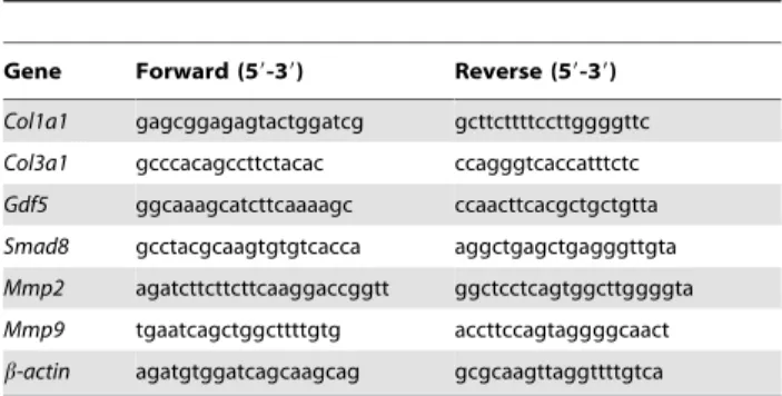
Tendons were harvested and RNA was extracted as previously described [7]. Briefly, five repaired tendons per time-point per experimental group were harvested and RNA was extracted using TRIzol (Invitrogen). cDNA was reverse transcribed using 1ug of RNA and the iScript cDNA synthesis kit (BioRad). Real-Time PCR was done using Absolute qPCR SYBR (ABgene) and gene specific primers (Table 5). Gene expression was normalized tob -actin, and expression on day three in WT tendons due the relative metabolic inactivity of un-injured tendons.
Histological Analysis of Healing
Whole hind limbs containing repaired tendons were harvested between seven and 28 days post-repair as previously described [49]. The intact foot and tibia were harvested by disarticulating the hind limb at the knee. Briefly, the hind limbs were fixed in 10% formalin with the tibia at 90urelative to the foot, and then decalcified in 10% EDTA at 4uC for 28 days. The decalcified tissues were dehydrated and embedded in paraffin to preserve the anatomical relationship between the repaired tendon and surrounding tissues. Serial three-micron sagittal sections through the FDL tendon plane were then cut and stained with Alcian Blue/Hematoxylin and Orange G. Four specimens per group per time-point were used, with approximately five sections per specimen analyzed.
Adhesion and Biomechanical Testing
Adhesion and biomechanical testing was completed as previ-ously described [7,49]. The hind limb was disarticulated at the knee, and the FDL tendon was released from the surrounding tissue proximal to the tarsal tunnel. The FDL tendon was secured between two pieces of tape using superglue. The limb was fixed in a custom apparatus, and a digital image was taken of the MTP joint in a neutral unloaded position with the digits extended. Loads were incrementally applied to the flexor tendon and the MTP joint range of motion relative to the unloaded position was measured from a digital image. MTP joint flexion angles were measured using ImageJ software (http://rsb.info.nih.gov/ij/), and plotted versus the applied loads. The gliding coefficient was determined by non-linear regression as a measure of resistance to MTP flexion to due impaired gliding.
Immediately following adhesion testing, the proximal aspect of the FDL tendon was released from the tarsal tunnel, the calcaneus and tibia were removed and the proximal end of the FDL was gripped in the Instron device as previously described [7,49]. The distal bones of the foot were also secured in the Instron device (Instron 8841 DynaMightTM axial servohydraulic testing system, Instron Corporation, Norwood, MA). The tendon was tested in tension in displacement control at a rate of 30 mm/minute until failure. Force-displacement data were automatically logged and plotted and the maximum tensile force and stiffness were determined. N = 7–10 animals/group/time-point.
Statistical Analysis
Biomechanical and Real-Time RT-PCR data were analyzed using a two-way analysis of variance (ANOVA) followed by Bonferroni’s multiple comparisons with a significance level of
a= 0.05. p values are reported for significance compared to gene
expression in WT day three samples; and biomechanical
Table 5.Primer sequences for Real-time RT-PCR.
Gene Forward (59-39) Reverse (59-39)
Col1a1 gagcggagagtactggatcg gcttcttttccttggggttc
Col3a1 gcccacagccttctacac ccagggtcaccatttctc
Gdf5 ggcaaagcatcttcaaaagc ccaacttcacgctgctgtta
Smad8 gcctacgcaagtgtgtcacca aggctgagctgagggttgta
Mmp2 agatcttcttcttcaaggaccggtt ggctcctcagtggcttggggta
Mmp9 tgaatcagctggcttttgtg accttccagtaggggcaact
b-actin agatgtggatcagcaagcag gcgcaagttaggttttgtca
significance is reported compared to control tendons, except where otherwise stated.
Bone Marrow Transplantation
C57BL/6 mice (Jackson Labs) were myeloablated and bone marrow transplantation was performed as previously described [50]. Briefly, an initial 5Gy whole body dose was given 24 hours prior to transplantation, and a second 5Gy dose was given two hours prior to transplantation. Whole bone marrow was asepti-cally isolated from mice expressing GFP in all tissues (C57BL/6-Tg (UBC-GFP) 30Scha/J, Jackson lab) and re-suspended in sterile PBS containing 2% FBS. Each mouse was given 56105GFP+cells via tail vein injection. Animals were preemptively given Sulfatrim antibiotic (Activas, Baltimore, MD) in the drinking water beginning seven days prior to irradiation, and were maintained on antibiotics for two weeks post-transplantation. Mice were allowed to recover and cells engrafted for two weeks post-transplant. At this time, standard flexor tendon repair was performed, and tendons healed for between three and 28 days. Tendons were harvested from the tarsal tunnel to the bifurcation at the digits, and placed in 4% Paraformaldehyde for one hour. Tissues were washed twice in PBS for 15 minutes, and placed in 15% sucrose for 1 hour until tissue was saturated. Tissues were then placed in 30% sucrose for 16 hours, and embedded in OCT media. Eight-10uM sections were cut using a cryostat. Sections were post-fixed with 0.2% Glutaraldehyde for 10 minutes, washed twice with PBS, counterstained and cover slipped using Vecta-shield Mounting Medium with DAPI (Vector Laboratories, Burlingame, CA) GFP was localized using fluorescent microscopy. N = 3 bone marrow transplant recipients/time-point.
WT and Mmp92/2 bone marrow transplants were conducted in the same manner as described above, with tissues harvested for
Real-time RT-PCR (N = 5/group/time-point), histology (N = 4/ group/time-point; 5 sections/specimen), in situ hybridization, as well as biomechanical and adhesion testing (N = 7–10 group/time-point).
Supporting Information
Figure S1 Cox-2 mRNA expression in WT (white bars) and Mmp92/2(black bars) flexor tendons from three to 28 days post-repair. Data were normalized to b-actin, and WT day three expression. Data are presented as mean6SEM.
(TIF)
Figure S2 Mmp9 mRNA expression in bone marrow from WT mice with Mmp92/2bone marrow (white bar), and Mmp92/2
mice with WT bone marrow (black bar) at seven days post-repair. Data were normalized tob-actin, and expression in WT mice with Mmp92/2 bone marrow at seven days. (*) Indicates p,0.05, data are presented as mean6SEM.
(TIF)
Acknowledgments
We would like to thank Ryan Tierney for technical assistance with histology, Dr. Tim Bushnell for assistance with flow cytometric analysis, and Dr. Matthew Hilton for helpful advice on this manuscript.
Author Contributions
Conceived and designed the experiments: AEL LMC EMS HAA RJO. Performed the experiments: AEL BJF MW JAJ. Analyzed the data: AEL HAA RJO. Contributed reagents/materials/analysis tools: LMC HAA. Wrote the paper: AEL HAA RJO.
References
1. Beredjiklian PK (2003) Biologic aspects of flexor tendon laceration and repair. J Bone Joint Surg Am 85-A: 539–550.
2. Lin T (2004) Biomechanics of tendon inury and repair. Journal of Biomechanics 37: 865–877.
3. Gelberman RH, Botte MJ, Spiegelman JJ, Akeson WH (1986) The excursion and deformation of repaired flexor tendons treated with protected early motion. J Hand Surg [Am] 11: 106–110.
4. Gelberman RH, Woo SL, Lothringer K, Akeson WH, Amiel D (1982) Effects of early intermittent passive mobilization on healing canine flexor tendons. J Hand Surg [Am] 7: 170–175.
5. Sharma P, Maffulli N (2005) Basic biology of tendon injury and healing. Surgeon 3: 309–316.
6. Strickland JW (2000) Development of flexor tendon surgery: twenty-five years of progress. J Hand Surg [Am] 25: 214–235.
7. Loiselle AE, Bragdon GA, Jacobson JA, Hasslund S, Cortes ZE, et al. (2009) Remodeling of murine intrasynovial tendon adhesions following injury: MMP and neotendon gene expression. J Orthop Res 27: 833–840.
8. Bi Y, Ehirchiou D, Kilts TM, Inkson CA, Embree MC, et al. (2007) Identification of tendon stem/progenitor cells and the role of the extracellular matrix in their niche. Nat Med 13: 1219–1227.
9. Jozsa L, Kannus P (1997) Human Tendons: Anatomy, Physiology, and Pathology. Champaign, IL.
10. Kannus P (2000) Structure of the tendon connective tissue. Scand J Med Sci Sports 10: 312–320.
11. Woo SL, Debski RE, Zeminski J, Abramowitch SD, Saw SS, et al. (2000) Injury and repair of ligaments and tendons. Annu Rev Biomed Eng 2: 83–118. 12. Coussens LM, Tinkle CL, Hanahan D, Werb Z (2000) MMP-9 supplied by bone
marrow-derived cells contributes to skin carcinogenesis. Cell 103: 481–490. 13. Heissig B, Nishida C, Tashiro Y, Sato Y, Ishihara M, et al. (2010) Role of
neutrophil-derived matrix metalloproteinase-9 in tissue regeneration. Histology and histopathology 25: 765–770.
14. Karousou E, Ronga M, Vigetti D, Passi A, Maffulli N (2008) Collagens, proteoglycans, MMP-2, MMP-9 and TIMPs in human achilles tendon rupture. Clinical orthopaedics and related research 466: 1577–1582.
15. Parkinson J, Samiric T, Ilic M, Cook J, Feller J, et al. (2010) Change in proteoglycan metabolism is a characteristic of human patellar tendinopathy. Arthritis Rheum 62: 3028–3035.
16. Voloshin I, Gelinas J, Maloney MD, O’Keefe RJ, Bigliani LU, et al. (2005) Proinflammatory cytokines and metalloproteases are expressed in the subacro-mial bursa in patients with rotator cuff disease. Arthroscopy 21: 1076. 17. Hsu JY, Bourguignon LY, Adams CM, Peyrollier K, Zhang H, et al. (2008)
Matrix metalloproteinase-9 facilitates glial scar formation in the injured spinal cord. J Neurosci 28: 13467–13477.
18. Corbel M, Belleguic C, Boichot E, Lagente V (2002) Involvement of gelatinases (MMP-2 and MMP-9) in the development of airway inflammation and pulmonary fibrosis. Cell Biol Toxicol 18: 51–61.
19. Busch SA, Horn KP, Silver DJ, Silver J (2009) Overcoming macrophage-mediated axonal dieback following CNS injury. The Journal of neuroscience : the official journal of the Society for Neuroscience 29: 9967–9976.
20. Lim DH, Cho JY, Miller M, McElwain K, McElwain S, et al. (2006) Reduced peribronchial fibrosis in allergen-challenged MMP-9-deficient mice. American journal of physiology Lung cellular and molecular physiology 291: L265–271. 21. Mukherjee R, Colbath GP, Justus CD, Bruce JA, Allen CM, et al. (2010)
Spatiotemporal induction of matrix metalloproteinase-9 transcription after discrete myocardial injury. The FASEB journal : official publication of the Federation of American Societies for Experimental Biology 24: 3819–3828. 22. Rosenberg GA, Cunningham LA, Wallace J, Alexander S, Estrada EY, et al.
(2001) Immunohistochemistry of matrix metalloproteinases in reperfusion injury to rat brain: activation of MMP-9 linked to stromelysin-1 and microglia in cell cultures. Brain research 893: 104–112.
23. Scabilloni JF, Wang L, Antonini JM, Roberts JR, Castranova V, et al. (2005) Matrix metalloproteinase induction in fibrosis and fibrotic nodule formation due to silica inhalation. American journal of physiology Lung cellular and molecular physiology 288: L709–717.
24. Tan RJ, Fattman CL, Niehouse LM, Tobolewski JM, Hanford LE, et al. (2006) Matrix metalloproteinases promote inflammation and fibrosis in asbestos-induced lung injury in mice. American journal of respiratory cell and molecular biology 35: 289–297.
27. Gieling RG, Wallace K, Han YP (2009) Interleukin-1 participates in the progression from liver injury to fibrosis. American journal of physiology Gastrointestinal and liver physiology 296: G1324–1331.
28. Natividad A, Cooke G, Holland MJ, Burton MJ, Joof HM, et al. (2006) A coding polymorphism in matrix metalloproteinase 9 reduces risk of scarring sequelae of ocular Chlamydia trachomatis infection. BMC medical genetics 7: 40. 29. Kajikawa Y, Morihara T, Watanabe N, Sakamoto H, Matsuda K, et al. (2007)
GFP chimeric models exhibited a biphasic pattern of mesenchymal cell invasion in tendon healing. J Cell Physiol 210: 684–691.
30. Kollet O, Shivtiel S, Chen YQ, Suriawinata J, Thung SN, et al. (2003) HGF, SDF-1, and MMP-9 are involved in stress-induced human CD34+stem cell recruitment to the liver. The Journal of clinical investigation 112: 160–169. 31. Wang L, Luo J, He S (2007) Induction of MMP-9 release from human dermal
fibroblasts by thrombin: involvement of JAK/STAT3 signaling pathway in MMP-9 release. BMC cell biology 8: 14.
32. Warner RL, Bhagavathula N, Nerusu KC, Lateef H, Younkin E, et al. (2004) Matrix metalloproteinases in acute inflammation: induction of MMP-3 and MMP-9 in fibroblasts and epithelial cells following exposure to pro-inflamma-tory mediators in vitro. Experimental and molecular pathology 76: 189–195. 33. Ingraham CA, Park GC, Makarenkova HP, Crossin KL (2011) Matrix
metalloproteinase (MMP)-9 induced by Wnt signaling increases the proliferation and migration of embryonic neural stem cells at low O2 levels. The Journal of biological chemistry.
34. Nee L, Tuite N, Ryan MP, McMorrow T (2007) TNF-alpha and IL-1 beta-mediated regulation of MMP-9 and TIMP-1 in human glomerular mesangial cells. Nephron Experimental nephrology 107: e73–86.
35. Turner NA, Aley PK, Hall KT, Warburton P, Galloway S, et al. (2007) Simvastatin inhibits TNFalpha-induced invasion of human cardiac myofibro-blasts via both MMP-9-dependent and -independent mechanisms. Journal of molecular and cellular cardiology 43: 168–176.
36. Wu CY, Hsieh HL, Sun CC, Yang CM (2009) IL-1beta induces MMP-9 expression via a Ca2+-dependent CaMKII/JNK/c-JUN cascade in rat brain astrocytes. Glia 57: 1775–1789.
37. Xia L, Shen H, Lu J, Xiao W (2011) TRAF2 and cIAP2 involve in TWEAK-induced MMP-9 production in fibroblast-like synoviocytes. Rheumatology international.
38. Muroski ME, Roycik MD, Newcomer RG, Van den Steen PE, Opdenakker G, et al. (2008) Matrix metalloproteinase-9/gelatinase B is a putative therapeutic
target of chronic obstructive pulmonary disease and multiple sclerosis. Current pharmaceutical biotechnology 9: 34–46.
39. Katzel EB, Wolenski M, Loiselle AE, Basile P, Flick LM, et al. (2011) Impact of Smad3 loss of function on scarring and adhesion formation during tendon healing. Journal of orthopaedic research : official publication of the Orthopaedic Research Society 29: 684–693.
40. Aslan H, Kimelman-Bleich N, Pelled G, Gazit D (2008) Molecular targets for tendon neoformation. J Clin Invest 118: 439–444.
41. Hoffmann A, Pelled G, Turgeman G, Eberle P, Zilberman Y, et al. (2006) Neotendon formation induced by manipulation of the Smad8 signaling pathway in mesenchymal stem cells. J Clin Invest 116: 940–952.
42. Shahab-Osterloh S, Witte F, Hoffmann A, Winkel A, Laggies S, et al. (2010) Mesenchymal stem cell-dependent formation of heterotopic tendon-bone insertions (osteotendinous junctions). Stem Cells 28: 1590–1601.
43. Wolfman NM, Hattersley G, Cox K, Celeste AJ, Nelson R, et al. (1997) Ectopic induction of tendon and ligament in rats by growth and differentiation factors 5, 6, and 7, members of the TGF-beta gene family. J Clin Invest 100: 321–330. 44. Beredjiklian PK, Favata M, Cartmell JS, Flanagan CL, Crombleholme TM, et
al. (2003) Regenerative versus reparative healing in tendon: a study of biomechanical and histological properties in fetal sheep. Ann Biomed Eng 31: 1143–1152.
45. Strickland JW (1995) Flexor Tendon Injuries: I. Foundations of Treatment. J Am Acad Orthop Surg 3: 44–54.
46. Kulick MI, Smith S, Hadler K (1986) Oral ibuprofen: evaluation of its effect on peritendinous adhesions and the breaking strength of a tenorrhaphy. J Hand Surg Am 11: 110–120.
47. Virchenko O, Skoglund B, Aspenberg P (2004) Parecoxib impairs early tendon repair but improves later remodeling. Am J Sports Med 32: 1743–1747. 48. Hu J, Van den Steen PE, Sang QX, Opdenakker G (2007) Matrix
metalloproteinase inhibitors as therapy for inflammatory and vascular diseases. Nature reviews Drug discovery 6: 480–498.
49. Hasslund S, Jacobson JA, Dadali T, Basile P, Ulrich-Vinther M, et al. (2008) Adhesions in a murine flexor tendon graft model: autograft versus allograft reconstruction. J Orthop Res 26: 824–833.
![Table 2. Stiffness [N/mm] of WT and Mmp9 2 / 2 mouse FDL tendon repairs over time up to 28 days.](https://thumb-eu.123doks.com/thumbv2/123dok_br/18304441.348042/6.918.94.445.136.275/table-stiffness-mmp-mouse-fdl-tendon-repairs-time.webp)