Brenda Paula Figueiredo de Almeida GOMES(a)
Daniel Rodrigo HERRERA(a)
(a) Universidade Estadual de Campinas –
Unicamp, Piracicaba Dental School, Department of Restorative Dentistry, Piracicaba, SP, Brazil.
Declaration of Interest: The authors certify that they have no commercial or associative interest that represents a conflict of interest in connection with the manuscript.
Corresponding Author:
Brenda Paula Figueiredo de Almeida Gomes [email protected]
https://doi.org/10.1590/1807-3107bor-2018.vol32.0069
Submitted: May 21, 2018
Accepted for publication: May 29, 2018 Last revision: June 12, 2018
Etiologic role of root canal infection in
apical periodontitis and its relationship
with clinical symptomatology
Abstract: Evidence shows the polymicrobial etiology of endodontic infections, in which bacteria and their products are the main agents for the development, progression, and dissemination of apical periodontitis. Microbial factors in necrotic root canals (e.g., endotoxin) may spread into apical tissue, evoking and supporting a chronic
inflammatory load. Thus, apical periodontitis is the result of the complex
interplay between microbial factors and host defense against invasion
of periradicular tissues. This review of the literature aims to discuss
the complex network between endodontic infectious content and host immune response in apical periodontitis. A better understanding of the relationship of microbial factors with clinical symptomatology is important to establish appropriate therapeutic procedures for a more predictable outcome of endodontic treatment.
Keywords: Periapical Periodontitis; Periapical Diseases; Lipopolysaccharides; Cytokines; Matrix Metalloproteinases.
Introduction
Apical periodontitis is mainly a consequence of root canal infection,
characterized by inflammation and destruction of periradicular tissues
resulting from the interaction between microbial factors and host immune response.1 Evidence has reinforced the microbial role in apical
periodontitis;2 however, given the diversity of the endodontic microbiota
and its different virulence factors, the exact pathogenic roles of microbial species have been under investigation to determine whether any particular
group of bacteria is associated with specific endodontic symptoms and
clinical signs.
Gram-negative bacteria predominate in root canals of teeth with pulp necrosis and periapical lesions.3,4,5,6,7,8,9,10,11,12 Among the virulence factors
of gram-negative bacteria, lipopolysaccharides (LPS/endotoxins) are especially important in endodontic infection because of their biological effects, which lead to a complex interplay with host factors, resulting
in clinical symptomatology, inflammatory reaction, and resorption of
mineralized tissues.4,11,12,13,14,15,16,17,18,19,20 On the other hand, lipoteichoic acid
(LTA), present in gram-positive bacteria, shares its pathogenic properties
with lipopolysaccharides (LPS),21,22 resulting in well-known injuries to
able to potently activate monocytes/macrophages, causing rapid release of cytokines at periradicular sites related to tissue destruction.21,22
Destruction of the periodontal ligament is triggered by degradation of the extracellular matrix by metalloproteinases (MMPs),23 involving periradicular
inflammation and bone destruction mediated by proinflammatory cytokines.24
LPS are the most potent stimuli for immune
cells regarding the release of several inflammatory
me d i ator s (e.g., IL -1α, IL -1β, TNF-α, IL - 6, PGE2, IL-10, a nd MMPs)11,12,17, 25, 26, 27, 28, 29, 30 a nd,
consequently, they are associated with clinical symptomatology.11,12,13,15,16,17,18,19,20,31
This review of the literature aims to discuss the
complex network between endodontic infectious content [(bacteria and virulence factors (endotoxins and
LTA)] and host immune response in apical periodontitis.
A better understanding of the relationship of microbial factors with clinical symptomatology is important to establish appropriate therapeutic procedures for a more predictable outcome of endodontic treatment.
Etiologic role of bacteria in root canals
The oral cavity has one of the highest rates of
microorganisms. Although viruses, fungi, yeasts, and protozoa can be found in this ecosystem, bacteria account for a larger number,10,32 about 10,10 distributed
in 700 species or phylotypes. Approximately 40 to 60% of these bacteria have not yet been cultured.10,32,33
The enclosed anatomy of the dental pulp provides
an effective primary barrier against its microbial colonization, once the teeth are part of the oral cavity. Actually, as long as the enamel layer is intact, bacteria will not reach the pulp through the crown.
Furthermore, root walls are similarly naturally impermeable. Nevertheless, it is clinically apparent
that the dental pulp does become infected.34
Pathways of infection
Under appropriate conditions, the normal oral microbiota may give rise to opportunistic pathogens if access to dental pulp tissues occurs. Openings in the physical barriers of dentin (enamel and cementum) by means of caries, cracks, or traumatic injuries create pathways for bacteria into the root canal system.2,32,35
Other routes are exposed dentinal tubules; direct pulpal exposure; restorative procedures; lateral canals of teeth with periodontal involvement; and entry into the systemic circulation, known as anachoresis.2,36
The most common route of contamination is dental caries, inducing successive inflammatory responses
in the pulp tissue, ending with pulp necrosis if appropriate therapeutic measures are not adopted.34
Having overcome either or both physical and/or biological barriers, establishment of infection will depend upon the survival of microorganisms within the pulp space.34
Pulpal response to injuries
The pulp tissue normally reacts to a biological,
physical, or chemical injury, provoking tertiary (reactionary or reparative) dentinal deposition, accompanied by moderate inflammatory cell
infiltration.37 Fibrosis and premature aging of the
pulp may accompany resolution.37
Once bacteria invade and colonize the dentin, their
removal is very difficult. Although pulpal infection
produces an immune response in the dental pulp, the immune response is not enough to eradicate the
pathogens. This occurs because immune cells and
molecules cannot reach into the dentin effectively, as the dental pulp tissues are entrapped by the dentin and also as a result of restricted vascularization.38 One should
recall that the pulp consists of a highly vascularized and highly innervated connective tissue, encased into rigid walls, in communication with the periodontium and with the rest of the body only through the apical
foramen, apical deltas, and accessory canals. Thus,
from a practical clinical point of view, the pulp is an end organ without collateral circulation.34
(and dentin) and breakdown of the alveolar bone ensues and all periodontal tissues end up affected.39
It is a cascade reaction which starts with dental caries and then progresses to pulpal disease, pulp
necrosis, and periapical disease. The latter may
have systemic presentation with clinical signs such as high temperature, malaise, and leukocytosis; in susceptible patients, for whom subacute bacterial endocarditis is a potential risk, there could be life-threatening implications.34,40,41
Bacteria colonizing the root canal damage
periradicular tissues; and periradicular inflammation
can be observed even before the entire root canal becomes necrotic.42,43 As the infection progresses, the
cellular infiltrate intensifies and tissue destruction
continues with the formation of small abscesses and necrotic foci in the pulp, which eventually leads to total pulp necrosis.44
After pulp necrosis, usually as a sequelae of dental caries, the root canal environment provides a selective habitat for the establishment of a mixed microbiota with predominance of anaerobic bacteria.45 To exert
its pathogenic effects, the root canal microbiota must either invade periradicular tissues or evoke (by their products and/or structural components) a defense response in the host for establishment of apical periodontitis.46
Microbial challenge emanating from the root
canal system elicits an inflammatory response in
periradicular tissues, in an attempt to prevent the
spread of the infectious process into bone tissue and beyond. Periradicular diseases can give rise to a multitude of clinical and radiographic presentations and can be regarded as infectious disorders caused by endodontic infections.38,45,46,47
Classification of bacteria based on Gram staining
Bacteria are traditionally classified as gram-positive
and gram-negative after Gram staining. Gram-positive bacteria are those whose cell walls have a single thick layer of peptidoglycans. By using Gram staining with crystal violet, the bacteria are stained purple or blue, as they retain the dye even when exposed to alcohol. Gram-negative bacteria are those with a thinner cell wall and a second lipid membrane outside the cell wall, which is exclusively found in these bacteria. In the staining process, the lipid in this outermost membrane is dissolved by alcohol and releases the
first dye, crystal violet. At the end of the staining
process, the cells are visualized by the second dye, safranin, which gives them a red color.48
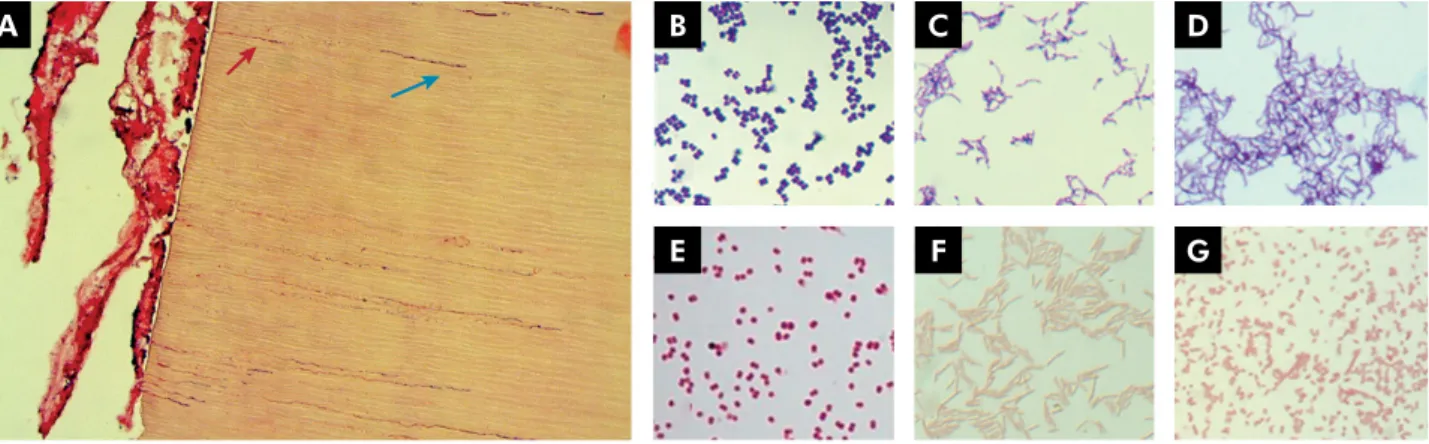
Figure 1 shows bacterial penetration into dentinal
tubules and examples of Gram staining morphology.
Table 1 shows the bacterial genera most commonly
found in endodontic infections according to their Gram staining characteristics and gaseous requirements. Some Gram-positive species detected in root canals are strict anaerobes, including Eubacterium, Filifactor, Parvimonas, Peptostreptococcus, Pseudoramibacter and
A B C D
E F G
Propionibacterium. Other Gram-positives arefacultative anaerobes, including Actinomyces*, Lactobacillus*, Streptococcus* (*some species can be strict anaerobes),
Enterococcus, Gemella and Staphylococcus. 9,10,32
Some members of the gram-negative community are strict anaerobes, including Fusobacterium, Porphyromonas, Prevotella, Tannerella and Treponema. Others are facultative anaerobes such as Capnocytophaga, Eikenella, Neisseria, among others.9,10,32
Escherichia coli is a gram-negative, facultatively anaerobic, rod-shaped, coliform bacterium found in the gastrointestinal tracts of humans and other vertebrates, and rarely isolated from root canals. However, it is widely used in laboratories because it is easy and inexpensive to grow, not particularly
virulent, and it is an excellent host for producing
several proteins. It was one of the first organisms to
have its genome sequenced, in 1997. More than 90% of endotoxin studies were conducted on E. coli LPS.49
Bacterial virulence factors
Bacterial virulence factors include the structural components and products of bacterial metabolism.
The latter are responsible for direct damage to pulp
tissue, whereas structural components of the bacterial cell, such as lipopolysaccharides (LPS) and lipoteichoic
acid (LTA), can injure tissues indirectly by activation
of an immune response.50
LPS, or endotoxin, is a major constituent of the cell wall of gram-negative bacteria and is secreted in vesicles by growing organisms or released during the disintegration of bacteria after their death. Endotoxin is one of the most important virulence factors involved in
the development of periapical inflammation and bone
destruction, activating immunocompetent cells and
leading to the release of a variety of proinflammatory
mediators. Lipid A is the bioactive component of LPS responsible for most of the host’s immune response.51,52,53,54
LTAs are present on the surface of gram-positive
cells, such as Enterococcus faecalis, and have adhesive properties, as they adsorb to hydroxyapatite, binding
through its lipid portion. They stimulate leukocytes, monocytes, and macrophages to release inflammatory
mediators.55 Thus, LTA may be associated with
resistance to medications used during endodontic treatment,56,57 also playing an important role in
biofilm formation, providing bacterial resistance
to antibiotics or disinfectants. It contributes to the virulence of E. faecalis by facilitating aggregation substance formation and plasmid transfer.56
LPS and LTA activate the immune system by similar mechanisms. Both bind to CD14, activating Toll-like
receptor signaling and inducing the production of
proinflammatory cytokines, such as tumor necrosis factor alpha (TNF-α), interleukin-1 (IL-1), interleukin-8
(IL-8), interleukin-12 (IL-12), and anti-inflammatory
cytokines such as interleukin-10 (IL-10).58,59 At low
concentrations, LPS and LTA stimulate the innate
response of the host defense system. At higher levels, they have been related to pain of pulp origin and to
periradicular inflammation.16,60
Table 1. Bacterial genera commonly occurring in endodontic infections according to the Gram-staining characteristics and gaseous requirements.
Bacteria morphology Obligate anaerobes Facultative anaerobes
Gram-positive cocci
Finegoldia Enterococcus
Parvimonas Gemella Peptoniphilus Staphylococcus
Peptostreptococcus Streptococcus*
Gram-negative cocci Veillonella Neisseria
Gram-positive rods
Actinomyces
Eggertella
Eubacterium Actinomyces*
Filifactor Corynebacterium Lactobacillus Lactobacillus*
Olsenella Propionibacterium* Propionibacterium
Pseudoramibacter
Gram-negative rods
Alloprevotella
Bacteroides Camphylobacter
Dialister Capnocytophaga Fretibacterium Eikenella
Fusobacterium Haemophilus Porphyromonas
Prevotella Tannerella
Treponema
Based on the facts and considerations above, it is clear that infection of the root canal system is the primary cause of apical periodontitis. In addition, virulence factors help the bacteria invade the host, cause disease, and evade host defenses, thereby worsening periradicular diseases.
The endodontic microbiota
Since Miller61 demonstrated the presence of bacteria
in necrotic pulp tissue, the role of oral microorganisms in the pathogenesis of pulpal and apical periodontitis has become increasingly evident. However, knowledge about the nature of the endodontic microbiota depends upon the recognition of the microorganisms present in the pulp space, which relies on contemporary knowledge and technology.34
Methods for microbial detection in root canals
Currently, there are several methods for microbial
identification, including culture- and non-culture-based techniques. Traditionally, microorganisms in endodontic samples have been identified by various
cultivation procedures, which rely on isolation, growth,
and laboratory identification, using morphology and
biochemical tests. However, the prevalence of some oral pathogens could have been underestimated by culture-based techniques as such approaches may fail to grow certain bacteria, especially fastidious anaerobic microorganisms such as spirochetes.4,9,10,62,63
Culture is a widely used method for evaluating the
antimicrobial efficacy of root canal procedures against
viable bacteria in root canal infection.2 Correlations
between absence of cultivable bacteria and a favorable treatment outcome have been reported.64 In order
to recover microorganisms from the necrotic pulp and from diseased periapical tissues and study their properties, stringent anaerobic sampling and cultivation techniques are necessary.2 Improvements in
anaerobic techniques have permitted a more detailed knowledge of the microbiota within the infected root canals and its association with periapical lesions.4,63
Adva nces i n mole cu la r te ch n iques h ave improved the identification of several novel and as-yet-uncultivated bacterial species. Molecular techniques include polymerase chain reaction
(PCR) and its variations (nested PCR, real-time quantitative PCR), cloning and sequencing, and
next generation sequencing (NGS). NGS is a high
throughput sequencing technology that has enabled the parallel sequencing of several microbiological samples by PCR amplification of a phylogenetic
marker, the 16S rRNA. It also provides the most
accurate detection of abundant and rare members of
the microbial community. Furthermore, an advantage
of this method is that it does not require laborious cloning to obtain microbial sequences.65 All of the
above-mentioned techniques are based on bacterial
identification through the 16S rRNA gene, a region of bacterial DNA present in all microorganisms that is well preserved and very specific to each species.63
Unlike the techniques based on the 16S rRNA gene, the checkerboard DNA hybridization or DNA-DNA hybridization was introduced for hybridizing large numbers of DNA samples against large numbers of DNA probes on a single support membrane.66
This method is fast, has adequate sensitivity, and
it is relatively inexpensive, overcoming several limitations of microbial culture, allowing the study of the communities present in clinical samples.66,67
Molecular methods overcome the limitations of culture-dependent methods; however, despite the inherent limitations of each method, the combined results of both cultural and molecular studies are necessary to improve the understanding of the endodontic microbiota.68 For example, the sensitivity
of microbial culture is approximately 104 to 105 cells
for target species using nonselective media and 103
using selective media, while for PCR it ranges from 10 to 102 cells, depending on the technique used. Nested
PCR brings the detection limit down to about 10 cells.69
The detection limits of the checkerboard DNA-DNA
hybridization method ranges from 103 to 104.63,66,70
Factors related to the composition of the root canal microbiota
The major ecological factors that define the
composition of the root canal microbiota are the availability of nutrients, redox potential variability, and bacterial interactions.5,6,32,34,71 Bacterial interactions
bacterial interaction can also have an effect on the microbial population itself, inhibiting the growth of particular species and perhaps increasing the chances of the host to cope with the infective agent and to restore health. Other factors involved are pH level, temperature, and host defense mechanisms.5,6,32,34,71
It has been reported that there is a difference between the microbiota of a root canal subjected to any source of oral contamination for some time and the microbiota of closed canals. In closed canals, the microbiota is predominantly anaerobic, while in open canals the recovered/detected microorganisms are mostly facultative anaerobes.34,45,47,72 Moreover, anaerobes are
also more frequently detected in root canal samples from clinically symptomatic teeth,4,7,9,10,32,45,47,67,72-78 which
have a bacterial community profile that is significantly different from asymptomatic (chronic) lesions. This
has also been observed for teeth with primary versus post-treatment apical periodontitis.7,9,10,32,77,79-81
In addition, the bacterial community profile exhibits a high interindividual variability and there are
significant geographic differences in the composition
of the endodontic microbiota, which may have
implications in terms of efficacy of antimicrobial
protocols used in different countries.10,32
Overall microbial composition of the root canal
Similarly to the oral cavity, over 500 different bacterial species or phylotypes have already been detected in infected root canals by culture or by
molecular methods such as 16S rRNA gene sequencing
or metagenomic sequencing. Bacteria are the most commonly found microorganisms in root canals, belonging to 20 phyla according to present knowledge.32
Table 2 shows some of the microorganisms isolated/
detected from root canals according to their phyla. Of
the major reported phyla, Firmicutes, Fusobacteria, and
Bacteroidetes were the most abundant in acute infections,
while Firmicutes, Bacteroidetes, and Actinobacteria
were the most abundant in chronic infections.76,77,81 In
addition, fungi, yeasts,10,80,82-86 viruses,87-90 and Archaea71,91
were also detected in root canals.
These microorganisms can be found suspended
in the lumen of the root canal (planktonic form) or
adhered to the walls of the root canal, forming a biofilm
(sessile form). They can penetrate into dentinal tubules
and into lateral, secondary, and accessory canals.
They can be found between the gutta-percha and the root canal walls, and they can also form biofilms in the extraradicular region. The colonization of these
sites is directly related to the time of infection and to the composition of the microbiota.32
Classification of endodontic infections
Endodontic infections can be classified according
to their anatomical location (intraradicular or extraradicular infection) and to how long it took microorganisms to reach the root canal (primary, secondary, or persistent infection). Usually, primary and secondary/persistent endodontic infections are located intraradicularly, and may originate in extraradicular infections if left untreated or inadequately treated.9,10,32
Primary infected root canals are untreated canals where microorganisms were able to access and colonize the pulpal tissue and impair its function.
Their microbial profile consists of 10–30 species per
canal,9,10,32,92 and the species present in the apical
region may have a major role in the pathogenesis of apical periodontitis.
Fusobacterium, Porphyromonas, Prevotella, Parvimonas, Tannerella, Treponema, Dialister, Filifactor, Actinomyces, Olsenella,and Pseudoramibacter predominated in the canals. Some facultative or microaerophilic streptococci are also commonly found in primary infections. 9,10,32
Persistent/secondary infected root canals are usually associated with post-treatment apical periodontitis, which indicates that there was a failure in endodontic treatment. In this case, microorganisms may have tolerated the chemomechanical procedures (persistent infection) or invaded the canal via coronal
leakage of the root filling (secondary infection).7,9,10,32,85
The microbiota found in cases of endodontic failure
is composed of a more restricted group of species when compared to primary infections. Canals apparently
well treated have been shown to harbor fewer than five
species. On the other hand, teeth with unsatisfactory root
filling may harbor 10 to 30 species, a number similar
to that of primary infections.9,32
Facultative anaerobic and gram-positive bacteria
to instrumentation and to antiseptic agents.5,6,82,85
Accord i ng to Mola nder e t a l.,82 fac u lt at ive
anaerobes, especially gram-positive ones, can survive in a quiescent phase with low metabolic activity for some time, and factors such as coronal leakage during or after root canal treatment can
change the nutritional conditions and contribute to bacterial growth.
E. faecalis, a facultative gram-positive bacterium, is capable of surviving in an environment with scant availabilit y of nut rients and m in imal commensality with other bacteria.83,93 It presents
Table 2. Microorganisms most commonly isolated/ detected from root canals distributed according to the phyla.
Firmicutes Actinobacteria Fusobacteria Proteobacteria Spirochaetes Bacteroidetes Synergistetes
Dialister pneumosintes Actinomyces gerencseriae
Fusobacterium necrophorum
Campylobacter gracilis
Treponema amylovorum
Alloprevotella tannerae
Dialister invisus Actinomyces israelii
Fusobacterium nucleatum ssp
nucleatum
Campylobacter rectus
Treponema. denticola
Porphyromonas endodontalis
Pyramidobacter piscolens
Eggerthella lenta Actinomyces naeslundii
Fusobacterium nucleatum ssp
polymorphum
Eikenella corrodens
Treponema lecithinolyticum
Porphyromonas gingivalis
Enterococcus. faecalis Actinomyces odontolyticus
Haemophilus aphrophilus
Treponema maltophilum
Prevotella denticola
Filifactor alocis Actinomyces viscosus
Treponema medium
Prevotella intermedia
Finegoldia magna Propionibacterium acnes
Treponema pectinovorum
Prevotella melaninogenica
Parvimonas micra Propionibacterium propionicum
Treponema socranskii
Prevotella nigrescens
Peptoniphilus asaccharolyticus Slackia exigua Treponema vincentii
Tannerella forsythia
Peptostreptococcus anaerobius
Pseudoramibacter alactolyticus
Selenomonas sputigena
Streptococcus anginosus
Streptococcus constellatus
Streptococcus intermedius
Streptococcus mitis
Streptococcus sanguis
different virulence and resistance mechanisms, which hinder its eradication from root canals.22
An important collagen-binding protein of E. faecalis
microbial surface components is Ace (adhesion of collagen from E. faecalis),93 which is related to its ability
to invade dentinal tubules and adhere to collagen in the presence of human serum.94,95
Streptococcalspecies are also frequently found in root canals of teeth with post-treatment disease. Other bacterial species, including anaerobic bacteria such as Parvimonas micra, Propionibacterium species and Pseudoramibacter alactolyticus are commonly reported in persistent intraradicular infections.
Candida species are more frequently found in root canals of teeth with endodontic failure than in those with primary infections .9,10,32,79,85
Extraradicular infections are mostly caused by
intraradicular infections. They are characterized by apical abscesses and extraradicular biofilms. However,
there are also independent extraradicular infections, such as apical actinomycoses, caused by Actinomyces
spp. and Propionibacterium spp., requiring periapical surgery for their resolution.32,36,96
Apical periodontitis may be acute or chronic, depending on several factors. Acute infection is usually caused by a community of highly virulent bacteria, probably due to the presence of species with greater virulence or to the synergism between species and is characterized by a high concentration of bacteria and tissue invasion, together with decreased host resistance.46 Chronic apical periodontitis, by contrast, is
usually associated with low virulence of the bacterial community involved. Bacterial persistence in the root canal system may be related to its organization
in biofilms, not allowing for host defense due to the
anatomical location of the infection.46
Acute apical abscess is the most common form of extraradicular infection. Microbial communities present in acute apical abscesses are complex, with a predominance of strict anaerobic microorganisms (approximately 90% of the isolates), mainly gram-negative bacilli, including Fusobacterium, Prevotella, Porphyromonas, Dialister,and Treponema, and
gram-positive cocci. They are polymicrobial infections where
the occurrence of strict anaerobes is 3 to 4 times greater than that of facultative anaerobes.76,78,97,98,99 However,
although the concentration of strict anaerobes is higher in periapical abscesses, their diversity is lower than that found in the root canals of periapical abscesses.98,99
Chronic apical abscess is characterized as an
inflammation in the periapical region with formation
and discharge of pus slowly through a fistula, recognized as a pathognomonic sign of this lesion. Radiographically, there are radiolucent signs of
apical bone destruction, however without significant
discomfort for the patient.100 The endodontic microbiota
of this pathology has been scarcely characterized in the literature, probably due to its low prevalence (between 9.7% and 18.1%).101 Rôças et al.75 used checkerboard
DNA-DNA hybridization to identify microorganisms
in the root canals of nine patients diagnosed with chronic apical abscess. Porphyromonas endodontalis (9/9 - 100%), Dialister invisus (8/9 - 89%), Parvimonas micra
(7/9 - 78%), and Solobacterium moorei (7/9 - 78%) were
the most prevalent species in these cases. Tennert
et al.,102 after sequencing the 16S rRNA genes of
isolated microorganisms, reported the presence of
Anaerococcus prevotii, Atopobium rimae, Dialistes invisus, and Fusobacterium nucleatum.
Bacterial biofilm is a community of microcolonies of bacterial cells involved in an extracellular matrix of polysaccharides, adhered to a solid substrate wet or liquid medium, from which bacteria can obtain their nutrients. Biofilm is a form of protection against bacterial development in hostile environments. It can be formed by one or multiple species.96,103,104,105,106,107
Periradicular dental biofilm is characterized by
microorganisms adhered to cementum and/or dentin in the apical portion of the root, surrounded by an external polysaccharide layer known as glycocalyx,
which forms an intermicrobial matrix. The structure of the polysaccharide matrix that surrounds the biofilm
limits the access of defense molecules (antibodies and complement) and of phagocytic cells (macrophages and neutrophils).108 The prevalence of intraradicular
bacterial biofilms is high, whereas extraradicular
biofilms are considered rare and are normally dependent on intraradicular infections.106
Bacteria in biofilm differ phenotypically from
a given environment.107 Clinically, one of the main
characteristics of bacterial biofilms is their greater
resistance to antimicrobial agents.109
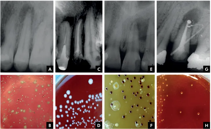
Figure 2 illustrates the radiographic aspects and microbiological findings observed in each type of infection. There is a great diversity of microbial
colonies in primary intraradicular infections
(Figure 2, A-B) and a lower diversity in secondary ones (Figure 2, C-D). A large diversity of microbial
colonies with a predominance of black-pigmented bacteria can be found in acute periapical abscesses
(extraradicular infections) (Figure 2, E-F), whereas
endodontic treatment failures may be due to the
presence of extraradicular apical biofilms, on which
microorganisms such as Actinomyces israelii may be
present (Figure 2, GH).
In summary, although the infectious nature of endodontic pathosis has been established for many decades, with the development of modern microbiological diagnostic techniques, the composition
of these infections has been improved and redefined.
The key role of endotoxin
All gram-negative bacteria, and only these microorganisms, have a differentiated external cell membrane with hydrophobic constituents composed of polysaccharides (sugar polymers), lipids (complexes
containing fatty acids), and proteins. This structure
is called LPS or endotoxin, highlighting the main components and biological effects of the molecule.3
Endotoxins are secreted into vesicles during the bacterial growth phase or released during cell death.
They are also released when the cell is chemically
treated to remove LPS. LPS represents the major antigenic surface of gram-negative bacteria, presenting
microbiological and immunological significance.51,52
Approximately 75% of the cell surface of gram-negative bacteria is composed of this molecule, which is essential for cell growth, decrease in membrane permeability, and structural integrity and stability, as well as for protection against external damage. 52,110
Endotoxins are heat-stable and, therefore, usual sterilization processes are ineffective for
A
B
C
D
E
F
G
H
their destruction.111 To degrade them chemically,
strong acids or bases or pyrolysis must be used, and different heating protocols are known: 180°C for 3 hours,111,112 200°C for 4 hours,113 or 250°C for 3 hours
according to the Cambrex/Lonza manual. Other processes that lead to the inactivation of endotoxins are ionizing radiation and treatment with polymyxin B. In endodontic therapy, hydroxyl ions present in the calcium hydroxide paste can hydrolyze LPS, degrading lipid A and neutralizing its residual effect after cell lysis.54,114
LPS is known to be the most toxic constituent of bacterial endotoxin, having no structural homologue among multicellular organisms.115
In general, bacterial LPS are composed of three structural domains: lipid A, core, and repeating O-antigen.116,117,118 The lipid A moiety exerts most of the
endotoxic activities, being regarded as the endotoxic principle of LPS.116 The chemical structure of lipid A
is composed of fatty acids with 15 to 17 carbon atoms, linked to two amino-sugar molecules (glycosamines), to which two phosphate radicals are bound, and a
protein residue bound to the phosphate radicals. The
positions of the phosphate radicals, as well as the number, type, and site of bonds seem to determine
the inflammatory potential of different LPS.92
Polysaccharide moiety is a potent antigen that can stimulate antibody formation even at submicrogram concentrations.119 The recognition of LPS occurs
through lipid A, which in turn activates different intracellular signaling pathways through its binding to proper receptors, according to the structure of the acyl chain.120 The major receptors are located
in the cell membrane of monocytes, called Toll-like receptors 4 (TLR4).121,122
Studies have shown that lipid A can vary among different bacterial species, depending on the number of phosphate groups and on the amount and position of fatty acids in the molecule.53,120,123 These variations
are closely related to the change in TLR4 signaling and,
consequently, to their immunostimulatory effects.118,120,124
TRL-4 mutations are likely to influence susceptibility to
gram-negative infection, or the course of infection once it is established, as they block LPS signaling, whereas overexpression greatly increases LPS signaling.115
Moreover, changes in the microenvironment, such as
hemin concentration and temperature, can structurally alter this bioactive portion.125
LPS structure is remarkably heterogeneous among bacterial species, thus evoking different patterns
of inflammatory response. The LPS molecule can
also vary among different strains of single species
and, consequently, exhibit different inflammatory
potentials.53,18,126
More than 90% of endotoxin studies have been conducted on enterobacterial LPS,49 of which
Escherichia coli LPS is the best known. LPS from gram-negative bacteria such as Prevotella and Porphyromonas isolated from the oral cavity are able to produce classic manifestations that are less damaging to host tissues than those from E. coli.127
On the other hand, Fusobacterium spp. LPS presents
a similar structure to that of gram-negative enteric bacilli,128 contributing to the high virulence of this
microorganism.127 According to Martinho et al.,126
Fusobacterium nucleatum induces a greater expression of IL-1β and TNF-α cytokines compared to Porphyromonas gingivalis. These two bacteria have different patterns
of macrophage activation, which may contribute to the immunopathogenesis of apical periodontitis.
LPS is essential for bacterial survival because it protects the bacterium from host defense cell stimuli.129 It has many biological activities including
fever induction, adjuvant activity, Schwartzman
reaction, cytotoxicity, blood clotting, and fibrinolysis,
among others. LPS can also stimulate production of bradykinin, which is a potent pain mediator.130
Siqueira Junior and Rôças46 mention several
biological effects of LPS, as follows:
a) Activation of macrophages/monocytes with
consequent synthesis and release of proinflammatory
cytokines (IL-1β, IL-6, CXCL8 or IL-8, TNF-α), prostaglandins, nitric oxide, and oxygen-derived
free radicals. These substances are chemical mediators of inflammation and most of them can stimulate
bone resorption;
b) Activation of the complement system. Some products of complement activation are chemotactic to
inflammatory cells (C5a), act as opsonins (C3b), and
can increase vascular permeability (C3a and C5a).
c) Activation of the Hageman factor, the first step
coagulation cascade or the production of bradykinin,
an important chemical mediator of inflammation;
d) Induction of the expression of leukocyte adhesion molecules in endothelial cells, which are important
in the early stages of inflammation;
e) Stimulation of osteoclast differentiation and bone resorption, particularly via interactions with
TLR4 in osteoblast lineage cells. LPS induces RANKL
expression in osteoblasts and stimulates these cells to secrete interleukin (IL)-1, IL-6, prostaglandin E2
(PGE2), and TNF-α, each of which is known to induce osteoclast activity and differentiation.
f) LPS may be mitogenic to B lymphocytes and epithelial cells.
g) LPS can stimulate naive B cells in the absence of
T helper cells. At low concentrations, LPS stimulates specific antibody production. At high concentrations,
this molecule can cause nonspecific polyclonal activation of B cells.
h) It has been recently demonstrated that trigeminal
afferent neurons express the TLR4 and CD14 receptor complex and that LPS activation of TLR4/CD14 may
trigger intracellular signaling cascades, leading to peripheral release of neuropeptides and central nociceptive neurotransmission. Hence, it is assumed that one of the pain mechanisms associated with bacterial infectious processes could result from direct
effects of LPS on sensory fibers via interaction and direct activation of the TLR4/CD14 complex.
LPS may evoke pain through activation of the Hageman factor or through neurotoxic properties when acting on presynaptic nerve terminals, direct sensitization of nociceptors, sensitization and up-regulation of the transient receptor potential
cation channel, subfamily V, member 1 (TRPV1).18
LPS concentrations found in infected root canals seem to promote a direct sensitization of receptors that activate the pain mechanism associated with bacterial infections, also sensitizing trigeminal sensory neurons.131,132
The presence of endotoxin has been reported in
samples taken from vital pulp,13 irreversible pulpitis,13
necrotic pulp,11,12,13,15,16,17,20,25,28,29,30,113,131,133,134,135,136,137,138,139
root canals of teeth with endodontic failure,19,20,26,132 and
Table 3. Total endotoxin levels in initial samples from root canal reported in previous studies.
Author Pulpal / Periodontal tissue status LAL Method Endotoxin
concentration
Schein and Schilder (1975)13 Vital pulp / healthy periodontium Gel clot 0.007 µg/mL
Irreversible pulpitis / healthy periodontium Gel clot 0.075 µg/mL
Schein and Schilder (1975)13
Pulp necrosis / apical periodontitis
Gel clot 0.192 µg/mL
Jacinto et al. (2005)15 QCL 18016.50 EU/mL
Vianna et al. (2007)133 QCL 151.61 EU/mL
Martinho and Gomes (2008)16 QCL 323.27 EU/mL
Gomes et al. 2009(113) QCL 212.23 EU/mL
Martinho et al. (2010a)11 TKA 9.19 EU/mL
Martinho et al. (2010b),134 (2012),17 (2014)25 TKA 7490.00 pg/mL
Martinho et al. (2011b)141 QCL 34.20 EU/mL
Martinho et al. (2011b)141 KQCL 7.49 EU/mL
Martinho et al. (2011b)141 TKA 9.19 EU/mL
Oliveira et al. (2011)135 KQCL 192.37 EU/mL
Gomes et al. (2012)20 TKA 7.49 EU/mL
Xavier et al. (2013)136 KQCL 153.13 EU/mL
Marinho et al. (2014)137 TKA 18.70 EU/mL
Marinho et al. (2015)29 TKA 32.43 EU/mL
Herrera et al. (2015)30 TKA 21.83 EU/mL
Cardoso et al. (2015)131 KQCL 10.92 EU/mL
Herrera et al. (2017)139 TKA 27.72 EU/mL
Endo et al. (2012),19 Gomes et al. (2012)20 Previous root canal treatment / apical periodontitis TKA 3.96 EU/mL
Duque et al. (2018)140 Vital pulp / chronic periodontal disease TKA 0.10 EU/mL
from root canals of teeth associated with periodontal disease.140 Table 3 shows some of the endotoxin levels
reported in initial samples collected from the root canals. LPS have been detected in 100% of root canals
with necrotic pulp, with significantly higher levels in
symptomatic teeth.12,15,20,141 Moreover, even though LPS
levels were higher in primary endodontic infections than in secondary/persistent infections, endotoxins were detected in all samples.20 This shows that
endotoxins are extremely strong stimulators of
inflammatory reactions, even at low concentrations. There is a correlation between higher levels of
endotoxins and a greater area of bone destruction in periapical tissues,142 as well as with the presence of
specific clinical features found in primary endodontic
infections.20,46 Increased endotoxin levels in infected
root canals may be associated with the severity of periapical disease, as well as with the development of clinical symptoms.20
Clinical endodontic researchers have investigated not only bacterial LPS in infected root canals, but also correlated higher endotoxin levels with clinical
signs, symptoms, and radiographic findings.
Interplay of infectious/endotoxic contents with inflammatory mediators and clinical symptomatology
The role of cytokines
Periapical immune response is a second line of defense that seeks to localize the infection of the
root canal system by confining it and preventing
its dissemination and systemic involvement.2 This
immune response is initially comparable to the pulpal response to microbial infection, characterized by a cell
infiltrate of polymorphonuclear neutrophils (PMNs)
and monocytes, with the subsequent additional
feature that the periradicular bone is destroyed. The
intensity of bacterial invasion of periradicular tissues depends on the number of pathogenic bacteria and on their degree of virulence.2 Once bacteria and their
virulence factors come into contact with periradicular tissues, they stimulate the synthesis and expression
of different mediators that will attract inflammatory
cells to the area.17 These factors, depending on host
resistance, may stimulate the development of an acute
inflammatory response (acute apical periodontitis or
acute periradicular abscess), or a chronic response (chronic apical periodontitis or chronic periradicular abscess).46 The destruction of periapical tissues seems
to be mainly indirect via host-derived stimuli rather than by the direct effects of bacteria on the bone.
Periapical immune response is predominantly a reaction to bacterial infection present in necrotic root canals.46,143,144 Immunocompetent cells settle in
periapical areas in an attempt to prevent the spread of the infectious microbiota.17 Among these cells,
macrophages are the defense cells responsible for the rapid recognition of pathogens and rapid presentation of lymphocytes and other cells of the immune system.17
Macrophages are stimulated predominantly by the bacterial endotoxin.12,132 Lymphocytes express different
sets of inflammatory cells, proinflammatory and
immunoregulatory cytokines and chemokines, which are considered important mediators in periapical immune response to infection.132,143
Among the various host degradative pathways, considerable interest has been focused on the study of cytokines, not only because of their role in regulating the humoral immune system and in cellular responses against invasive bacteria, but also as mediators of periapical tissue destruction.145
Inflammatory cytokines produced from host cells (e.g., monocytes/ macrophages) reflect root canal conditions and determine the local immune process within the periapical environment.29 After
the macrophage and PMN activation by bacterial components, a cascade of proinflammatory cytokines,
including IL-1α, IL-1-β, and TNFα, is triggered.144
Cytokines are small signaling molecules that
mediate host responses to infection, inflammation, and trauma. Proinflammatory cytokines initiate or enhance
systemic inflammation while anti-inflammatory
cytokines reduce inflammation and promote healing.144
Examples of proinflammatory cytokines include
IL-1 and TNF-α, whereas IL-10 is an important
anti-inflammatory cytokine. Some cytokines can have both proinflammatory and anti-inflammatory properties, such as IL-6, as it can inhibit TNF-α and IL-1 and activate IL-10 at the same time. Cytokines work with each other in a homeostatic network regulation to
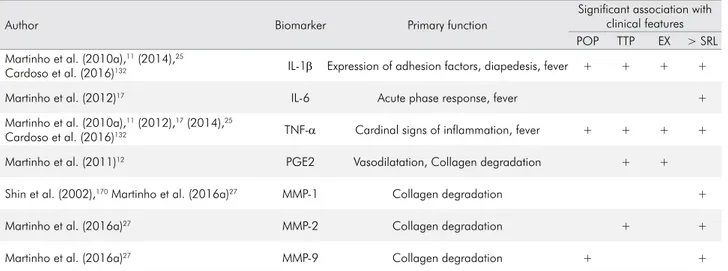
Interleukin-1β (IL-1β) and interleukin-6 (IL-6) act as
proinflammatory cytokines during apical periodontitis, initiating or intensifying systemic inflammation.144,146 In
addition, they also stimulate osteoclast differentiation and bone resorption in chronic apical periodontitis, inducing the secretion of chemokines during the destruction of periodontal tissues.146
On the other hand, the reactions provoked in the
host organism by proinflammatory cytokines can be
prevented by suppressing the activity of these cytokines
through the activity of anti-inflammatory cytokines,
such as interleukin 4 (IL-4), interleukin 10 (IL-10), IL-13,
and the transforming growth factor beta (TGF-β).146
Proinflammatory cytokines, such as IL-1β, IL-2,
IL-8, IL-17, IFN-ɣ, and TNF-α, are detected in the interstitial fluid of periapical lesions, where the high concentration of bacteria found in root canal infection was correlated with a higher rate of detection
of proinflammatory cytokines.147,148
Gram-negative bacterial species tend to induce a
greater proportion of TNF- α, but other cytokines, for example IL-1β, IL-6, IL-8, and IL-10, increase their levels during the course of endodontic infection.144
TNF-α, IL-1β, and IL-6 are examples of important
cytokines in the acute phase of inflammation.144,148 On
the other hand, LTA, present in gram-positive bacteria,
has pathogenic properties similar to those of LPS,21,22
resulting in well-known injuries to the dental pulp and
periapical tissues. Overall, both LPS and LTA are able
to potently activate monocytes/macrophages, causing rapid release of cytokines at periradicular sites related to tissue destruction.21,22 However, by comparing the
cytokines released by LTA in immune cells with the
cytokine released by LPS of gram-negative bacteria,
LTA has a power that is 100 to 1,000 times lower
than that of LPS.144 While LPS is a potent inducer of
proinflammatory cytokines and IL-10, LTA exhibits less induction of proinflammatory cytokines and does not induce IL-12 and the subsequent formation of
IFN-ɣ,149,150 which can be explained by the use of different
TRLs.149 TRL2 appears to be the primary mediator of
the innate immune response to the LTA of several
gram-positive bacteria151 and is highly induced in
inflamed dental pulps.152,153
TNF-α is the cytokine most abundantly detected in endodontic infection.29,144 TNF-α is a potent immune
mediator of acute and chronic inflammatory responses,
with the potential to increase bone resorption.154
It is considered the main mediator of the acute
inflammatory response induced by gram-negative
bacteria and other infectious microorganisms. LPS is the most important stimulus to activate the production
of TNF by macrophages, although activated T cells, NK, and mast cells can also secrete this cytokine. IFN-γ produced by T and NK cells increases the synthesis of TNF by LPS-stimulated macrophages.155
TNF-α stimulates the production of collagenase and prostaglandin E2 (PGE2), factors related to the
induction of chemokines, cytokines, cell adhesion molecules, and bone resorption.17 PGE
2 can induce
or inhibit IL-6, another proinflammatory cytokine
that stimulates osteoclast differentiation and bone resorption in chronic apical periodontitis.17 IL-6 can
also induce or inhibit PGE2, depending on the duration
of the lesion, bacterial load, stimulated signaling pathways, and cytokines.17 PGE
2 was directly and
indirectly related to most of the inflammatory and
destructive alterations in apical lesions, such as vasodilation, increased vascular permeability, and collagen degradation.17 In the same study, PGE
2 was
also positively related to TNF-α and IL-1β.17
IL-1 has the main function of mediating the
host’s inflammatory response to infections and to other stimuli, similarly to the effects of TNF. It acts
on endothelial cells by inducing the expression of surface molecules that mediate leukocyte adhesion. Macrophages are the main source of IL-1 production, with neutrophils and endothelial cells involved in their
production. There are two forms of IL-1, alpha (α) and beta (β), the latter of which is the most commonly found in the human circulatory system.155 IL-1α plays a critical
role in protecting the body from external invaders such as bacteria and viruses, and it is also involved in bone resorption.156 IL-1β has been correlated with clinical
signs/symptoms and with greater bone resorption.157
IL-6 is a cytokine that mediates the host’s response to infection and has been observed in exudates (EX) associated with endodontic apical lesions.160
In addition, IL-6 was positively correlated with the area of the radiographic lesion in the study by Martinho et al.,17 thus confirming its role in bone
resorption in chronic inflammatory periodontitis.
However, the same study demonstrated that PGE2
and IL-6 together were negatively correlated with the
size of the radiographic lesion, confirming that the
relationship between these cytokines is complex and
difficult to establish in many respects.17 IL-6 has potent
proinflammatory effects at local and systemic levels, including the acute-phase inflammatory response,
in which C-reactive protein (CRP) can be detected in response to IL-6 action.160 Although IL-6 is detected in
all analyzed samples of root canals, whether healthy or with apical periodontitis, in the latter cases its
expression is significantly increased.160 IL-6 expression
is induced by IL-1β and TNF-α during the early
stages of inflammation, and all act synergistically, promoting the recruitment of PMNs and monocytes, shifting from acute to chronic inflammation, which
induces activation of MMPs and stimulation of osteoclastogenesis and bone resorption.160
IL-8 is mainly produced by monocytes/macrophages
and in smaller amounts by fibroblasts, endothelial
cells, keratinocytes, melanocytes, hepatocytes, and
chondrocytes. Its stimuli are usually IL-1, TNF-α, and
IFN-g. IL-8 can be inhibited by corticosteroids and
cyclosporine A. It is a chemokine, thus increasing chemokinesis and acting as a chemiotactic factor.
The term ‘chemokine’ comes from the contraction of ‘chemotactic cytokines’. Other chemokines include MIP-1a, MIP-1b, MCP, eotaxin, and RANTES. IL-8
provides potent migratory stimulus for the cells of the immune system, mainly neutrophils, also determining an increase in the expression of adhesion molecules
by endothelial cells. It also activates PMNs, increasing
oxidative metabolism. It antagonizes the production of IgE stimulated by IL-4, but it does not affect the production of other immunoglobulins.150
IL-10 is produced by monocytes, macrophages, and lymphocytes,161,162 originally identified for
their ability to antagonize cellular immunity.163
Among its characteristics, immunosuppression
is noteworthy, as it depresses the activation of mononuclear cells and prevents the production of
mediators of inflammation.163,164 IL-10 inhibits antigen
presentation in monocytes by negative regulation of MHC class II163and the in vitro expression of
costimulatory molecules.165 Its biological effects
would be a consequence of its ability to inhibit many of the functions of activated macrophages, such as
the production of IL-12 and TNF-α.155
Although many of the cytokines involved in the
immunoregulatory network were identified in apical
periodontitis, the longitudinal expression of this
network and its effects on proinflammatory cytokines
still need to be further investigated.
The role of matrix metalloproteinases
Matrix metalloproteinases are members of an enzyme family that require a zinc ion in their active site for catalytic activity. MMPs are critical for maintaining tissue allostasis. MMPs are active at neutral pH and can therefore catalyze the normal turnover of extracellular matrix (ECM) macromolecules such as interstitial and basement membrane collagens, proteoglycans such as
aggrecan, decorin, biglycan, fibromodulin, and versican, as well as accessory ECM proteins such as fibronectin.166
The degradation of the ECM by MMPs seems to
be an important trigger for the progression of the
inflammatory process.148,167
Proinflammatory cytokines, such as IL-1β and
TNF-α, are able to stimulate, either directly or indirectly, the release of MMPs in the periapical/periodontal region,
maintaining a persistent inflammatory process.30,168
MMPs are deeply involved in the pathogenesis of pulp, periodontal, and periapical tissue destruction.168
MMPs are commonly organized in groups, based partly on historical assessment of the substrate
specificity of the MMP and partly on the cellular localization of the MMP. These groups are the
collagenases [MMP 1 (interstitial collagenase), MMP 8 (neutrophil collagenase), MMP 13 (collagenase
3), MMP18 (collagenase 4)], the gelatinases [MMP 2 (gelatinase A), 9 (gelatinase B)], the stromelysins (MMP 3, 10, 11), membrane-type MMPs (MT-MMPs
14, 15, 16, 17, 23A, 23B, 24, 25), and other MMPs.169
evidence that these MMPs play an important role in the pathogenesis of pulp, periodontal, and periapical tissue destruction.170,171 They are responsible for the degradation
of gelatin (denatured collagen) and type IV collagen, the major component of basement membranes.172
MMP-2 can also degrade collagens V, VII, and X,
decorin, elastin, and fibronectin.172,173 Its collagenolytic
activity is shown by its action on fibroblasts, the main
constituent cells of connective tissue of the periodontal ligament, responsible for collagen production.30
Fibroblasts secrete MMP-2 when induced by endodontic
content in primary endodontic infections, and this process contributes to the progression of periapical
inflammation and tissue destruction.30
It has been reported that the expression of MMP-9
in inflamed pulps has shown higher levels than those
recorded in clinically healthy pulps.167 MMP-9 was
detected in endothelial cells, osteoblasts, fibroblasts, and inflammatory cells, and could be released during
the inflammatory reaction induced directly by
bacteria or indirectly by proinflammatory cytokines,
evidencing the role of MMPs in the pathogenesis of
inflammation,27 where its synthesis is controlled by
proinflammatory cytokines.167
Ahmed et al.167 showed a correlation between
gram-negative bacteria and MMP-9 hyperactivity in symptomatic periapical lesions. MMPs also act in the processes of bone resorption and destruction of periapical tissues, promoting direct degradation of the ECM, exhibiting a correlation between the concentration of gram-negative bacteria and MMP-9 expression in symptomatic periapical lesions.167
The destruction of the periodontal ligament is
initiated by the degradation of extracellular membrane and serine proteases.174 Connective tissue destruction
is essentially controlled by MMPs, which contributes to the destruction of gingival tissue and alveolar bone surrounding the teeth.23 MMP activity requires
a balance with the intrinsic inhibitors known as
tissue inhibitors of MMPs (TIMPs),27,175 because an
excessive production of MMPs leads to accelerated matrix degradation and tissue destruction, which is associated with pathological conditions such as periodontitis and apical periodontitis.168,176
Specific Quantikine ELISA kits (R&D Systems,
Min neapolis, MD, USA) have been used for
measurement of both cytokines (IL-1α, IL-1β, TNF-α, and PGE2) and MMPs (2, 3, 8,
MMP-9, and MMP-13). Cytokines and MMPs are measured indirectly after stimulation of host cells (macrophages and fibroblasts) with infectious contents,17,29,30 or
directly from samples of the periapical region.27,140
Regardless of the method used, the data obtained in the studies mentioned above reveal that MMPs are involved in apical periodontitis because they interact with complex networks, which include cytokines, in the development of clinical features and severity of bone destruction.
Association of microorganisms with endodontic clinical features
The microbiota of infected root canals consists
of a complex polymicrobial population. In such heterogeneous community, interactions among
several microbial species may play a significant role
in the balance between individual microorganisms. Combinations of bacteria are more potent at inducing pathological state in the host (e.g., apical periodontitis) than are single strains. Moreover, complex interactions of species result in characteristic clinical pictures that cannot be achieved by individual species alone.6
Gram-negative species, such as Fusobacterium, Prevotella, and Porphyromonas, are likely to have some
clinical significance due to the presence of endotoxin.
LPS has many biological activities, including fever induction, adjuvant activity, Schwartzman
reaction, cytotoxicity, blood clotting, fibrinolysis,
and production of bradykinin, which is a potent pain mediator.4-7,130 However, the cell walls of gram-positive
bacteria such as Peptostreptococcus and Eubacterium
spp. can also produce inflammatory reactions due to the presence of peptidoglycans and LTA. They
enhance the pathogenicity of “black-pigmented
Bacteroides” and are also related to acute symptoms
and destruction of periapical tissues. The combination
of P. micros and Prevotella spp. was associated with clinical features such as pain and swelling.4-7 One
explanation for the synergy between these species is the known enhancement of the endotoxin effect by gram-positive superantigens.7 Superantigens
interact with antigen-presenting cells (APCs), such as
and massive cytokine production, which leads to clinical symptomatology.177
A long-held desire in endodontic microbiology has
been to find a single or at least a group of bacterial
species that is responsible for acute symptoms.32,78
However, while several bacterial species seem to be more prevalent when associated with pain, the very same species have also been encountered in
asymptomatic cases. The possibility exists that some
of these species really play a role in making the bacterial mixed community more virulent. Several
other factors can be regarded as influential to the
development of symptoms, including differences in virulence among clonal types of the same species, bacterial interactions in the multispecies community,
resulting in collective pathogenicity, total and specific
bacterial counts, and host-related factors.32
Endodontic symptomatology includes history of pain, spontaneous pain, pain on palpation (POP),
and tenderness to percussion (TTP). Clinical signs
mean presence of sinus swelling in periodontal tissues, presence of apical periodontitis, status of the root canal such as dry canal, and presence of clear, hemorrhagic, or purulent exudate.
Several works usi ng the st rict a naerobic culture technique reported an association between speci fic bacteria a nd endodont ic sig n s a nd symptoms.4-7,72-74,85,178-181
Nowadays, with the advance of molecular
microbiology, thanks to which more than 500 species or phylotypes have been detected in root canals, it seems even harder to associate a species or group of species
with clinical symptoms and signs. Nevertheless, the bacterial community profiles associated with teeth with symptomatic apical periodontitis are significantly different from asymptomatic lesions. The same has
been observed for teeth with primary versus post-treatment apical periodontitis.32
Association of LPS with clinical features
A n ae robic g ra m-neg at ive b ac te r i a h ave been frequently isolated from root canals of endodontically involved teeth; consequently, their endotoxins may affect the periapical tissues and
exert a role in the pathogenesis of inflammatory
lesions of pulpal origin.15
Since the work by Schein and Schilder,13 the
relationship between endotoxin levels and presence of endodontic clinical signs and symptoms has been
investigated. These authors found a significant
correlation between endotoxin levels and presence of exudate and radiolucent areas.13
Horiba et al.31 showed that teeth with clinical
symptoms contained higher levels of endotoxin than those that were asymptomatic. Jacinto et al.15 reported a
positive association between endotoxin and symptomatic
cases (e.g., spontaneous pain, TTP, POP, swelling,
and purulent exudate), which exhibited higher levels of endotoxin than asymptomatic cases. A negative association was reported between the endotoxin present in the root canals and asymptomatic teeth.15
Martinho and Gomes16 found a positive correlation
between LPS and TTP in primary infected root canals.
Higher levels of endotoxin were found in teeth with clinical symptomatology.
Martinho et al.11 reported that larger areas of bone
destruction, identified by the size of the radiolucent
area, were related to higher levels of endotoxin. Additionally, a correlation was found between levels of endotoxins and the number of gram-negative bacterial species. Moreover, higher levels of endotoxin were detected in teeth with exudate.12
Endo et al.,19 after investigating endotoxin levels
in teeth with post-treatment apical periodontitis, reported that higher levels of endotoxin are related to a larger radiolucent area (> 5 mm).
Gomes et al.20 compared root canal samples
collected from primary and secondary infections with median levels of endotoxins found in primary and secondary endodontic infections with apical periodontitis by correlating LPS contents with clinical/
radiographic findings. Endotoxins were detected in
100% of the values of 7.49 EU/mL and 3.96 EU/mL,
respectively (p < .05). The median value of endotoxins
found in the presence of clinical symptoms was
significantly higher than in asymptomatic teeth with
primary infections (p < .05). A positive correlation was found between endotoxin contents and a larger radiolucent area (> 3 mm) (p < .05).20
Martinho et al.,18 in their systematic review and
symptoms and radiographic features in patients with endodontic infection. Among the 385 articles
identified in their initial search, 30 were included for
full-text appraisal and only eight studies11-13,15,16,19,20,31
met the inclusion criteria for the systematic review.
The meta-analysis revealed that individuals with teeth with TTP (p = 0.04; I2 57%)11-13,15,16,19,20 and previous
episode of pain (PEP, p = 0.001; I2 81%)15,16,31 had higher
levels of endotoxin than their counterparts. These
correlations are consistent with the hypothesis that LPS in clinical infections is related to the production of pain and mechanical allodynia.18
Size of radiographic lesion > 2 mm (SRL, p =
0.02; I2 68%) was also associated with higher levels
of endotoxin.11,13,19,20,31 Previous studies have also
demonstrated this association, where the endotoxin
content of teeth with radiolucent areas is five times
as great as that of teeth without them.183
Presence of root canal exudation (p = 0.0007; I2 0%)
was associated with higher levels of endotoxins,11,12,31
indicating acute inflammation in a periapical lesion.
Overall, the meta-analysis provided strong evidence that endotoxin is related to the presence of clinical signs/symptoms and radiographic features in patients with endodontic infection.18
It is important to highlight that not only the levels of endotoxin are implicated in the presence/development of symptoms and severity of bone destruction, but also the bacterial community involved in the infection, its interplay (synergism/antagonism), and consequently, the type of bacterial LPS and its lipid A structure.18
Since a more complex gram-negative bacterial community is associated with a primary endodontic infection, it is clear that higher levels of endotoxins will be present in these teeth compared with teeth with secondary infections.20 However, both situations
have been associated with the relation between endotoxin levels and bone destruction in periapical tissues as well as with the development of clinical
features. Thus, it is important to correlate endotoxin
levels with immune stimuli by the expression of
inflammatory mediators.
Association of LPS with cytokines
LPS is considered the major etiologic component
responsible for pathophysiology of inflammation
and post-infectious sequelae. It is able to potently activate monocytes/macrophages, causing rapid release of cytokines in periradicular sites related to tissue destruction.21,22
Gram-negative species tend to induce a higher
ratio of cytokines, particularly TNF-α. However, interleukins such as IL-1β, IL-6, IL-8, and IL-10 are also increased during the course of endodontic infection. Chemokines such as CXCL2 and CXCL10 are also detected in endodontic infections. Chemokines activate
inflammatory cells and also influence angiogenesis.144
Positive correlations have been established between the number of gram-negative bacteria, endotoxin
contents, and the levels of TNF-α, IL-1β, and PGE2
found in primarily infected root canals.11,12,144
Although LPS is known to induce cytokine production, the amount of cytokines can be lowered
to control inflammation by providing immune cells with ‘LPS tolerance.’ This can be done by exposing immune cells to low doses of LPS. The ability to develop
tolerance is also better in young than in older adults.144
Cytokines/MMPs and clinical symptomatology
The virulence factors present in gram-negative- (LPS) and in gram-positive bacteria (LTA) are
responsible for stimulating host cells to express various cytokines, most of which are harmful to
the host organism. Specific functions are assigned
to each cytokine and the effects of their interactions are implicated in the development of clinical signs and symptoms, as well as in the mechanisms of bone resorption exhibited by apical periodontitis.
The study of Martinho et al.26 showed several
correlations between clinical symptomatology and
cytokine expression. The presence of POP has been positively associated with TNF-α and IFN-γ, whereas higher levels of IL-4 and IL-13 decreased the chances
of POP. The chance of having TTP is increased in the presence of higher levels of TNF-α and IFN-γ, whereas elevated levels of IL-4, IL-5, and IL-13 were considered a protective factor. Larger size of bone destruction
(>3 mm) was positively associated with TNF-α and negatively associated with higher levels of IL-4.