Genara Brum Gomes(a)
Rafael Sarkis-Onofre(b)
Maria Laura Menezes Bonow(c)
Adriana Etges(b)
Rogério Castilho Jacinto(b)
(a) Department of Orthodontics and Pediatric Dentistry, School of Dentistry, Univ Federal de Minas Gerais - UFMG, Belo Horizonte, MG, Brazil.
(b) Department of Semiology and Clinics, School of Dentistry, Univ Federal de Pelotas - UFPel, Pelotas, RS, Brazil.
(c) Department of Social and Preventive Dentistry, School of Dentistry, Univ Federal de Pelotas - UFPel, Pelotas, RS, Brazil.
Corresponding Author: Rogério Castilho Jacinto
E-mail: [email protected]
An investigation of the presence of
specific anaerobic species in necrotic
primary teeth
Abstract: Different microbial identiication methods have shown that the microbial community proiles in endodontic infections are diverse and assorted. The aim of this study was to evaluate the frequency of selected endodontic pathogens in the pulp chambers (PCs) and root ca-nals (RCs) of infected primary teeth using PCR methods. Paired PC and RC samples were collected from 15 subjects and analyzed by PCR for the presence of Filifactor alocis, Fusobacterium nucleatum, Parvimo-nas micra, PorphyromoParvimo-nas endodontalis, PorphyromoParvimo-nas gingivalis, Prevotella intermedia, Prevotella nigrescens, Prevotella tannerae, Tan-erella forsythia, Treponema denticola, and Treponema socranskii. The frequency of each species was determined in the PC and RC of each case. The species most frequently detected in PCs were P. nigrescens (86.7%),
P. gingivalis (73.3%), and F. alocis (73.3%). Of the PC samples, 13.3% contained P. micra and T. denticola, and 6.7% contained T. forsythia. The species most frequently detected in RCs were P. gingivalis (100%) and P. nigrescens (93.3%). P. tannerae,P. micra, and T. denticola were found in 40% of the RC samples; T. forsythia was found in 26.7% of the RC samples. The “red complex”, which comprises P. gingivalis, T. den-ticola, and T. forsythia, was not found in the PC of any tooth but was found in 30% of the RC samples. The detection of P. nigrescens in the PC was statistically associated with the presence of P. nigrescens in the RC (p = 0.04). The results suggest high heterogeneity among the samples, even among those from the same subject.
Descriptors: Tooth, Deciduous; Endodontics; Bacteria, Anaerobic; Polymerase Chain Reaction.
Introduction
Endodontic infections of primary teeth are related to bacterial incur-sion and multiplication in the pulp chamber (PC) and root canals (RCs). Depending on the virulence and number of microorganisms present in RCs, acute or chronic inlammation may be established in the periapical region.1,2
In infected primary teeth, lesions normally develop in the furcation area instead of around the tooth apex, which could be related not only to the high incidence of accessory furcation canals3 but also to
infec-tions with certain bacteria at these sites. However, few studies have been performed using molecular methods to identify the presence of
anaero-Declaration of Interests: The authors certify that they have no commercial or associative interest that represents a conflict of interest in connection with the manuscript.
Submitted: Sep 26, 2012
bic bacteria in RCs of necrotic primary teeth.4,5 In
addition, there are no reports in the literature that compare the microbial composition of PCs with that of RCs of infected primary teeth. Many anaerobic microorganisms are dificult to culture and identify precisely; thus, molecular methods have been used to identify microorganisms from endodontic infec-tions.6
The aim of this study was to detect the presence of Filifactor alocis, Fusobacterium nucleatum, Par-vimonas micra, Porphyromonas endodontalis, Por-phyromonas gingivalis, Prevotella intermedia, Pre-votella nigrescens, PrePre-votella tannerae, Tanerella forsythia, Treponema denticola, and Treponema socranskii in the PCs and RCs of primary teeth, as well as to compare the prevalence of these species in both environments.
Methodology
Study sampleThe present research was approved by the Re-search Ethics Committee of the Pelotas Dental School (protocol no. 126/2009, Federal University of Pelotas, Brazil), and informed consent agreements were obtained from the parents of the children in-volved in this study. Fifteen subjects ranging from 3 to 8 years of age who presented with pulp necrosis in primary molars were selected at the Department of Pediatric Dentistry (UFPel). The following infor-mation was recorded for each patient, according to Jacinto et al.:7
• age, • gender,
• previous episodes of pain, • presence of tooth mobility, • sinus tract,
• presence of swelling on periodontal tissues, • presence or absence of periapical/interradicular
bone resorption,
• RC status during sampling (such as dry or wet canals), and
• presence or absence of a foul odor.
The selected teeth had not received previous end-odontic treatment. Children who received antibiotic treatment within the preceding 3 months or who
presented with systemic diseases were excluded from the study. Teeth with exposure of the PC to the oral cavity before pulp management and teeth with inter-nal resorption were excluded from the study. All of the teeth had intact roots or resorption of less than one-third of the physiological root. In addition, all selected molars had clinical crowns that permitted effective rubber dam isolation and only occlusal car-ies, without involvement of proximal surfaces, and to an extent that did not expose the PC to the oral cavity. There was an absence of a history of trauma associated with the selected teeth and an absence of periodontal involvement.
Clinical procedures
Clinical procedures and sample collection pro-cedures were adapted from Jacinto et al.7 After
ap-plication of local anesthesia, antisepsis of the child’s oral cavity was performed with 0.12% chlorhexidine gluconate. The involved tooth received coronary polishing and was isolated with a rubber dam. The tooth surfaces, clamp, rubber dam, and arch were disinfected with sterile swabs soaked irst in 30% hydrogen peroxide followed by 2.5% sodium hypo-chlorite for 30 seconds each, and then were neutral-ized with a sterile 5% sodium thiosulfate solution.8
After disinfection, a sample was taken with a sterile swab from the operational ield and was analyzed by PCR. Carious tissues were removed using sterile burs, and preparation for complete access was car-ried out with an Endo-Z stainless steel bur (Dentsp-ly Maillefer, Ballaigues, Switzerland) at high speed under manual irrigation with sterile 0.9% (w/v) so-dium chloride until coronal access was gained.
Sample collection
gene, were used to verify the presence of bacterial DNA in the samples, and speciic primers were used to investigate speciic species: F. alocis, F. nuclea-tum, P. micra, P. endodontalis, P. gingivalis, P. in-termedia, P. nigrescens, P. tannerae, T. forsythia, T. denticola, and T. socranskii (Table 1). For each sam-ple from the PC and RC, the PCR reaction was per-formed with 1.5 µL of extracted DNA added to the reaction mixture. The mixture consisted of 2.5 µL 10× reaction buffer, 0.75 µL 100 mM forward prim-er, 0.75 µL 100 mM reverse primer, 0.5 µL 25 mM dNTPs, 1.25 µL 25 mM MgCl2, 0.125 µL 5 U/mL Taq polymerase, and 17.625 µL milli-Q water.
The steps of the PCR cycle included an initial denaturation (95°C, 2 minutes); 36 cycles of dena-turation (94°C, 30 seconds), annealing (tempera-ture according to Table 1, 1 minute), and extension (72°C, 2 minutes); and a inal extension (72°C, 10 minutes). PCR reactions were performed in a ther-mocycler (Mastercycler Family; Brazil Eppendorf; the full length of the largest canal (palatal canal of
maxillary molars and distal canal of mandibular molars)9 for 60 seconds and were then transferred to
an empty sterile 1.5-mL tube. If the canal was dry, the paper point was moistened in sterile saline solu-tion before transfer to the tube to ensure viable sam-ple acquisition. The samsam-ples were stored at −80°C. All teeth involved in the study were endodontically treated after sampling. The technique for primary tooth endodontic treatment followed the American Association of Pediatric Dentistry guidelines,10 and
all teeth were restored with modiied glass ionomer resin (Vitremer; 3M-ESPE, St. Paul, USA).
Detection of target species by PCR
PCR reactions were conducted according to Montagner et al.11 Bacterial DNA was extracted
with the PureLink Genomic DNA Mini kit (Invitro-gen, Carlsbad, USA). The universal bacterial primers EuF and EuR, which are directed to the 16S rRNA
Species Sequence Size (bp) A.T.
Non-specific
(EuF/EuR) 5’ GGA CTA CCA GGG TAT CTA ATC CTG TT 3’5’ TCC TAC GGG AGG CAG CAG T 3’ 466 60°C
F. alocis 5’ AAA CCC ATC TCT GAG TTC TTC TTC 3’5’ ATG CCA ACT TGA CGT TAA AT 3’ 594 60°C
F. nucleatum 5’ ACC CTC ACT TTG AGG ATT ATA G 3’5’ ATT GTG GCT AAA AAT TAT AGT T 3’ 1000 55°C
P. micra 5’ ATA TCA TGC GAT TCT GTG GTC TC 3’5’ AGA GTT TGA ATC CTG GCT CAG 3’ 207 60°C
P. endodontalis 5’ GCT GCA GCT CAA CTG TAG TC 3’5’ CCG CTT CAT GTC ACC ATG TC 3’ 672 58°C
P. gingivalis 5’ AGG CAG CTT GCC TAG AGT CGG 3’5’ ACT GTT AGC AAC TAC CGA TGT 3’ 404 58°C
P. intermedia 5’ TTT GTT GGG GAG TAA AGC GGG 3’5’ TCA ACA TCT CTG TAT CCT GCG T 3’ 575 58°C
P. nigrescens 5’ ATG AAA CAA AGG TTT TCC GGT AAG 3’5’ CCC ACG TCT CTG TGG GCT GCG A 3’ 804 58°C
P. tannerae 5’ CTT AGC TTG CTA AGT ATG CCG 3’5’ CAG CTG ACT TAT ACT CCC G 3’ 550 55°C
T. forsythia 5’ TGC TTC AGT GTC AGT TAT ACC T 3’5’ TGC TTC AGT AGT TAT ACC T 3’ 641 56°C
T. denticola 5’ TCA AAG AAG CAT TCC CTC TTC TTC TTA 3’5’ TAA TAC CGA ATG TGC TCA TTT ACA T 3’ 316 60°C
T. socranskii 5’ GAT CAC TGT ATA CGG AAG GTA GAC A 3’5’ TAC ACT TAT TCC TCG GAC AG 3’ 288 56°C Table 1 - Primer sequences,
according to Montagner et al.,11
and annealing temperature of each primer set used to detect 11 bacterial species.
São Paulo, Brazil). Aliquots of extracted DNA from ATCC strains and aliquots of ultrapure water were used as positive and negative controls, respectively. The reference strains were as follows:
• F. alocis ATCC 35896,
• F. nucleatum ATCC 25586,
• P. micra ATCC 33270,
• P. endodontalis ATCC 35406,
• P. gingivalis ATCC W83,
• P.intermedia ATCC 25611,
• P. nigrescens ATCC 33563,
• P. tannerae ATCC 51259, and
• T. forsythia ATCC 43037.
PCR products were analyzed by 1% agarose gel electrophoresis stained with GelRed (Biotium, Hayward, USA) and viewed under ultraviolet light transillumination (Major Science, Saratoga, USA). Detection was based on the presence of clear bands of the expected molecular size using a 50-bp DNA ladder (Invitrogen Corporation).
Statistical analysis
The data collected for each subject were entered into a spreadsheet and statistically analyzed us-ing SPSS for Windows (SPSS Inc., Chicago, USA). Fisher’s exact test was used to test the null
hypoth-esis that there was no statistically signiicant asso-ciation between the presence of speciic species in the PCs and RCs. The level of signiicance was 5% (p < 0.05).
Results
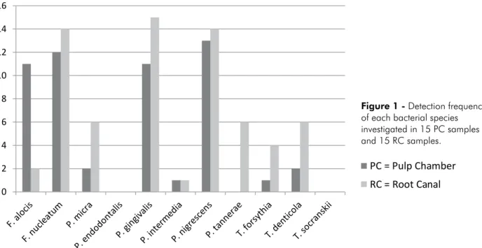
All samples were positive for the presence of bac-terial DNA when the primers directed to the 16S rRNA gene were used. Figure 1 shows the frequen-cy of the 11 species found by species-speciic 16S rRNA–directed PCR among the 15 samples from both the PCs and RCs. In general, more species were detected in the RC (mean, 3.6) than in the PC (mean, 2.7). The highest number of species found in the same tooth was seven, and all teeth presented at least one of the target species, either in the PC or in the RC. The species most frequently detected in the PC were P. nigrescens (86.7%), P. gingivalis
(73.3%), and F. alocis (73.3%). Of the PC samples, 13.3% contained P. micra and T. denticola, and 6.7% contained T. forsythia. The species most fre-quently detected in RC samples were P. gingivalis
(100%) and P. nigrescens (93.3%). P. tannerae, P. micra, and T. denticola were found in 40% of RC samples; T. forsythia was found in 26.7%, F. alocis
in 13.3%, and P. intermedia in 6.7% of RC samples. The species T. socranskii and P. endodontalis were
PC = Pulp Chamber RC = Root Canal
0 2 4 6 8 10 12 14 16
not detected in RC or PC samples.
The concomitant presence of microbial species in PC and RC paired samples occurred in some cases. The detection of P. nigrescens in the PC was statisti-cally associated with the presence of P. nigrescens
in the RC (p = 0.04). Interradicular bone resorption was detected in nine cases; the most frequent species that were associated with these cases were F. alocis
and P. nigrescens in the PCand P. gingivalis and P. nigrescens in the RC.
There was no statistically signiicant association between the presence of bacteria and clinical signs and symptoms.
The simultaneous presence of P. gingivalis, T. forsythia, and T. denticola, which together form the “red complex”,12 was not found in the PC of any
teeth but was found in the RC in three cases.
Discussion
The RCs of primary teeth with necrotic pulp and chronic interradicular/apical lesions present a high number of bacterial species and often exhibit poly-microbial infection with a high prevalence of an-aerobic bacteria.2,13,14 In the present study, bacterial
DNA was detected in all PC and RC samples inves-tigated. Samples collected from the operational ield were DNA-free, conirming that the DNA detected in the PC and RC samples did not result from trace external contamination.
The most commonly isolated genera in endodon-tic infections include Fusobacterium, Prevotella, and Porphyromonas spp.15 In the present study,
P. gingivalis was detected in all RCs investigated, and species such as P. nigrescens were also highly detected. Tavares et al.4 detected P. intermedia in
96.9% of the samples from the RC system of pri-mary teeth exhibiting pulp necrosis with or without radiographically detectable interradicular bone re-sorption, followed by other obligate anaerobes, such as P. nigrescens, T. forsythia, P. denticola, and F. nucleatum ss vincenti. In a study by Ruviere et al.,16
Campylobacter rectus, T. denticola, and Gemella morbillorum were the most prevalent taxa, whereas Cogulu et al.5 found that T. denticola and P.
gingi-valis were the most prevalent species.
The most commonly isolated Gram-positive
coc-ci from RCs of primary teeth include Peptostrepto-coccus spp.17 The present study found the presence
of P. micra, which formerly belonged to the genus
Peptostreptococcus, in 40% of the RCs. P. micra is associated with the pathogenesis of P. gingivalis in endodontic abscesses.18 Ruvieri et al.16 detected P.
micra in 26% of the primary teeth examined, where-as Cogulu et al.5 detected this species only in 3% of
primary teeth exhibiting periapical/interradicular radiolucency. The differences in these results could be attributed to the different sets of primers used or the different features of the samples.
Socransky et al.12 stratiied the periodontal
mi-crobiota into groups or complexes. The “red com-plex” is part of the climax community in bioilms and comprises species that are considered oral pathogens, namely P. gingivalis, T. denticola, and T. forsythia. These species were found in association in three RC samples in this study.
The occurrence of endodontic symptomatol-ogy may be the result of an increased virulence of microorganisms. Cogulu et al.5 found that T.
den-ticola and Enterococcus faecalis are highly associ-ated with previous pain and that P. gingivalis is as-sociated with tenderness to percussion in primary teeth. However, the different incidences of the mi-croorganisms detected in the present report, as com-pared with those of other studies that used different methods for microbial identiication, suggest that, similar to infections in RCs of permanent teeth, the microbial composition of endodontic infections of primary teeth is heterogeneous. This inding con-irms that it is not possible to attribute the etiology of these pathologies to speciic microorganisms.
Even though DNA sequence analysis is the gold standard in microbial identiication, PCR also rep-resents a sensitive, fast, and accessible method for the study of endodontic bacteria.19 Therefore, PCR
possi-bly because of factors such as the gradual decrease in oxygen tension in RCs, together with the nutritional needs of microorganisms and the food chain that in-luence the process of bacterial succession from the saccharolytic cariogenic lora in the PCs to a more anaerobic and proteolytic lora in the RCs.
The ample medullar bone spaces of children favor infection dissemination. In rare situations, bacteria and their toxic products that enter the tra-becular bone in the interradicular area of primary teeth20 and induce acute inlammation and pus
for-mation21 might lead to a spread of these dental
ab-scesses, threatening the life of the child. Another and more likely consequence of pulp necrosis in pri-mary teeth is that these infections may potentially affect the permanent tooth germ.22 In the present
study, no statistical association was found between speciic species and the presence of radiolucent in-terradicular areas. When analyzing 79 patients with infected primary teeth, Cogulu et al.5 found that T.
denticola and E. faecalis are highly associated with periapical radiolucency. These differences in results could be attributed to sample size. Furthermore, in cases with interradicular radiolucency in the present
study, P. gingivalis was found in ive PC and nine RC samples, and P. nigrescens was found in six PC and eight RC samples. The pathogenesis of these species, especially when associated with other anaerobes, may inluence bone loss in the interradicular area.
Conclusion
The role of bacteria in lesion pathogenesis is un-deniable, although modern diagnostic techniques have not identiied a single causative pathogen. The results suggest a high bacterial heterogeneity among PCs and RCs. Strict anaerobes were frequently de-tected in PC and RC samples, but speciic anaero-bic species were not associated with interradicular radiolucent lesions or other signs and symptoms of infection. Therefore, studies comprising a larger number of cases and a wider range of species are necessary to investigate these associations.
Acknowledgements
We would like to thank the CCDB - Pelotas Den-tal School, Federal University of Pelotas and CNPq (483809/2007-1).
References
1. Tani-Ishii N, Wang CY, Tanner A, Stashenko P. Changes in root canal microbiota during the development of rat periapical lesions. Oral Microbiol Immunol. 1994 Jun;9(3):129-35. 2. Toyoshima Y, Fukushima H, Inoue JI, Sasaki Y, Yamamoto K,
Katao H, et al. [A bacteriological study of periapical pathosis on deciduous teeth]. Shoni Shikagaku Zasshi. 1988;26(3):449-58. Japanese.
3. Wrbas KT, Kielbassa AM, Hellwig E. Microscopic studies of accessory canals in primary molar furcations. ASDC J Dent Child. 1997 Mar-Apr; 64(2):118-22.
4. Tavares WL, Brito LCN, Teles RP, Massara ML, Ribeiro Sobrin-ho AP, Haffajee AD, et al. Microbiota of deciduous endodontic infections analysed by MDA and Checkerboard DNA-DNA hybridization. Int Endod J. 2011 Mar;44(3):225-35.
5. Cogulu D, Uzel A, Oncag O, Eronat C. PCR-based identifica-tion of selected pathogens associated with endodontic infec-tions in deciduous and permanent teeth. Oral Surg Oral Med Oral Pathol Oral Radiol Endod. 2008 Jun;106(3):443-9. 6. Fouad AF, Barry J, Caimano M, Clawson M, Zhu Q, Carver
R, et al. PCR-based identification of bacteria associated with endodontic infections. J Clin Microbiol. 2002 Sep;40(9):3223-31.
7. Jacinto RC, Montagner F, Signoretti FG, Almeida GC, Gomes BP. Frequency, microbial interactions, and antimicrobial sus-ceptibility of Fusobacterium nucleatum and Fusobacterium necrophorum isolated from primary endodontic infections. J Endod. 2008 Dec;34(12):1451-6.
8. Moller AJ. Microbiological examination of root canals and periapical tissues of human teeth. Methodological studies. Odontol Tidskr. 1966 Dec 20;74(5):Suppl:1-380.
9. Gomes BP, Jacinto RC, Pinheiro ET, Sousa EL, Zaia AA, Ferraz CC, et al. Porphyromonas gingivalis, Porphyromonas endodontalis, Prevotella intermedia and Prevotella nigrescens in endodontic lesions detected by culture and by PCR. Oral Microbiol Immunol. 2005 Aug;20(4):211-15.
10. American Academy of Pediatric Dentistry. Guideline on pulp therapy for primary and young permanent teeth. Pediatr Dent. 2004;26(7 Suppl):115-9.
11. Montagner F, Jacinto RC, Signoretti FG, Sanches PF, Gomes BP. Clustering behavior in microbial communities from acute endodontic infections. J Endod. 2012 Feb;38(2):158-62. 12. Socransky SS, Haffajee AD, Cugini MA, Smith C, Kent RL Jr.
13. Pazelli LC, Freitas AC, Ito IY, Souza-Gugelmin MC, Me-deiros AS, Nelson-Filho P. Prevalence of microorganisms in root canals of human deciduous teeth with necrotic pulp and chronic periapical lesions. Pesqui Odontol Bras. 2003 Oct-Dec;17(4):367-71.
14. Silva LA, Nelson-Filho P, Faria G, Souza-Gugelmin MC, Ito IY. Bacterial profile in primary teeth with necrotic pulp and periapical lesions. Braz Dent J. 2006 Jul;17(2):144-48. 15. Sundqvist G, Johansson E, Sjogren U. Prevalence of
black-pigmented bacteroides species in root canal infections. J En-dod. 1989 Jan;15(1):13-9.
16. Ruviere DB, Leonardo MR, Silva LA, Ito IY, Nelson-Filho P. Assessment of the microbiota in root canals of human primary teeth by checkerboard DNA-DNA hybridization. J Dent Child (Chic). 2007 May-Aug;74(2):118-23.
17. Yang QB, Fan LN, Shi Q. Polymerase chain reaction-denatur-ing gradient gel electrophoresis, clonreaction-denatur-ing, and sequence analysis of bacteria associated with acute periapical abscesses in chil-dren. J Endod. 2010 Feb;36(2):218-23.
18. Jacinto RC, Gomes BP, Ferraz CC, Zaia AA, Souza Filho FJ. Microbiological analysis of infected root canals from symp-tomatic and asympsymp-tomatic teeth with periapical periodontitis and the antimicrobial susceptibility of some isolated anaerobic bacteria. Oral Microbiol Immunol. 2003 Oct;18(5):285-92. 19. Siqueira Jr JF, Rocas IN. PCR methodology as a valuable
tool for identification of endodontic pathogens. J Dent. 2003 Jul;31(5):333-9.
20. Tannure PN, Barcelos R, Portela MB, Gleiser R, Primo LG. Histopathologic and SEM analysis of primary teeth with pulp-ectomy failure. Oral Surg Oral Med Oral Pathol Oral Radiol Endod. 2009 Jul;108(1):e29-33.
21. Nair PN. Pathogenesis of apical periodontitis and the causes of endodontic failures. Crit Rev Oral Biol Med. 2004 Nov 1;15(6):348-81.