Authors
Patricia Milhoransa1
Carolina Caruccio Montanari1 Rosangela Montenegro2 Roberto Ceratti Manfro1,2
1 Universidade Federal do Rio Grande do Sul, Programa de Pós-Graduação em Medicina: Ciências Médicas, Porto Alegre, RS, Brasil. 2 Hospital de Clínicas de Porto Alegre, Unidade de Transplante Renal, Divisão de Nefrologia, Porto Alegre, RS, Brasil.
Submitted on: 04/24/2018. Approved on: 08/06/2018.
Correspondence to: Patricia Milhoransa. E-mail: milhoransa@ig.com.br
with delayed graft function
Expressão do Micro RNA 146a-5p em pacientes transplantados
renais com disfunção inicial do enxerto
Introdução: O desenvolvimento de novos biomarcadores não invasivos para dis-função do enxerto renal, especialmente no decurso da disfunção inicial do enxer-to, seria de enorme valia para a prática clínica do transplante renal. Métodos: A técnica de RT-PCR foi utilizada para avaliar a expressão de microRNA 146a-5p no sangue periférico e no tecido renal de receptores de transplante submetidos a biópsia renal de controle no decurso de disfunção inicial do enxerto. Resultados: A expressão de miR-146a-5p estava sig-nificativamente aumentada nas amostras de biópsia do grupo de pacientes com dis-função inicial do enxerto (DIE) (n = 33) em relação aos pacientes estáveis (n = 13) e aos com rejeição aguda (RA) (n = 9) (p = 0,008). Foi detectado aumento não sig-nificativo da expressão de miR-146a-5p nas amostras de sangue periférico do gru-po com DIE em comparação aos pacien-tes estáveis e com RA (p = 0,083). Não foi identificada correlação significativa entre os níveis de expressão no plasma e na bi-ópsia. A análise da curva COR revelou uma ASC de 0,75 (IC 95%: 0,62-0,88) para a expressão no tecido renal e de 0,67 (IC 95% 0,52-0,81) no sangue pe-riférico. Conclusão: A expressão de miR-146a-5p tem um padrão distinto no teci-do renal e talvez no sangue periférico em cenários de DIE. Maiores refinamentos e estratégias adicionais de estudo devem ser desenvolvidos na área do diagnóstico molecular não invasivo da disfunção do enxerto renal.
R
ESUMOPalavras-chave: Transplante renal; Rejeição de Enxerto; Função Retardada do Enxerto; Sangue; Biópsia; MicroRNAs; Biomarcado-res; Técnicas de Diagnóstico Molecular.
Introduction: The development of novel non-invasive biomarkers of kidney graft dysfunction, especially in the course of the delayed graft function period would be an important step forward in the clini-cal practice of kidney transplantation.
Methods: We evaluated by RT-PCR the expression of miRNA-146 to -5p ribo-nucleic micro-acids (miRNAs) in the pe-ripheral blood and renal tissue obtained from kidney transplant recipients who un-derwent a surveillance graft biopsy dur-ing the period of delayed graft function.
Results: In biopsy samples, the expres-sion of miR-146a-5p was significantly increased in the group of patients with delayed graft function (DGF) (n = 33) versus stables patients (STA) (n = 13) and patients with acute rejection (AR) (n = 9) (p = 0.008). In peripheral blood samples, a non-significant increase of miR-146a-5p expression was found in the DGF group versus STA and AR groups (p = 0.083). No significant correlation was found be-tween levels of expression in biopsy and plasma. ROC curve analysis revealed an AUC of 0.75 (95% CI: 0.62-0.88) for the renal tissue expression and 0.67 (95% CI 0.52-0.81) for the peripheral blood ex-pression. Conclusion: We conclude that miR-146a-5p expression has a distinct pattern in the renal tissue and perhaps in the peripheral blood in the setting of DGF. Further refinements and strategies for studies should be developed in the field of non-invasive molecular diagnosis of kidney graft dysfunction.
A
BSTRACTKeywords: Kidney transplantation; Graft Rejection; Delayed Graft Function; Blood; Biopsy; MicroRNAs; Biomarkers; Molec-ular Diagnostic Techniques.
I
NTRODUCTIONRenal transplantation is the treatment of choice for many patients with end-stage renal impairment1,2. It offers a significant increase in life expectancy and qua-lity of life of patients with end-stage renal function3. However, as evidenced since the beginning of organ transplants, tissues and organs of genetically distinct individuals lose their functions through a rejection process that is mediated by the immune system. Such process is only partially controlled by modifying the receptor immune response with immunosuppressive drugs and biological agents3-5.
In the last century, the mechanisms of alloimmune response were elucidated and immunosuppressive drugs capable of preventing rejection were developed turning organ transplantation into a clinical reality6,7.
In kidney transplantation, injury due to ischemia and reperfusion is an inevitable process resulting many times in delayed graft function (DGF) that is currently characterized by the need of dialysis within the first week after transplantation. Ischemia and re-perfusion injury (IRI) also promotes activation of the innate and adaptive responses of the immune system, leading to processes with great potential to produce significant graft harm. It is believed that these inju-ries also facilitate mechanisms of acute rejection (AR) and act by programming gene, metabolic, and tissue changes that culminate in tissue graft fibrosis and chronic loss of function8,9.
AR, a frequent and ominous complication of or-gan transplantation, is still considered a risk factor for early and late graft loss10. Currently, the clinical phenotypes of rejection are well elucidated in clinical practice. Rejection is usually evidenced by the organ dysfunction, which leads to a graft biopsy that is clas-sified by histological alterations11. Its evolution is dif-ficult to predict and the histological findings observed in renal tissue obtained are still considered the best predictors11,12. Biopsy on the other hand is associ-ated with a variety of complications. In addition, it is costly, has representativeness issues, and is subject to interpretation variability. Nevertheless, in current practice, it is still the gold standard for the diagnosis of renal graft dysfunctions12,13.
Graft damage classification is done through the Banff classification, a standard international con-sensus of nomenclatures and specific criteria for the histological characterization of organ rejection, in which T cell mediated rejection or antibody mediated
rejection are diagnosed based on empirical rules and the lesions are graded semi-quantitatively14. Briefly, diagnoses according to this classification may be grouped as borderline rejection, acute, tubulointersti-tial or vascular cell rejections of different severities, or as antibody-mediated acute rejection, characterized by histological findings, presence of donor antibodies anti-HLA and by the labeling of C4d in the peritubu-lar capilperitubu-laries14.
Accurate non-invasive biomarkers are an unmet need in the clinical practice of organ transplantation. Most of the work with molecular biomarkers has been done analyzing messenger RNA expression15,16. An important discovery of molecular biology in re-cent years is the micro-RNAs (miRNAs)17,18. They consist of small conserved and non-RNA coding frag-ments of approximately 25 nucleotides, which inhibit transcription of mRNA, are induced by translational depression or degradation of mRNA, and are respon-sible for regulating gene expression18-20.
Many microRNAs are involved in the develop-ment or progression of chronic or acute kidney dis-ease in patients or animal models. hsa-miR-146a-5p was upregulated in patients with FSGS (focal seg-mental glomerulosclerosis) and MPGN (membrano-proliferative GN) compared with patients with DN (diabetic nephropathy). miR-146a was modulated in an experimental model of renal I/R in mice and in pa-tients with IgA nephropathy, where its levels in renal tissue and urine were correlated with injury severity. It is important to emphasize that miR-146a-5p, which demonstrated a very high diagnostic value in ICU (intensive care unit) patients, presents a strong and significant downregulation during early AKI (acute kidney injury). Thus, this miRNA could be consid-ered a precise and early AKI diagnostic tool in several clinical contexts18-21.
MicroRNA 146a-5p acts as a mediator of the re-nal tubular response to the ischemia-reperfusion inju-ry, limiting the inflammatory process in this setting21. In the present study, we quantitatively assessed miR-146a-5p in the renal tissues and peripheral blood lym-phocytes of kidney transplant recipients with delayed graft function. We hypothesized that miR-146a-5p would present enhanced transcription signaling in re-cipients with DGF compared to patients with stable function and those with AR at two compartments, peripheral blood and renal graft tissue.
M
ETHODSPATIENTS
In order to obtain adequate statistical power, with an estimated AR incidence of 40% in patients with acu-te graft dysfunction and of 20% in patients without acute graft dysfunction, a proposed sample size of 55 patients that underwent renal graft biopsies was cal-culated and enrolled in the study. The sample size was calculated following the parameters: a) study power of 80%; b) Pα = 0.05; c) Pβ: 0.20; d) magnitude of the difference: 50%.
Peripheral blood was also obtained from these pa-tients for miRNA analysis. Thirty-three papa-tients had DGF when the biopsies were performed (surveillance biopsies), nine had acute graft dysfunction (indica-tion biopsies) that was attributed to AR, and 13 were normal protocol biopsies obtained at three months after transplantation. The study was conducted at the Renal Transplantation Unit, Division of Nephrology, Hospital de Clínicas de Porto Alegre, Brazil, between May 2013 and April 2017. All patients provided writ-ten informed consent for their participation.
All patients were on immunosuppression con-sisting of a combination of corticosteroids, sodium mycophenolate, and calcineurin inhibitors. Either anti-IL2 receptor antibodies (Basiliximab®) or rabbit anti-thymocyte globulin (Thymoglobulin®) induction therapy was used in all deceased-donor graft recipi-ents and for all living-donor graft recipirecipi-ents consid-ered at increased risk of rejection.
The ethical and methodological aspects of this study were approved by the Hospital de Clínicas de Porto Alegre Research Ethics.
SAMPLES
Biopsies were performed percutaneously, under real--time ultrasound guidance, using a semi-automatic
biopsy gun with a 16G needle. At the time of biopsy, all patients had well-controlled blood pressure and all parameters of the coagulation panel (obtained no mo-re than 24 hours befomo-re) within normal limits. Befomo-re renal biopsy, a comprehensive workup was perfor-med to rule out obstructive or vascular issues, urinary fistula, infection, or drug toxicity as causes of graft dysfunction.
SPECIMENCOLLECTIONANDPREPARATION
Two renal cortex fragments were collected during each biopsy procedure. One-third of one of these fragments was placed in a microtube, flash-frozen by submerging in liquid nitrogen, and stored at -80°C. Peripheral blood samples (5 mL collected into EDTA-containing tubes) were obtained immediately before the biopsies.
For cell separation, both sample types were rinsed and processed to concentrate the cells of interest (blood). In the case of blood samples, 2-mL aliquots were transferred to sterile, 12-mL Falcon tubes to which 10 mL of erythrocyte-lysing Buffer EL (Qiagen Inc., Chatsworth, CA, USA) were added, followed by 21 minutes of incubation on ice with intermittent vor-texing every 7 minutes. After this step, the samples were centrifuged at 1800 rpm for 10 minutes, leaving a pellet that contained the cells of interest at the bot-tom of the tube. The pellet was preserved and the su-pernatant discarded. The pellet was then resuspended in 1.5 mL of Buffer EL, transferred to microtubes, further centrifuged for 10 minutes at 10,000 rpm, the supernatant discarded, and the resulting cell concen-trate frozen at -80°C.
MICRO RNA PROCESSING
Micro RNAs were extracted from samples using the mirVana™ PARIS™ commercial kit (Ambion®, Life Technologies Corporation). Briefly, cell concentrate/ sediment was dissolved or fragmented with 500 μL of buffer in a dispersing machine (ULTRA-TURRAX T 10 basic - IKA, Campinas, SP, Brazil) and eluted in 60 μL of water for injection preheated to 95°C, in accor-dance with manufacturer instructions.
lower phase of the acid-phenol:chloroform provided in the kit was added, the lysate vigorously vortexed for 1 minute, and then centrifuged at 13,000 rpm for 5 minutes for organic phase separation. The phase of interest (the upper phase) was then collected, mea-sured (maximum volume 600 μL), and transferred to a fresh microtube, to which was added 100% ethanol at room temperature corresponding to one-third of the obtained volume. The resulting solution was ho-mogenized and 700 μL transferred to the filtering ap-paratus provided in the kit. This apap-paratus was then centrifuged at 10,000 rpm for 30 seconds. The result-ing filtrate contained the desired miRNAs. Ethanol (466 μL) was added to this fluid, which was passed through a second filter, and the washing process be-gun. This process consisted of the addition of 700 μL of Wash Solution 1, centrifuging for 15 seconds at 10,000 rpm, addition of 500 μL of Wash Solution 2/3, and repeating the two preceding steps. After discarding the flow-through, the apparatus is centri-fuged for 1 minute to remove any residual reagent, the column is transferred into a fresh collection tube, and the miRNAs are collected by eluting with 60 μL of water for injection heated to 95ºC. The eluate was then centrifuged at 10,000 rpm for 50 seconds and stored at -80ºC until the next stage.
The concentration of extracted miRNA was quan-tified in a full-spectrum spectrophotometer (220-750nm) with sample retention technology (Nanodrop 1000, Thermo Fischer Scientific, Wilmington, DE, USA). The nucleic acid concentration is expressed in ng/μL based on optical density at 260 nm, and purity is calculated based on the A260/280 and A260/230 ratios. A ratio of approximately 2.0 is generally ac-cepted as “pure” RNA. Samples were considered vi-able if they had a concentration of at least 2 ng/μL. All samples with a higher concentration were diluted to this concentration in a 50 μL volume of nuclease-free water.
AMPLIFICATIONANDDETECTION
The following specific TaqMan primers (Applied Biosystems®) were used for real-time reverse transcrip-tion polymerase chain reactranscrip-tion (RT-PCR): miR146a--5p 4427975/000468; RNU-48 4427975/001006 and Cel-miR-39-5p 4427975/464312. The endoge-nous control used for sample normalization was syn-thetic exogenous control Cel-miR-39 from C. elegans (Qiagen, catalog number MSY0000010), which was
spiked into samples before the reverse transcription stage in a 0.5 μL volume at a 50 pM concentration.
COMPLEMENTARY DNA
The complementary DNA (cDNA) formation stage was carried out with the TaqMan MicroRNA RT kit (Applied Biosystems®) as per manufacturer instruc-tions, using 8 μL of reaction mix: 0.12 μL of 100mM dNTPs, 0.8 μL of MultiScribe™ reverse transcripta-se (50 U/μL), 1.2 μL of enzyme buffer, 0.144 μL of RNase inhibitor (20 U/μL), 3.40 μL of nuclease-free water, and 2.4 μL of target-specific primer, to which 4 μL of miRNA were added, finalizing a total volume of 12 μL. For synthesis, the samples were incubated at 16ºC for 30 minutes, at 42ºC for 30 minutes, and at 85ºC for 5 minutes, and then stored at -20ºC until the time for RT-PCR.
REVERSETRANSCRIPTIONPOLYMERASECHAINREACTION
RT-qPCR which consisted of the amplification of 2μL cDNA using 5.0μL of TaqMan Universal PCR Master Mix (Applied Biosystems®), 0.5 μL of specific primers, and 2.5 μL of nuclease-free water, in a final reaction volume of 10 μL. Analyses of miRNA ex-pression were performed using TaqMan MicroRNA Assay and individual TaqMan MicroRNA Assays (TaqManMicroRNA Assay, Applied Biosystems, Foster City CA, USA) for miR-146a-5p a Step-One Real Time PCR 48-welloptical plate (Applied Biosystems, Foster City CA, USA)22.
One endogenous miRNAs (RNU48) was ana-lyzed, and the expression biopsy data were normal-ized against RNU48.The data were presented as the relative quantity of target miRNA normalized to endogenous. The exogenous control used was a cel-miR-39 of blood samples.
The controls and normalizers were selected ac-cording to literature and with the help of the scien-tific advice of Thermo Fischer Scienscien-tific (https://www. thermofisher.com/br).
The cycle threshold (Ct) was calculated automati-cally using software. miRNAs expression was nor-malized using the 2-∆∆Ct method described by Livak and Schmittgen22.
STATISTICALANALYSES
means ± standard deviations. The Kruskal–Wallis and Mann-Whitney U tests were used for paired--samples analysis of variance and for between-group analysis. Spearman’s correlation was used to see the association between two variables. Qualitative data are expressed as absolute and relative counts, and the chi-square or Fisher’s exact tests were used for betwe-en-group analyses. All tests were two-tailed and a p--value < 0.05 was defined as statistically significant. All analyses were carried out in PASW Statistics 21.0 (SPSS Inc., Chicago, IL, USA).
R
ESULTSDiagnostic classification was achieved by a combi-nation of the clinical assessment, response to specific therapy and biopsy findings, as interpreted per the Banff 2013 classification14. Fifty-five patients were divided between three groups according to the esta-blished diagnosis. Thirty-three patients were on DGF,
nine were having an AR episode, and thirteen were patients with stable graft function that underwent a protocol biopsy. For all patients, a biopsy and a peri-pheral blood sample were obtained and processed for miRNA 146a-5p expression. Demographic data are shown in Table 1. Only serum creatinine levels (p = 0.022) and time elapsed from transplant to biopsy (p = 0.001) differed significantly between groups. Serum creatinine was significantly elevated in the DGF group and time to biopsy was longer in the DGF group com-pared to the stable group. As expected, time to biopsy was also longer in the stable group compared to the other two groups. The differences observed in the ini-tial immunosuppression regimens did not reach statis-tical significance (p = 0.069).
In the renal tissue, microRNA 146a-5p was differ-entially expressed between groups. It was enhanced in the DGF group (median [IQR], 3.23 [1.46-5.74]) compared to the stable group (median [IQR], 0.78
Variables Stable n = 13 DGF n = 33 AR n = 9 p
Age (years; mean±SD) 48.3±12.2 46.8±14.5 42.8±11.6 0.632*
Sex (male/female) 9/4 15/18 4/5 0.318**
Race (white/nonwhite) 11/2 28/5 8/1 0.950**
Donor age (years; mean+SD) 45.6±10.4 46±16.9 42.8±17 0.864*
Donor sex (male/female) 4/9 17/16 4/5 0.444**
Early graft dysfunction (yes/group total) 9/4 29/4 7/2 0.434**
HLA mismatches (A, B, DR; mean+SD) 2.53±0.5 2.2±0.8 2.5±0.5 0.252*
Last PRA (%; median (IQR))
Class I 1 (0-23.5)a 7.0 (0-21.5)a 11 (0-24.1)a 0.430*
Class II 2 (0-28.5)a 1.5 (0-22)a 2.5 (0-24)a 0.930*
Initial immunosuppression (n; %) 0.069**
No induction 6 (46.2) 10 (30.3) 4 (44.4) 0.518*
Rabbit anti-thymocyte globulin 1 (7.7.) 16 (50) 4 (44.4) 0.029*
Basiliximab 6 (46.2) 6 (18.8) 1 (11.1) 0.091*
Cold ischemia time (h; mean+ SD) 20+5.1 25.2+7.7 21.8+7.4 0.196*
Underlying renal disease (n; %) 0.509**
Unknown 6 (46.2) 7 (22.6) 4 (50) 0.165*
HTN 5 (38.5) 13 (39.4) 4 (44.4) 0.955*
DM 3 (23.1) 8 (24.2) 0 (0) 0.259*
HTN+DM 1 (8.3) 4 (12.5) 0 (0) 0.554*
APKD 2 (16.7) 4 (12.5) 1 (12.5) 0.934*
Other 0 (0) 8 (25.8) 2 (25) 0.147*
Serum Creatinine at Biopsy (mg/dL; mean ± SD) 2.5±1.9a 5.7±4b 3.8±3.3ab 0.022*
Time to biopsy (days; median [IQR]) 99 [86-116]b 14 [12-26]a 58 [14-331]ab 0.001***
TABLE 1 DEMOGRAPHICPROFILEOFSTUDIEDPATIENTSANDTRANSPLANTVARIABLES
SD: standard deviation; HLA: human leukocyte antigen; HTN: hypertension; DM: diabetes mellitus; APKD: autosomal dominant polycystic kidney disease; *Analysis of variance (ANOVA); ** Pearson’s chi-square; *** Kruskal-Wallis test; a,b Equal letters do not differ by the Tukey’s (ANOVA) or
STABLE (n = 13) DGF (n = 33) AR (n = 9) p-value
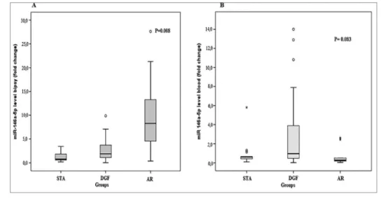
Biopsy miR-146a-5p 0.78 [0.57 - 1.99] 3.23 [1.46 - 5.74] 1.07 [0.43 - 2.11] 0.008
Peripheral Blood miR-146a-5p 0.62 [0.31 - 0.90] 0.96 [0.46 - 3.88] 0.27 [0.15 -1.47] 0.083
TABLE 2 MIR-146A-5PEXPRESSION* INTHERENALTISSUEANDPERIPHERALBLOODINTHEGROUPSOFPATIENTS
*Median [IQR]; P-values determined by Kruskal-Wallis/Mann-Whitney U test.
Figure 1. (A) Comparison of expression levels of miR-146a-5p in the renal tissue in the diagnostic categories (B) Comparison of the expression of the miR-146a-5p in peripheral blood in diagnostic categories.
Figure 2. Correlation between expression of miR-146a-5p levels at the renal tissue and pheripheral blood.
[0.57-1.99]; p = 0.019). The differences observed in the comparisons between the DGF group (median [IQR], 3.23 [1.46-5.74]) and the AR group (median [IQR], 1.07 [0.43-2.11]; p = 0.106) and the compari-son between the AR group and the stable group (me-dian [IQR], 0.78 [0.57-1.99]; p = 1.0) were not statis-tically significant (Table 2 and Figure 1A).
In the peripheral blood, the microRNA 146a-5p expression was heightened in the DGF group (median [IQR], 0.96 [0.46-3.88]) compared to AR group (me-dian [IQR], 0.27 [0.15-1.47]) and the stable group (median [IQR], 0.62 [0.31-0.90]), however the dif-ferences were not statistically significant (p = 0.083) (Table 2 and Figure 1B).
As illustrated in Figure 2, no significant correlation was found between expression level of miR-146a-5p in different compartments, biopsy, and peripheral blood (r=0.084; p = 0.541).
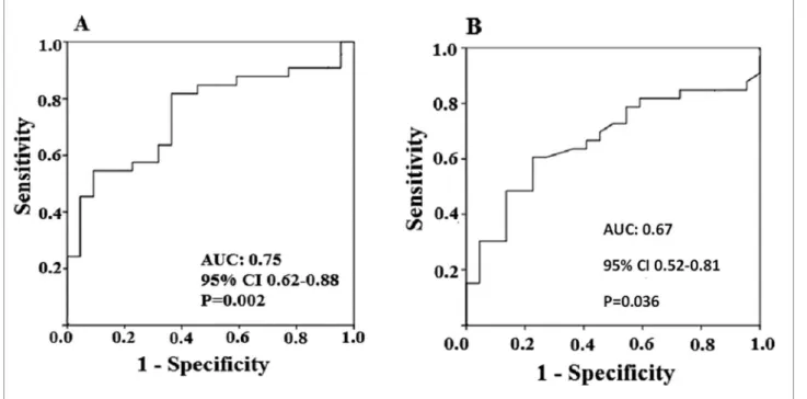
Receiver operating characteristic (ROC) curve of the biopsy analysis was plotted for assessment of diagnostic parameters of miRNA 146a-5p gene expression for the DGF diagnosis. The area under the curve (AUC) was 0.75 (95% CI: 0.62-0.88). Using a cutoff point of 1.64 at the ROC curve, i.e., a 64% increase in gene expres-sion relative to controls, the obtained parameters were: sensitivity of 67.0%; specificity of 64.0%; positive pre-dictive value of 73.3%; and negative prepre-dictive value of 56% (p = 0.002, Pearson’s chi-square test) (Figure 3A).
ROC curve of the peripheral blood analysis was plotted for assessment of diagnostic parameters of miRNA 146a-5p gene expression for the DGF diagnosis. The AUC for miR-146a-5p was 0.67 (95% CI 0.52-0.81). Using a cutoff point of 0.63 on the ROC curve, i.e., a 63% in-crease in gene expression relative to controls, the obtained parameters were: sensitivity of 64%; specificity of 64%; positive predictive value of 72.4%; and negative predic-tive value of 53.8% (p = 0.036, Pearson’s chi-square test) (Figure 3B).
Figure 3. (A) ROC curve of the miR-146a-5p expression levels do the diagnoses of delayed graft function at the renal tissue; (B) ROC curve of the miR-146a-5p expression levels do the diagnoses of delayed graft function at the peripheral blood.
heart failure and neoplasms. The utility of miRNAs as biomarkers depends on several factors related to care during sample collection, processing, and storage. The conditions involved in the processing and stor-age of the miRNAs are of extreme importance for the integrity of these molecules be maintained until the moment of their analysis. Thus, the results obtained will not change due to methodological problems. Although the need for sample handling care is well known in the literature, it is still necessary to establish standard protocols for the collection, processing, and storage of this type of sample. In addition, the stabil-ity of the miRNAs during the storage period and in different conditions also does not have a consensus in the scientific community. This heterogeneity of pro-cedures may be an important source of disagreement and questioning regarding the utility of miRNAs as disease biomarkers.
D
ISCUSSIONDelayed graft function is a peculiar and very fre-quent form of acute kidney injury (AKI) that occurs immediately after renal transplantation. Its incidence is of around 25% in the United Network for Organ Sharing (UNOS) in North America but is substan-tially higher in Brazil8. Besides, Brazil carries a worse graft prognosis and perhaps patient survival. Similar
to native kidney AKI survivors, DGF exhibits a signi-ficant risk of developing chronic kidney graft disease (CKD), with a course to accelerated end-stage renal disease8,23-26.
The ischemia and reperfusion injury (IRI) that oc-curs after renal transplantation is the main driver of the DGF clinical phenotype and perhaps, in its more serious forms, may lead to graft primary non-func-tion. IRI also promotes activation of the innate and adaptive responses of the immune system, leading to processes that are potentially harmful to the kidney graft. It is believed that these injuries may also facili-tate or trigger mechanisms of acute rejection (AR) and later, by gene reprogramming, metabolic and tis-sue changes may culminate in tistis-sue graft fibrosis and chronic loss of function8,9.
The search for noninvasive biomarkers reflecting intra-graft events opens important avenues for diag-nosis, progdiag-nosis, and therapeutic monitoring in trans-plantation science. In the present study miR-146a-5p was assessed as potential biomarker of IRI that occurs in DGF after renal transplantation21,27.
The precursor pri-miR, miR-146a has been shown to be modulated in an experimental model of renal IRI in mice and in patients with IgA nephropathy. In this study, the miR levels in the renal tissue were found correlated with injury severity21. This precur-sor is processed into mature miR-146a-5p, which has been shown to be related to ischemia and renal IRI.
In present study, expression of miR-146a-5p in the renal tissue was significantly increased in biopsies of patients with DGF. In the sub-group analyses, the DGF group presented higher expression compared with patients with stable graft function. A non-sig-nificant difference was found in the comparison be-tween the DGF and AR groups and very similar levels of expression were found in the comparison between the AR and stable groups. In the analysis of the miR-146a-5p obtained from the peripheral blood samples, the levels of expression were also higher in the DGF group, in comparison with the group of stable pa-tients. In this analysis, the differences were of bor-derline statistical significance. Numerically, the DGF group presented higher levels of expression.
The miRNAs are involved in a variety of physi-ological and pathphysi-ological processes, including stress response, inflammation, heart disease, neurodegen-erative diseases (e.g. Alzheimer’s and Parkinson’s disease), autophagy, apoptosis, and various types of cancer. In the specific case of miRNAs exerting their regulatory role in cancer cells, these small RNA mol-ecules can act as both oncogenes (activating the cell cycle) and tumor suppressor genes (inhibiting cell di-vision), depending on the nature of the miRNA and the metabolic pathway in which they are involved. The association of characteristics such as biological function, presence in biological fluids, and stability places the miRNAs as promising biomarkers for the diagnosis and prognosis of various diseases.
Amrouche and colleagues found that this molecule might act as general regulator of the innate immune re-sponse in not only immune cells but also cells that are targeted by inflammation in human renal tissue and urine. They observed an increase in expression of this
biomarker in patients with acute tubular necrosis early after transplantation compared to those who displayed normal allograft biopsy results21. Experimentally, they were able to demonstrate that renal ischemia induces tubular miR-146a expression in mouse kidneys after unilateral IRI and that, in comparison with the con-tralateral kidneys, the heightened levels of expression were still demonstrable up to 7 days after IRI. They also found that miR-146a was predominantly overex-pressed in tubular cells. These results emphasize miR-146a as an important effector of the pathogenesis of the renal response to IRI injury21.
Baker and collaborators studied the relative contri-butions of micro RNAs to the establishment of kidney disease. Analyzing human renal biopsies of patients with diabetic nephropathy, focal and segmental glo-merulosclerosis, IGA nephropathy, membrane prolif-erative GN and controls they found that miR-146a-5p distinguished diabetic nephropathy from the other conditions and concluded that this molecule may be used as a biomarker of kidney diseases and is perhaps involved in disease mechanisms28. Fraile and collabo-rators analyzed miR-146a-5p expression levels in the sera of AKI and control individuals. They found that this microRNA is overexpressed in the serum of pa-tients with AKI compared to healthy controls. It was also found that its expression levels in the renal tissue and urine are correlated with injury severity29-31.
Tang et al. identified a miRNAs as regulator of tar-get gene expression involved in shaping the immune re-sponse. These authors investigated the role of miR-146a in the pathogenesis of systemic lupus erythematosus and found that this microRNA, among others, act as a negative regulator of innate immunity in these patients. Further analysis showed that underexpression of miR-146a negatively correlates with clinical disease activity and with interferon (IFN) scores in patients with sys-temic lupus erythematosus. The microRNA miR-146a is a negative regulator of the IFN pathway; underexpres-sion of miR-146a contributes to alterations in the type I IFN pathway in lupus patients by targeting key signal-ing proteins. They suggested that their findsignal-ings provide potential novel strategies for therapeutic intervention37.
Previous research has shown that miRNAs can ex-it cells through exosomes and be transported in body fluids to other compartments where they may act as local regulators38,39. This biological property might, at least in part, explain the discrepancies between tissue and peripheral blood expression found in the pres-ent study. Alternatively, and perhaps more plausibly, the higher amounts found in tissue allows a better and more reliable detection of the microRNA. It is also possible that the evaluation in the urine would provide interesting results in terms of applicability of this molecule expression as a non-invasive biomarker. However, DGF recipients are frequently anuric ant that would be a relevant limitation for its application as a non-invasive biomarker in renal transplantation.
Among the weaknesses of the present study, per-haps the most important is the restricted number of individuals evaluated, which may have contributed to the negative results in the sub-group comparisons, more importantly in the peripheral blood analysis.
Although a miRNA panel capable of distinguish-ing among the various etiologies of dysfunction that can affect renal allografts has yet to be established, we identified miR-146a-5p as a potential biomarker for differentiating IRI, with the DGF clinical phenotype, from other conditions such as acute rejection. We sug-gest that the combined analysis of micro RNAs may lead to an accurate non-invasive diagnosis of kidney graft injuries.
In summary, further studies with other potential biomarkers of IRI and acute rejection, perhaps involv-ing already surveyed microRNAs such as miR-146a-5p and miR-142-3 in different non-invasive samples might contribute to the development of accurate non-invasive biomarkers(s) for use in clinical organ transplantation40.
R
EFERENCES1. Harmath CB, Wood CG 3rd, Berggruen SM, Tantisattamo E. Renal Pretransplantation Work-up, Donor, Recipient, Surgical Techniques. Radiol Clin North Am 2016;54:217-34.
2. Wilflingseder J, Regele H, Perco P, Kainz A, Soleiman A, Mühlbacher F, et al. miRNA profiling discriminates types of rejection and injury in human renal allografts. Transplantation 2013;95:835-41.
3. Muduma G, Shupo FC, Dam S, Hawken NA, Aballéa S, Odeye-mi I, et al. Patient survey to identify reasons for non-adherence and elicitation of quality of life concept associated with immu-nosuppressant therapy in kidney transplant recipients. Patient Prefer Adherence 2016;10:27-36.
4. Doi K, Rabb H. Impact of acute kidney injury on distant organ function: recent findings and potential therapeutic targets. Kid-ney Int 2016;89:555-64.
5. Saat TC, van den Akker EK, IJzermans JN, Dor FJ, de Bruin RW. Improving the outcome of kidney transplantation by ame-liorating renal ischemia reperfusion injury: lost in translation? J Transl Med 2016;14:20.
6. Denecke C, Tullius SG. Innate and adaptive immune responses subsequent to ischemia-reperfusion injury in the kidney. Prog Urol 2014;24:S13-9.
7. Menon MC, Keung KL, Murphy B, OʼConnell PJ. The Use of Genomics and Pathway Analysis in Our Understanding and Prediction of Clinical Renal Transplant Injury. Transplantation 2016;100:1405-14.
8. Helfer MS, Vicari AR, Spuldaro F, Gonçalves LF, Manfro RC. Incidence, risk factors, and outcomes of delayed graft function in deceased donor kidney transplantation in a Brazilian center. Transplant Proc 2014;46:1727-9.
9. Nankivell BJ, Alexander SI. Rejection of the kidney allograft. N Engl J Med 2010;363:1451-62.
10. Bandari J, Fuller TW, Turner Іі RM, D'Agostino LA. Renal biopsy for medical renal disease: indications and contraindica-tions. Can J Urol 2016;23:8121-6.
11. Broecker V, Mengel M. The significance of histological diagnosis in renal allograft biopsies in 2014. Transpl Int 2015;28:136-43. 12. Loupy A, Lefaucheur C, Vernerey D, Chang J, Hidalgo LG,
Beuscart T, et al. Molecular microscope strategy to improve risk stratification in early antibody-mediated kidney allograft rejection. J Am Soc Nephrol 2014;25:2267-77.
13. Bhatt K, Kato M, Natarajan R. Mini-review: emerging roles of microRNAs in the pathophysiology of renal diseases. Am J Physiol Renal Physiol 2016;310:F109-18.
14. Haas M, Sis B, Racusen LC, Solez K, Glotz D, Colvin RB, et al.; Banff meeting report writing committee. Banff meeting re-port writing committee. Banff 2013 meeting rere-port: inclusion of c4d-negative antibody-mediated rejection and antibody-as-sociated arterial lesions. Am J Transplant 2014;14:272-83. 15. Aquino-Dias EC, Joelsons G, da Silva DM, Berdichevski RH,
Ribeiro AR, Veronese FJ, et al. Non-invasive diagnosis of acute rejection in kidney transplants with delayed graft function. Kid-ney Int 2008;73:877-84.
16. Suthanthiran M, Schwartz JE, Ding R, Abecassis M, Dadhania D, Samstein B, et al.; Clinical Trials in Organ Transplantation 04 (CTOT-04) Study Investigators. Urinary-cell mRNA profile and acute cellular rejection in kidney allografts. N Engl J Med 2013;369:20-31.
17. Liu J. Control of protein synthesis and mRNA degradation by microRNAs. Curr Opin Cell Biol 2008;20:214-21.
18. Bartel DP. MicroRNAs: genomics, biogenesis, mechanism, and function. Cell 2004;116:281-97.
19. Ambros V, Bartel B, Bartel DP, Burge CB, Carrington JC, Chen X, et al. A uniform system for microRNA annotation. RNA 2003;9:277-9. 20. Wei Q, Mi QS, Dong Z. The regulation and function of
mi-croRNAs in kidney diseases. IUBMB Life 2013;65:602-14. 21. Amrouche L, Desbuissons G, Rabant M, Sauvaget V, Nguyen
22. Livak KJ, Schmittgen TD. Analysis of relative gene expression data using real-time quantitative PCR and the 2(-Delta Delta C(T)) Method. Methods 2001;25:402-8.
23. Bonventre JV, Yang L. Cellular pathophysiology of ischemic acute kidney injury. J Clin Invest 2011;121:4210-21.
24. Srisawat N, Kellum JA. Acute kidney injury: definition, epide-miology, and outcome. Curr Opin Crit Care 2011;17:548-55. 25. de Sandes-Freitas TV, Felipe CR, Aguiar WF, Cristelli MP,
Te-desco-Silva H, Medina-Pestana JO. Prolonged Delayed Graft Function Is Associated with Inferior Patient and Kidney Allo-graft Survivals. PLoS One 2015;10:e0144188.
26. Haase M, Shaw A. Acute kidney injury and cardiopulmonary bypass: special situation or same old problem? Contrib Ne-phrol 2010;165:33-8.
27. Li JY, Yong TY, Michael MZ, Gleadle JM. Review: The role of microRNAs in kidney disease. Nephrology (Carlton) 2010;15:599-608.
28. Baker MA, Davis SJ, Liu P, Pan X, Williams AM, Iczkowski KA, et al. Tissue-Specific MicroRNA Expression Patterns in Four Types of Kidney Disease. J Am Soc Nephrol 2017;28:2985-92. 29. Aguado-Fraile E, Ramos E, Conde E, Rodríguez M, Martín--Gómez L, Lietor A, et al. A Pilot Study Identifying a Set of microRNAs As Precise Diagnostic Biomarkers of Acute Kidney Injury. PLoS One 2015;10:e0127175.
30. Wang G, Kwan BC, Lai FM, Chow KM, Li PK, Szeto CC. Elevated levels of miR-146a and miR-155 in kidney biopsy and urine from patients with IgA nephropathy. Dis Markers 2011;30:171-9.
31. Godwin JG, Ge X, Stephan K, Jurisch A, Tullius SG, Iacomini J. Identification of a microRNA signature of renal ischemia re-perfusion injury. Proc Natl Acad Sci U S A 2010;107:14339-44.
32. Dziedzic M, Powrózek T, Orłowska E, Koch W, Kukula-Koch W, Gaweł K, et al. Relationship between microRNA-146a ex-pression and plasma renalase levels in hemodialyzed patients. PLoS One 2017;12:e0179218.
33. Sonkoly E, Ståhle M, Pivarcsi A. MicroRNAs and immunity: novel players in the regulation of normal immune function and inflammation. Semin Cancer Biol 2008;18:131-40.
34. Quinn SR, O'Neill LA. A trio of microRNAs that control Toll--like receptor signalling. Int Immunol 2011;23:421-5.
35. Zbroch E, Małyszko J, Małyszko J, Koc-Żórawska E, Myśliwiec M. Renalase, kidney function, and markers of endothelial dys-function in renal transplant recipients. Pol Arch Med Wewn 2012;122:40-4.
36. Kalyani A, Sonawane PJ, Khan AA, Subramanian L, Ehret GB, Mullasari AS, et al. Post-Transcriptional Regulation of Renala-se Gene by miR-29 and miR-146 MicroRNAs: Implications for Cardiometabolic Disorders. J Mol Biol 2015;427:2629-46. 37. Tang Y, Luo X, Cui H, Ni X, Yuan M, Guo Y, et al.
MicroR-NA-146A contributes to abnormal activation of the type I in-terferon pathway in human lupus by targeting the key signaling proteins. Arthritis Rheum 2009;60:1065-75.
38. Gibbings DJ, Ciaudo C, Erhardt M, Voinnet O. Multivesicular bodies associate with components of miRNA effector complexes and modulate miRNA activity. Nat Cell Biol 2009;11:1143-9. 39. D'Alessandra Y, Devanna P, Limana F, Straino S, Di Carlo
A, Brambilla PG, et al. Circulating microRNAs are new and sensitive biomarkers of myocardial infarction. Eur Heart J 2010;31:2765-73.