Upregulation of microRNA 142-3p in the peripheral
blood and urinary cells of kidney transplant recipients
with post-transplant graft dysfunction
T.D. Domenico
1, G. Joelsons
1, R.M. Montenegro
2and R.C. Manfro
1,2 1Programa de Pós-Graduac¸ão em Medicina: Ciências Médicas, Faculdade de Medicina, Universidade Federal do Rio Grande do Sul,
Porto Alegre, RS, Brasil 2
Unidade de Transplante Renal, Servic¸o de Nefrologia, Hospital de Clínicas de Porto Alegre, Porto Alegre, RS, Brasil
Abstract
We analyzed microRNA (miR)-142-3p expression in leucocytes of the peripheral blood and urinary sediment cell samples obtained from kidney transplant recipients who developed graft dysfunction. Forty-one kidney transplant recipients with kidney graft dysfunction and 8 stable patients were included in the study. The groups were divided according to histological analysis into acute rejection group (n=23), acute tubular necrosis group (n=18) and stable patients group used as a control for gene expression (n=8). Percutaneous biopsies were performed and peripheral blood samples and urine samples were obtained. miR-142-3p was analyzed by real-time polymerase chain reaction. The group of patients with acute tubular necrosis presented significantly higher expressions in peripheral blood (Po0.05) and urine (Po0.001) compared to the stable patients group. Also,
in the peripheral blood, miR-142-3p expression was significantly higher in the acute tubular necrosis group compared to the acute rejection group (Po0.05). Urine samples of the acute rejection group presented higher expression compared to the stable
patients group (Po0.001) but the difference between acute tubular necrosis and acute rejection groups was not significant
in the urinary analyzes (P=0.079). miR-142-3p expression has a distinct pattern of expression in the setting of post-operative acute tubular necrosis after kidney transplantation and may potentially be used as a non-invasive biomarker for renal graft dysfunction.
Key words: microRNA; Graft dysfunction; Renal transplantation; Gene expression; Biomarker
Introduction
Renal transplantation is currently the treatment of choice for many patients with end-stage kidney disease providing significant improvements in life quality and expectancy (1–3). However, despite the advances in the
field, acute rejection (AR) remains a significant cause of graft damage and can lead to its failure (4–6). Also, as a
result of ischemia and reperfusion injuries, post-transplant acute tubular necrosis (ATN) occurs frequently post-operatively and creates difficulties in the diagnosis of AR and of other causes of graft dysfunction (7).
The diagnosis of post-operative ATN requires a graft biopsy and other conditions, including mainly AR, infec-tions, and drug toxicities, which may occur concomitantly. The ATN diagnosis may be complicated by the presence of a non-specific tubulo-interstitial cell infiltrate and a lack of synchrony between structure and function is frequently observed in this setting (8,9). Additionally, the incidence of AR during the period of delayed graft function (DGF) has been shown to be higher. A meta-analysis of 34 studies
from 1988 through 2007 concluded that patients with DGF had a 49% pooled incidence of acute rejection compared to 35% incidence in non-DGF patients (10). The kidney biopsy, however, has a number of shortcomings including its invasiveness, risk, sampling error, poor interpretation reproducibility and high costs. As a consequence, the development of non-invasive biomarkers for the different causes of graft injury is highly desired for aiding clinical practice.
MicroRNAs (miRNAs) are small fragments of non-coding DNA that are highly conserved across species (11). A single miRNA may negatively regulate hundreds of messenger RNAs (mRNAs). In turn, the expression of one mRNA may be regulated by several miRNAs acting in tandem. These small RNA fragments control processes such as cell development, proliferation, differentiation, apoptosis and metabolism, and their deregulation may lead to derangement and suppression of genes that play a role in intracellular cascade signaling, leading to disease
Correspondence: R.C. Manfro:<[email protected]>
onset or progression (12–15). miRNAs are actively involved
in the regulation and development of cells of the immune system. It has been recently demonstrated that specific miRNAs have a significant impact on B and T-cell differen-tiation and on a variety of other processes related to innate and adaptive immunity, including inflammation, signaling, cytokine production, regulatory T-cells (Treg) function, and antigen presentation, as well as a role on hypoxia- and reperfusion-related injuries (13,16).
MicroRNA-142-3p (miR-142-3p) is directly involved in the suppression of immune system functions, particularly in the differentiation and suppression of CD4+CD25+ Tregs (13,17,18). It also plays a role in the stimulation of transforming growth factor-b (TGF-b) (19) and acts as a cyclic AMP regulator (18,19). Also, miR-142-3p has consid-erable specificity for hematopoietic lineages whose compo-nents infiltrate allografts both in rejection and in cell necrosis processes (20,21), and elevated levels of this miRNA may suggest the presence of an inflammatory response (20). In the present study, we analyzed miR-142-3p expression in the peripheral blood and urinary sediment cells of kidney transplant recipients with graft dysfunction. Our aim was to verify whether this miRNA would be differentially express-ed in non-invasive samples of kidney transplant recipients with ATN or with acute rejection.
Patients and Methods
Patients
Forty-one kidney transplant recipients with kidney graft dysfunction and 8 stable patients were included in the study. All patients were on immunosuppressive drug therapy with a combination of corticosteroids, sodium mycophenolate, and tacrolimus. Induction with either Basiliximab or rabbit anti-thymocyte globulin was used for all deceased-donor graft recipients and living-donor graft recipients at higher risk of rejection. Dysfunctional grafts were submitted to indication biopsies and the patients were classified into three groups, acute rejection (AR) 23 patients, acute tubular necrosis (ATN) 18 patients, according to the Banff 07 classification (22). A group of 8 stable patients who had a normal protocol biopsy at three months after transplantation, was used as a control group (STA).
All patients provided written informed consent for participating. The study was approved by the hospital’s research Ethics Committee.
Methods
Percutaneous biopsies were performed under real-time ultrasound guidance, using a semi-automatic biopsy gun with a 16G needle. Peripheral blood samples and urine samples were obtained immediately before the renal biopsy. For cell separation, both sample types were rinsed and processed so as to concentrate peripheral blood mononuclear cells or to obtain urinary sediment cells. For the EDTA-blood samples, the erythrocyte-lysing buffer EL
(Qiagen Inc., USA) was used and the resulting cell con-centrate wasflash frozen using liquid nitrogen and stored at–80°C. For urine samples, the entire volume was
centri-fuged and the sediment of the tubes resuspended in 750mL of phosphate-buffered saline and after another centrifugation the supernatant was discarded and the cell sediment was flash frozen using liquid nitrogen and stored at–80°C.
miRNAs were extracted from samples using the mirVanatPARIStcommercial kit (Ambions, Life
Technol-ogies Corporation, USA). Briefly, cell concentrate/sediment was dissolved and tissue was fragmented with 500 mL of buffer in a dispersing machine (Ultra-Turrax T 10 basic - IKA, Brazil) and eluted in 60mL of water for injection preheated to 95°C, in accordance with manufacturer’s instructions.
Residual contaminating DNA was removed using the DNA-freet kit (Applied Biosystemss, Life Technologies
Corporation, USA) and the purified samples were stored at–80°C until the next stage. The concentration of extracted
miRNA was quantified in a full-spectrum spectrophotometer (220–750 nm) with sample retention technology (Nanodrop
1000, Thermo Fischer Scientific, USA). The nucleic acid concentration is reported in ng/mL, based on absorbance at 260 nm, and purity was calculated on the basis of the A260/ 280 and A260/230 ratios. A ratio of approximately 2.0 is generally accepted as "pure" RNA. Samples were consid-ered viable if they had a concentration of at least 2 ng/mL. All samples with a higher concentration were diluted to this concentration in a 50-mL volume of nuclease-free water.
Specific miR-142-3p TaqMan primers (Applied Biosys-tems" catalog number 4427975/000464) were used for real-time reverse transcription polymerase chain reaction (RT-PCR). Sample normalization was performed with a synthetic exogenous control Cel-miR-39 from C. elegans (Qiagen, catalog number MSY0000010), which was spiked into samples before the reverse transcription stage in a 0.5 mL volume at a 50 pM concentration. Complementary DNA (cDNA) synthesis was carried out with the TaqMan MicroRNA RT kit (Applied Biosystemss) as per manufac-turer instructions, then stored at–20°C until the time of
RT-PCR, which consisted of the amplification of 2 mL cDNA using 5mL of TaqMan Universal PCR Master Mix (Applied Biosystemss), 0.5 mL of specific primers, and 2.5 mL of nuclease-free water, in afinal reaction volume of 10mL. The reaction was run on an ABI Prism 7000 system (Applied Biosystemss). Samples were incubated for 10 min at 95°C, followed by 40 cycles at 95°C for 15 s and 60°C for 1 min. The cycle threshold (Ct) was calculated automatically by the machine software. miRNA expression was quanti-fied using the 2-DDCtmethod, as described by Livak and Schmittgen (23).
Statistical analyses
paired-samples analysis of variance and for between-group analysis. Qualitative data are reported as absolute and relative counts, and the chi-square or Fisher’s exact tests were used for between-group analyses. All tests were two-tailed, and a P-valueo0.05 was defined as
sta-tistically significant. All analyses were carried out in PASW Statistics 18 (SPSS Inc., USA).
Results
Demographic data of the patients are reported in Table 1. DGF occurred differently among groups and the highest incidence was observed in the ATN group, as expect-ed (Po0.05). DGF was present in the ATN group, and
some patients in the AR and STA group presented pre-vious DGF. Induction with polyclonal anti-T cell antibodies was more frequent in the ATN group as well (Po0.05).
Lower serum creatinine at biopsy (P=0.07) and longer time between transplantation and biopsy (P=0.06) were
observed in the STA group with borderline statistical significance.
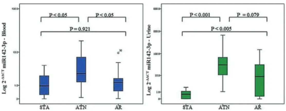
miR-142-3p expression levels are shown in Figure 1. In the peripheral blood analysis, substantially higher expres-sions were found in the comparisons of the ATN with the STA group (Po0.05) and with the AR group (Po0.05)
and no difference was found in the comparison between the STA and AR groups (P=0.921). The analysis of the miR-142-3p from the urinary sediment cells showed signifi -cantly higher expression in the ATN group (Po0.001) and
AR group (Po0.005) in comparison with the STA group.
Also, the expression was higher, with a borderline signifi -cance level, in the ATN group compared with the AR group (P=0.079).
Receiver operating characteristics (ROC) curves were built for the assessment of the diagnostic parameters for ATN and are shown in Figure 2. In the peripheral blood analysis, the area under the curve (AUC) was 0.75 (95% CI=0.56–0.94; P=0.016). In the urinary cells analysis, the
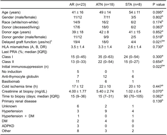
Table 1.Demographic data of the groups.
AR (n=23) ATN (n=18) STA (n=8) P value
Age (years) 41±16 49±14 54±11 0.095a
Gender (male/female) 11/12 7/11 3/5 0.802b
Race (white/non-white) 14/9 16/2 6/2 0.174b
Donor (deceased/living) 17/6 18/0 6/2 0.063b
Donor age (years) 39±18 42±8 41±15 0.852a
Donor gender (male/female) 11/12 9/9 2/5 0.519b
Delayed graft function (yes/no)c 19/4 18/0 4/4 0.021bn
HLA mismatches (A, B, DR) 3.5±1.4 3.3±1.4 2.6±1.4 0.730a Last PRA (%; median [IQR])
Class I 15 (0–45) 35 (0–63) 24 (0–54) 0.300a
Class II 13 (0–33) 22 (0–54) 15 (0–27) 0.654
a
Initial immunosuppression (n) 0.022bn
No induction 5 0 1
Anti-thymocyte globulin 7 12 6
Basiliximab 11 6 1
Cold ischemia time (h) 17±12 22±10 20±10 0.447a
Creatinine at biopsy (mg/dL) 4.00±1.77 5.40±2.74 1.02±0.4 0.070a Time to biopsy (days; median [IQR]) 15 (9–36) 15 (7–31) 90 (70–105) 0.062
a
Primary renal disease 0.139b
Unknown 6 2 1
Hypertension 3 9 4
Hypertension+DM 1 0 1
DM 2 4 0
ADPKD 3 0 0
Other 8 3 2
Data are reported as means±SD, unless otherwise indicated. AR: acute rejection; ATN: acute tubular necrosis; STA: stable renal function; HLA: human leukocyte antigen; PRA: panel reactive antibodies; DM: diabetes mellitus; ADPKD: autosomal dominant polycystic kidney disease; IQR: interquartile range. a
ANOVA;bPearson’s chi-square/Fisher’s exact test;bn
AUC was 0.77 (95%CI=0.62–0.92, P=0.010). The cut-off
selected for the blood samples analysis was 1.59 and the resulting parameters were sensitivity=75%; specificity=63%; positive predictive value=81%, and a negative predictive value=50% (Po0.05). For the urinary samples analysis, the
cut-off selected was 2.03 and the resulting parameters were sensitivity=92%; specificity=87%; positive predictive value= 90% and a negative predictive value=87% (Po0.001).
Discussion
The understanding of the molecular basis of allograft injuries remains an important and incompletely solved issue in organ transplantation. The molecular mechanisms appear to be highly regulated and the interplay between mRNAs
and miRNAs are probably crucial (24). Moreover, the dis-covery of noninvasive biomarkers that reflect accurately graft-related events is an unmet need in the clinical practice of organ transplantation, since the current available methods are either invasive or lack accuracy. In the present study, we evaluated the expression of miR-142-3p as a biomarker of injury in the immediate post-renal transplantation period and found that its expression was significantly increased in the peripheral blood and in the urinary sedimentary cells of patients with kidney grafts undergoing post-transplant ATN. In the last decade, miRNAs were studied as biomark-ers in renal transplantation (25). Over time, a number of reports have suggested their usefulness as potential non-invasive biomarkers of acute rejection in the peripheral blood and in the urine (26). Also, miRNAs expression has Figure 1.Logarithmic transformation of microRNA 142-3p expression in the peripheral blood and urine of renal transplanted recipients. STA: stable group of patients; ATN: group of patients with acute tubular necrosis; AR: group of patients with acute rejection. Data are reported as medians and interquartile range. Statistical analysis was performed with Mann-WhitneyUtest.
been studied in other post-transplant conditions, such as chronic graft dysfunction, acute pyelonephritis, BK virus nephropathy and operational tolerance (27–29). In many
of these studies, specific miRNAs were uncovered by analysis in high throughput platforms and validated by PCR techniques. The above studies lead to the notion that these molecules might become useful biomarkers of specific clinical conditions after organ transplantation.
Cloning studies allowed the determination of miRNAs from opposite arms of the hairpin precursor. As for miR-142, it is processed into two mature miRNAs: miR-142-3p and miR-142-5p. Merkerova and colleagues examined their expression in hematopoietic cell lineages and found that miR-142-3p was approximately 10-fold more expressed than miR-142-5p in this compartment, whose compo-nents infiltrate allografts both in rejection and in cell necrotic events (30). Therefore, its increased expression in these conditions might be expected. miR-142-3p expression has been demonstrated in normal human T cells and granulo-cytes and weak levels of expression occur in monogranulo-cytes and B cells. Increased levels of expression suggest infl am-matory processes within allografts that could be due to either rejection or necrosis (20). This miRNA has been reported to be more expressed in naive T cells than in differentiated Th1 and Th2 cells (31). However, it has been shown that the transcription factor FoxP3 is one of the mediators of transcriptional repression of miR-142-3p and increases in this messenger RNA expression are associated with acute rejection, and thus can lead to decrease in the expression of the miR-142-3p (3,25,32). In support, we found in the present study significant increases of this miRNA in the peripheral blood and urine of kidney graft patients under-going ATN compared to those with stable grafts and AR, suggesting a more relevant role for the cell necrosis processes in the increase of this miRNA.
Previous research has reported on the miR-142-3p ex-pression on graft tissue and non-invasive samples (periph-eral blood or urine) from kidney transplant recipients. Danger et al. (33) verified the expression of miR-142-5p (50arm of
miR-142) in the peripheral blood and renal graft tissue of patients with chronic antibody-mediated rejection and found it to be over expressed in comparison with patients with stable graft function. The authors reinforced the participation of this miRNA in immunological disorders, and similar with our findings, did notfind a significant increase in the expression of this biomarker in patients with acute rejection (33).
Scian et al. (34) evaluated miRNAs expression in allo-graft tissue and paired urine samples of kidney transplant recipients with chronic allograft dysfunction and interstitial fibrosis and tubular atrophy (IF/TA). They reported that the miR-142-3p expression is increased in patients with IF/TA, both in renal graft tissue and urine and suggested the potential use of miRNAs as noninvasive markers of IF/TA and for monitoring graft function. Ben-Dov et al. (27) found that this miRNA is overexpressed in biopsies of patients with IF/TA in comparison with normal allograft biopsies.
Maluf et al. (35) described that in the early period after kidney transplantation urinary miR-142-3p, along with other four miRNAs, were differentially expressed in the group of patients that developed IF/TA in the transplant course. Interestingly, overexpression was detectable before histol-ogical allograft injury was evident suggesting that miRNAs are potential biomarkers for monitoring graft function in the anticipation of progression to chronic graft dysfunction. Samples of patients with IF/TA were not included in our study therefore we could not confirm or deny the above findings. However, IF/TA may often be related to infl am-matory injuries and increased expression of miR-142-3p might therefore be expected.
Anglicheau et al. (13) demonstrated elevated intra-graft miRNA expression in both stable allointra-grafts and those undergoing acute rejection. The authors found that 17 miRNAs, including miR-142-5p, which is functionally related to miR-142-3p, could individually distinguish biopsy samples from allografts with acute rejection from those without rejection. Soltaninejad and colleagues analyzed the expression levels of miR-142-3p, miR-142-5p and others in paired biopsy and peripheral blood mononu-clear cell samples of renal allograft recipients with acute T-cell mediated rejection, comparing with normal allografts. These authors found elevated levels of miR-142-3p in blood samples of acute T-cell mediated rejection and reported that analyzes of miR-142-3p expression in the peripheral blood could predict acute T-cell mediated rejection. Interestingly a correlation could not be observed between miR-142-3p expressions in biopsy tissue and peripheral blood mono-nuclear cell (36). Inflammatory infiltrates are present in acute cellular rejection and ATN, therefore an increase in miR-142-3p might be expected in both situations. Neither of the above studies included patients with ATN; thus, comparisons with the present study are difficult to make and further research may be necessary to clarify thesefindings.
Finally, studies in which miR-142-3p was tested in drug-free tolerant kidney transplant recipients identified this molecule as possibly involved in tolerance mech-anisms, probably related to the negative regulation of TGF-b signaling. Danger et al. reported an increased expression of miR-142-3p in B cells purified from operationally toler-ant kidney graft recipients in comparison with stable graft recipients under immunosuppressive therapy (19). It has been suggested that this miRNA might be a prom-ising predictor of patients eligible for immunosuppression weaning or withdrawal (19,37).
the graft response may lead to a better understanding of pathophysiology and mechanisms of graft injuries (24,39). In this research, we described that miR-142-3p is over-expressed in non-invasive samples of kidney transplant recipients with ATN and may became an useful biomarker of such condition. However, appropriate validation of the molecular approaches in adequately designed longitudinal studies is necessary before clinical applicability.
Acknowledgements
Financial support for this study was provided by the Brazilian Research Council (CNPq) and Research and Event Incentive Fund from Hospital de Clínicas de Porto Alegre. T.D. Domenico received a scholarship from CAPES Foundation, Brazil, and R.C. Manfro received a scholarship from CNPq.
References
1. Meier-Kriesche HU, Ojo AO, Port FK, Arndorfer JA, Cibrik DM, Kaplan B. Survival improvement among patients with end-stage renal disease: trends over time for transplant recip-ients and wait-listed patrecip-ients. J Am Soc Nephrol2001; 12: 1293-1296.
2. Nankivell BJ, Alexander SI. Rejection of the kidney allograft. N Engl J Med2010; 363: 1451-1462, doi: 10.1056/NEJMra 0902927.
3. Muthukumar T, Dadhania D, Ding R, Snopkowski C, Naqvi R, Lee JB, et al. Messenger RNA for FOXP3 in the urine of renal-allograft recipients.N Engl J Med2005; 353: 2342–
2351, doi: 10.1056/NEJMoa051907.
4. Gupta G, Womer KL. Profile of belatacept and its potential role in prevention of graft rejection following renal transplan-tation.Drug Des Devel Ther2010; 4: 375–382.
5. Geddes CC, Woo YM, Jardine AG. The impact of delayed graft function on the long-term outcome of renal transplanta-tion.J Nephrol2002; 15: 17–21.
6. Spiegel JC, Lorenzen JM, Thum T. Role of microRNAs in immunity and organ transplantation.Expert Rev Mol Med 2011; 13: e37, doi: 10.1017/S1462399411002080. 7. Rosen S, Stillman IE. Acute tubular necrosis is a syndrome
of physiologic and pathologic dissociation. J Am Soc Nephrol2008; 19: 871–875, doi: 10.1681/ASN.2007080913. 8. Colvin RB. Antibody-mediated renal allograft rejection: diag-nosis and pathogenesis.J Am Soc Nephrol2007; 18: 1046–
1056, doi: 10.1681/ASN.2007010073.
9. Solez K, Racusen LC, Marcussen N, Slatnik I, Keown P, Burdick JF, et al. Morphology of ischemic acute renal failure, normal function, and cyclosporine toxicity in cyclosporine-treated renal allograft recipients.Kidney Int1993; 43: 1058–
1067, doi: 10.1038/ki.1993.148.
10. Yarlagadda SG, Coca SG, Formica RN, Poggio ED, Parikh CR. Association between delayed graft function and allograft and patient survival: a systematic review and meta-analysis. Nephrol Dial Transplant2009; 24: 1039–1047, doi: 10.1093/
ndt/gfn667.
11. Reinhart BJ, Slack FJ, Basson M, Pasquinelli AE, Bettinger JC, Rougvie AE, et al. The 21-nucleotide let-7 RNA reg-ulates developmental timing in Caenorhabditis elegans. Nature2000; 403: 901–906, doi: 10.1038/35002607. 12. Harris A, Krams SM, Martinez OM. MicroRNAs as immune
regulators: implications for transplantation.Am J Transplant 2010; 10: 713–719, doi: 10.1111/j.1600-6143.2010.03032.x.
13. Anglicheau D, Sharma VK, Ding R, Hummel A, Snopkowski C, Dadhania D, et al. MicroRNA expression profiles pre-dictive of human renal allograft status. Proc Natl Acad Sci U S A2009; 106: 5330–5335, doi: 10.1073/pnas.08131
21106.
14. Hartono C, Muthukumar T, Suthanthiran M. Noninvasive diagnosis of acute rejection of renal allografts. Curr Opin Organ Transplan. 2010; 15: 35–41, doi: 10.1097/MOT.0b013
e3283342728.
15. O’Connell RM, Taganov KD, Boldin MP, Cheng G, Baltimore D. MicroRNA-155 is induced during the macrophage infl am-matory response.Proc Natl Acad Sci USA2007; 104: 1604– 1609, doi: 10.1073/pnas.0610731104.
16. Anglicheau D, Muthukumar T, Hummel A, Ding R, Sharma VK, Dadhania D, et al. Discovery and validation of a molec-ular signature for the noninvasive diagnosis of human renal allograftfibrosis.Transplantation2012; 93: 1136–1146, doi:
10.1097/TP.0b013e31824ef181.
17. Huang B, Zhao J, Lei Z, Shen S, Li D, Shen GX, et al. miR-142-3p restricts cAMP production in CD4+ CD25-T cells and CD4+CD25+ TREG cells by targeting AC9 mRNA.EMBO Rep2009; 10: 180–185, doi: 10.1038/embor.
2008.224.
18. Risøe PK, Ryg U, Wang YY, Rutkovskiy A, Smedsrød B, Valen G, et al. Cecal ligation and puncture sepsis is asso-ciated with attenuated expression of adenylyl cyclase 9 and increased miR142-3p.Shock2011; 36: 390–395, doi: 10.1097/SHK.0b013e318228ec6f.
19. Danger R, Pallier A, Giral M, Martínez-Llordella M, Lozano JJ, Degauque N, et al. Upregulation of miR-142-3p in peripheral blood mononuclear cells of operationally tolerant patients with a renal transplant.J Am Soc Nephrol2012; 23: 597–606, doi: 10.1681/ASN.2011060543.
20. Chen CZ, Li L, Lodish HF, Bartel DP. MicroRNAs modulate hematopoietic lineage differentiation. Science 2004; 303: 83–86, doi: 10.1126/science.1091903.
21. Zhou Q, Haupt S, Prots I, Thummler K, Kremmer E, Lipsky PE, et al. miR-142-3p is involved in CD25+ CD4 T cell proliferation by targeting the expression of glycoprotein A repetitions predominant.J Immunol2013; 190: 6579–6588,
doi: 10.4049/jimmunol.1202993.
22. Solez K, Colvin RB, Racusen LC, Haas M, Sis B, Mengel M, et al. Banff 07 classification of renal allograft pathology: updates and future directions. Am J Transplant 2008; 8: 753–760, doi: 10.1111/j.1600-6143.2008.02159.x.
23. Livak KJ, Schmittgen TD. Analysis of relative gene expres-sion data using real-time quantitative PCR and the 2(-Delta Delta C(T)) Method.Methods2001; 25: 402–408, doi: 10.1006/
meth.2001.1262.
24. Nankivell BJ. microRNA in transplantation: small in name only.Transplantation2015; 99: 1754–1755, doi: 10.1097/TP.
0000000000000807.
renal transplantation. Transpl Immunol 2008; 19: 81–85,
doi: 10.1016/j.trim.2008.01.007.
26. Lorenzen JM, Volkmann I, Fiedler J, Schmidt M, Scheffner I, Haller H, et al. Urinary miR-210 as a mediator of acute T-cell mediated rejection in renal allograft recipients.Am J Trans-plant2011; 11: 2221–2227, doi: 10.1111/j.1600-6143.2011.
03679.x.
27. Ben-Dov IZ, Muthukumar T, Morozov P, Mueller FB, Tuschl T, Suthanthiran M. MicroRNA sequence profiles of human kidney allografts with or without tubulointerstitial fibrosis. Transplantation2012; 94: 1086–1094.
28. Oghumu S, Bracewell A, Nori U, Maclean KH, Balada-Lasat JM, Brodsky S, et al. Acute pyelonephritis in renal allografts: a new role for microRNAs?Transplantation2014; 97: 559–568,
doi: 10.1097/01.TP.0000441322.95539.b3.
29. Yap M, Boeffard F, Clave E, Pallier A, Danger R, Giral M, et al. Expansion of highly differentiated cytotoxic terminally differentiated effector memory CD8+T cells in a subset of clinically stable kidney transplant recipients: a potential marker for late graft dysfunction.J Am Soc Nephrol2014; 25: 1856–1868, doi: 10.1681/ASN.2013080848.
30. Merkerova M, Belickova M, Bruchova H. Differential expres-sion of microRNAs in hematopoietic cell lineages. Eur J Haematol 2008; 81: 304–310, doi: 10.1111/j.1600-0609.
2008.01111.x.
31. Monticelli S, Ansel KM, Xiao C, Socci ND, Krichevsky AM, Thai TH, et al. MicroRNA profiling of the murine hemato-poietic system.Genome Biol2005; 6: R71, doi: 10.1186/gb-2005-6-8-r71.
32. Aquino-Dias EC, Joelsons G, da Silva DM, Berdichevski RH, Berdichewski RH, Ribeiro AR, et al. Non-invasive diagnosis of acute rejection in kidney transplants with
delayed graft function.Kidney Int2008; 73: 877–884, doi:
10.1038/sj.ki.5002795.
33. Danger R, Paul C, Giral M, Lavault A, Foucher Y, Degauque N, et al. Expression of miR-142-5p in peripheral blood mononuclear cells from renal transplant patients with chronic antibody-mediated rejection. PLoS One 2013; 8: e60702, doi: 10.1371/journal.pone.0060702.
34. Scian MJ, Maluf DG, David KG, Archer KJ, Suh JL, Wolen AR, et al. MicroRNA profiles in allograft tissues and paired urines associate with chronic allograft dysfunction with IF/ TA. Am J Transplant 2011; 11: 2110–2122, doi: 10.1111/
j.1600-6143.2011.03666.x.
35. Maluf DG, Dumur CI, Suh JL, Scian MJ, King AL, Cathro H, et al. The urine microRNA profile may help monitor post-transplant renal graft function.Kidney Int2014; 85: 439–449,
doi: 10.1038/ki.2013.338.
36. Soltaninejad E, Nicknam MH, Nafar M, Ahmadpoor P, Pourrezagholi F, SharbafiMH, et al. Differential expression of microRNAs in renal transplant patients with acute T-cell mediated rejection. Transpl Immnol. 2015; 33: 1–6, doi:
10.1016/j.trim.2015.05.002.
37. van den Akker EK, Dor FJ, IJzermans JN, de Bruin RW. MicroRNAs in kidney transplantation: living up to their expec-tations?J Transplant2015; 2015: 354826, doi: 10.1155/2015/ 354826.
38. Trionfini P, Benigni A, Remuzzi G. MicroRNAs in kidney physiology and disease.Nat Rev Nephrol2015; 11: 23–33, doi: 10.1038/nrneph.2014.202.
39. Vitalone MJ, Sigdel TK, Salomonis N, Sarwal RD, Hsieh SC, Sarwal MM. Transcriptional perturbations in graft rejection. Transplantation2015; 99: 1882–1893, doi: 10.1097/TP.00