ACTA RADIOLÓGICA PORTUGUESA Janeiro-Abril 2017 Vol 29 nº 1 27-33
Benign Mesenchymal Breast Tumours – A Series of 39 Cases
Tumores Mesenquimatosos Benignos da Mama – Uma Série de 39 Casos
Inês Dias Marques
1, António Guimarães dos Santos
2, Ana Teresa Aguiar
21 Interna de Radiologia no Serviço de Imagiologia do Centro Hospitalar de Vila Nova de Gaia/Espinho, Vila Nova de Gaia, Portugal 2 Radiologista no Serviço de Radiologia do Instituto Português de Oncologia do Porto FG, EPE, Porto, Portugal
Correspondência Inês Dias Marques Serviço de Imagiologia
Centro Hospitalar de Vila Nova de Gaia/ Espinho
Rua Conceição Fernandes 4434-502 Vila Nova de Gaia Portugal
email: [email protected]
Resumo
Os tumores mesenquimatosos da mama originam-se do estroma mamário e incluem lesões benignas, malignas e pseudotumorais compostas principalmente por células mesenquimatosas. Encontrámos 39 lesões mamárias classificadas como tumores mesenquimatosos benignos entre Janeiro de 2010 e Julho de 2014 e que preenchiam os nossos requisitos. Entre elas incluem-se hemangioma, hiperplasia estromal pseudoangiomatosa (PASH), miofibroblastoma, fibromatose (tipo desmóide), angiolipoma e tumor de células granulares. Apesar desta série não ser um reflexo da população geral, uma vez que está baseada num centro de referência oncológico, permite-nos mostrar lesões raras nas suas apresentações habituais e atípicas. Descrevemos os nossos achados e realizamos uma breve revisão da literatura, incluindo aspectos imagiológicos e orientação. Apesar do seu aspecto imagiológico variável, o radiologista dedicado a patologia mamária deve estar familiarizado com estas lesões para poder oferecer o melhor cuidado e orientação na decisão entre seguimento ou necessidade de excisão.
Palavras-chave
Doenças mamárias; Neoplasias da mama; Mamografia; Ecografia mamária.
Abstract
Mesenchymal breast tumours arise in the stro-ma of the breast and comprise benign, stro- malig-nant and tumour-like lesions composed mainly of mesenchymal cells. We found 39 lesions that were classified as benign mesenchymal breast tumours from January 2010 until July 2014 and that met our criteria. They include haemangioma, pseudoangiomatous stromal hyperplasia (PASH), myofibroblastoma, desmoid-type fibromatosis, angiolipoma and granular cell tumour. Although our series does not reflect the general popula-tion because it is based at an oncologic referral center, it allows us to describe some rare lesions in their typical and unusual presentations. We define their imaging appearances and provide a short review of the literature, including imaging features and management. Despite their variable appearance, the radiologist must be familiar with these entities to provide the best care regarding the decision to maintain imaging follow up or the need for excision.
Keywords
Breast diseases; Breast neoplasms; Mammography; Breast ultrasound.
Artigo de Revisão / Review Article
Introduction
Mesenchymal breast tumours arise in the stroma of the breast.
Their latest classification was revised and published by the
WHO in 2012, and comprises benign, malignant and
tumour-like lesions composed mainly of mesenchymal cells(Table 1).
1It incorporates lesions of fibro-epithelial, fibroblastic and
myoblastic, vascular, lipomatous, neural, myogenic, and osseous
origin.
2While some of these tumours are fairly common, most
Nodular fasciitis Benign vascular lesions
Pseudoangiomatous stromal hyperplasia Myofibroblastoma
Desmoid-type fibromatosis Inflammatory myofibroblastic tumour Lipoma
Granular cell tumour and benign peripheral nerve-sheath tumour Angiosarcoma
Results
We found 50 lesions (49 patients) that were classified as
benign mesenchymal breast tumours according to our
criteria. Eight lesions were excluded because images were not
available and 3 others because PASH was a secondary finding
(Table 2). We retrospectively reviewed the records of these
patients, and described the imaging findings on ultrasound
and mammography. (Table 3 – Appendix).
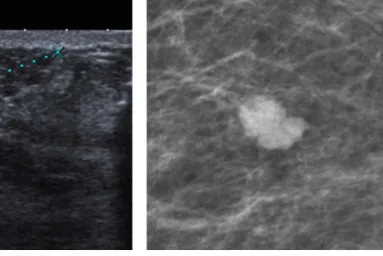
hemangiomas (Fig 1) in our series were masses in superficial
location, faintly seen on ultrasound, whose margins ranged
from circumscribed to microlobulated on ultrasound
and mammography. None of our hemangiomas showed
calcifications. These findings are in concordance with the
literature.
4In our series only one was excised, due to discrete
atypia.
Pseudoangiomatous stromal hyperplasia (PASH)
PASH is defined as a benign lesion comprising stromal
myofibroblastic proliferation and having the appearance
of anastomosing slit-like pseudovascular spaces lined by
spindle-shaped cells. It must be histologically distinguished
from angiosarcoma and might resemble myofibroblastoma
1.
PASH is especially frequent in premenopausal women or
women taking hormone therapy. Although most often stable
over time, it may increase in size or recur.
5It is a common
incidental finding in breast biopsies but its nodular form is
rare.
We examined the imaging appearances of cases where
PASH was considered a primary finding. From 25 lesions in
our series, 3 were occult on ultrasound and 7 where occult
on mammography (mammograms were not available in 8
patients). Most masses were hypoechoic with circumscribed
or microlobulated margins but there were 2 cases of masses
with indistinct margins and posterior shadowing (Fig 2).
We also found 4 cases of architectural distortion without
a clear mass on ultrasound. Mammogram findings were
nonspecific, including masses with circumscribed, obscured
or indistinct margins. Pathologically, most cases were defined
solely as PASH, one case was categorized as nodular PASH
(Fig 3), four cases were categorized as either nodular PASH
or fibroadenoma with superimposed PASH, and there was
one case where a differential diagnosis between PASH and
myofibroblastoma could not be made.
Myofibroblastoma
Myofibroblastomas are benign tumours of the mammary
stroma composed of fibroblasts and myofibroblasts.
1Unfortunately, there are several unusual morphologic
variants that might be difficult to correctly diagnose on core
biopsy. While initially described in older men, they have
been increasingly recognized in postmenopausal women,
probably due to screening.
6They do not have a tendency
for local recurrence.
1In the literature they are described as
well circumscribed, homogeneous, hypoechoic masses on
ultrasound, commonly evocative of fibroadenomas, whereas
Lesions Sex (F/M) Excluded Included Haemangioma 3 3/0 0 3 PASH 30 30/0 5 25 Myofibroblastoma 2 2/0 0 2 Desmoid-type fibromatosis 6 5/1 2 4 Lipoma 1 1/0 1 0 Angiolipoma 4 3/0 2 2
Granular cell tumour 4 3/1 1 3
Table 2 – Results - Histologic subtypes found in our series.
Discussion
Our results are based in a referral oncologic centre; while
some of the lesions were the reason for referral, others
were incidental findings in the imaging workup of oncologic
patients. Besides, some studies were done in outpatient care
and the images were not available. So, although our series
has to be interpreted in its context and does not reflect
the general population, it allows us to describe the imaging
appearances of some rare tumours and uncommon imaging
features of more frequent lesions.
Haemangioma
Haemangiomas are a benign proliferation of mature vessels.
According to the WHO, the finding of a haemangioma in
core biopsy specimens should prompt surgical excision
to exclude well-differentiated angiosarcoma.
1However, in
clinical practice, masses with classic imaging and pathologic
features are often followed up with imaging.
3The three
Figure 1 – Haemangioma. Subcutaneous, oval, hypoechoic mass with microlobulated margins, seen as a
mammography descriptions reveal well circumscribed,
round to oval masses without calcifications.
6We found
two myofibroblastomas in our database (there is another
lesion that could not be confidently distinguished from
PASH included in that group). Both lesions occurred in
postmenopausal women, and were moderately worrisome for
malignancy. One was isoechoic with circumscribed margins
on ultrasound but was irregular with obscured margins on
mammography, and the other (Fig 4) was hypoechoic with
indistinct margins on ultrasound and showed obscured
margins on mammography. They were both excised.
Desmoid-type fibromatosis
Desmoid-type fibromatosis, also known as extra-abdominal
desmoid tumour, is a locally infiltrative lesion without
metastatic potential that originates from fibroblasts or
myofibroblasts. Frequently it extends from the pectoral fascia
into the breast. There is an association with trauma, including
surgery.
1Some of these lesions are occult on mammography,
but they can appear as irregular lesions with spiculate or
indistinct margins, mimicking tumour. They usually do not
have calcifications or adenopathy. Ultrasound appearances
are variable, but hypoechoic masses with irregular, spiculate,
Figure 2 – PASH. Irregular mass with heterogeneous echo pattern, indistinct margins and posterior shadowing. On mammography a subtle round mass with obscured margins can be seen. These imaging findings are atypical for PASH.
Figure 3 – Nodular PASH. Rare case of “pure” nodular PASH, appearing as a hypoechoic oval mass, parallel to the surface, with circumscribed margins. The mass also appears circumscribed on mammography and magnified view.
Figure 4 – Myofibroblastoma in a 73-year old female. Hypoechoic mass with indistinct margins on ultrasound and obscured margins on mammography. Pathology showed myofibroblastoma.
or microlobulate borders have been described.
2Due to its
aggressive growth pattern and tendency for recurrence,
management consists of wide excision with clear margins.
2Recurrences usually occur within 3 years of excision and
commonly require radical surgery.
7In our series all of the
lesions were hypoechoic and had other findings suspicious
for malignancy on ultrasound, such as irregular shape and
indistinct margins; two of them also showed posterior
shadowing (Fig. 5). On the other hand, two of them
appeared as focal asymmetries on mammography, and only
one appeared as a spiculate mass. All of the four patients in
our series were treated with lumpectomy.
Figure 5 – Fibromatosis (desmoid-type). Hypoechoic, irregular mass with posterior shadowing. Another mass with spiculate margins on mammography.
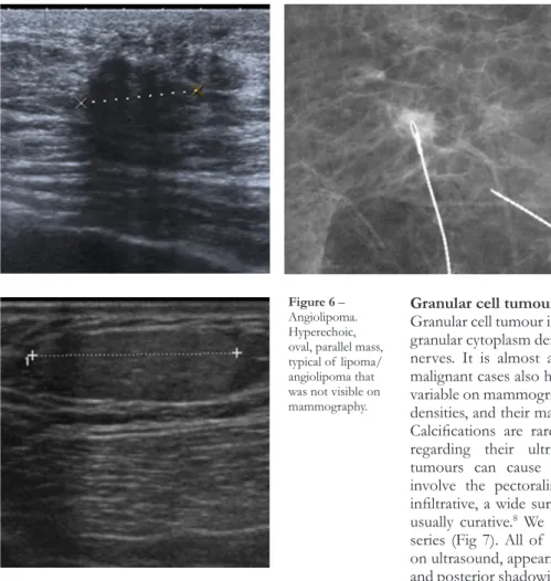
Angiolipoma
Angiolipomas are considered by the WHO under the
lipoma category, and are characterized by nodules of
mature fat incorporating small vessels containing fibrin
thrombi.
1Angiolipomas are traditionally described as
hyperechoic, circumscribed masses on ultrasound with
variable mammographic appearances.
3,4The angiolipomas
in our series appeared as iso or hyperechoic masses, both
oval and parallel with circumscribed margins. One was not
seen on mammography, while the other presents as a focal
asymmetry. They were all excised because of their individual
clinical context(Fig. 6).
Figure 6 – Angiolipoma. Hyperechoic, oval, parallel mass, typical of lipoma/ angiolipoma that was not visible on mammography.
Granular cell tumour
Granular cell tumour is defined as a tumour with eosinophilic
granular cytoplasm derived from Schwann cells of peripheral
nerves. It is almost always benign (>99%).
1Nevertheless,
malignant cases also have been reported.
8The appearance is
variable on mammography, ranging from masses to indistinct
densities, and their margins from spiculate to circumscribed.
Calcifications are rare. The reports are also confounding
regarding their ultrasound appearance.
2Granular cell
tumours can cause skin retraction, nipple inversion or
involve the pectoralis fascia.
1Since they may be locally
infiltrative, a wide surgical excision is recommended, and is
usually curative.
8We found 3 granular cell tumours in our
series (Fig 7). All of them were very suspicious for cancer
on ultrasound, appearing hypoechoic with indistinct margins
and posterior shadowing or with combined posterior pattern.
On mammography, the masses were round with indistinct or
obscured margins. They were all treated with lumpectomy.
One of them showed atypical findings on pathology.
Figure 7 – Granular cell tumour. Hypoechoic mass with indistinct margins and posterior shadowing. On
Conclusion
Benign mesenchymal breast tumours have variable imaging
appearances that range from benign to overtly malignant.
Radiologists working in breast imaging must be familiar with
these entities to provide the best care, regarding the decision
to maintain imaging follow up or the need for excision.
References
1. Lakhani SR, Ellis IO, Schnitt SJ, Tan PH, van de Vijver MJ. (Eds.): WHO classification of tumours of the breast, 4th Ed., IARC: Lyon, 2012. 2. Schickman R, Leibman AJ, Handa P, Kornmehl A, Abadi M. Mesenchymal breast lesions. Clin Radiol. 2015;70:567-75.
3. Jesinger RA, Lattin GE, Ballard EA, et al. Vascular abnormalities of the breast: arterial and venous disorders, vascular masses, and mimic lesions with radiologic-pathologic correlation. RadioGraphics. 2011;31: E117-E136. 4. Glazebrook KN, Morton MJ, Reynolds C. Vascular tumors of the breast: mammographic, sonographic, and MRI appearances. AJR Am J Roentgenol. 2005;184:331-8.
5. Jones KN, Glazebrook KN, Reynolds C. Pseudoangiomatous stromal hyperplasia: imaging findings with pathologic and clinical correlation. AJR Am J Roentgenol. 2010;195:1036-42.
6. Magro G. Mammary Myofibroblastoma: a tumor with a wide morphologic spectrum. Arch Pathol Lab Med. 2008;132:1813-20.
7. Erguvan-Dogan B, Dempsey PJ, Ayyar G, Gilcrease MZ. Primary desmoid tumor (extraabdominal fibromatosis) of the breast. AJR Am J Roentgenol. 2005;185:488-9.
8. Irshad A, Ackerman SJ, Pope TL, et al. Panzegrau, B. Rare breast lesions: Correlation of imaging and histologic features with WHO classification. RadioGraphics. 2008;28:1399-414.
Recebido / Received 14/11/2016 Aceite / Acceptance 29/12/2016 Divulgações Éticas / Ethical disclosures
Conflitos de interesse: Os autores declaram não possuir conflitos de interesse. Conflicts of interest: The authors have no conflicts of interest to declare. Suporte financeiro: O presente trabalho não foi suportado por nenhum
subsídio ou bolsa.
Financing Support: This work has not received any contribution, grant or
Age/Sex Ultrasound Mammography Pathology Record Haemangioma
58/F Mass: round, isoechoic,
circumscri-bed, superficial Mass: round, obscured margins Lumpectomy
46/F N/A Mass: round, microlobulated margins Core Biopsy
54/F Mass: oval, isoechoic,
microlobula-ted, superficial Mass: oval, microlobulated margins Core Biopsy PASH
77/F Not visible Mass: irregular, obscured margins Core Biopsy 45/F Mass: oval, hypoechoic, parallel,
circumscribed N/A Core Biopsy
47/F Not visible Mass: oval, low density,
circumscri-bed Core Biopsy
43/F Mass: oval, hypoechoic, parallel,
circumscribed Not visible Mastectomy
32/F Mass: round, parallel,
microlobula-ted margins Mass: round, obscured margins Core Biopsy Nodular PASH vs. Fibroadenoma 43/F Mass: irregular, parallel, indistinct
margins, shadowing N/A Lumpectomy PASH vs. Myofibroblastoma 47/F Mass: oval, hypoechoic parallel,
circumscribed Not visible Core Biopsy
40/F Mass: oval, isoechoic, parallel,
circumscribed N/A Core Biopsy
46/F Architectural distortion with
combi-ned posterior pattern Not visible (dense breast) Lumpectomy 46/F Not visible Mass: irregular, indistinct margins Core Biopsy 52/F Mass: oval, hypoechoic, parallel,
microlobulated margins Not visible Core Biopsy 30/F Mass: oval, parallel, hypoechoic,
circumscribed Not visible Core Biopsy
46/F Mass: oval, hypoechoic,
microlobu-lated margins N/A Core BiopsyNodular PASH vs. Fibroadenoma 66/F Mass: oval, hypoechoic, parallel,
circumscribed N/A Core Biopsy
43/F Architectural distortion N/A Core Biopsy
50/F Mass: oval, hypoechoic, parallel,
circumscribed Mass: oval, circumscribed Lumpectomy Nodular PASH 47/F Mass: irregular,heterogeneous echo
pattern, indistinct margins, poste-rior shadowing
Mass: round, obscured margins Lumpectomy 47/F Architectural distortion with
combi-ned posterior pattern Focal asymmetry Core Biopsy 39/F Mass: oval, hypoechoic,
microlobu-lated margin N/A Core Biopsy Nodular PASH vs. Fibroadenoma 37/F Mass: oval, hypoechoic,
circums-cribed Not visible Biopsy
Appendix
Myofibroblastoma
67/F Mass: oval, isoechoic, circumscribed
margins Mass: irregular, obscured margins Lumpectomy 73/F Mass: hypoechoic, round, indistinct
margins Mass: round, obscured margins Lumpectomy Desmoid-type fibromatosis
68/F Mass: irregular, hypoechoic,
poste-rior shadowing Focal asymmetry Lumpectomy
54/F Mass: round, hypoechoic indistinct
margins Focal asymmetry Lumpectomy
46/F N/A Mass: round, spiculated Lumpectomy
39/F Mass: irregular, hypoechoic,
poste-rior shadowing N/A Lumpectomy
Angiolipoma
49/F Mass: isoechoic, oval, parallel,
circumscribed Focal asymmetry Mastectomy
51/F Mass: hyperechoic, oval, parallel,
circumscribed Not visible Lumpectomy
Granular cell tumor
50/F Mass: irregular, hypoechoic,
indis-tinct margins, shadowing Mass: round, indistinct margins LumpectomyBenign features 50/F Mass: irregular, hypoechoic,
indis-tinct margins, shadowing Mass: round, obscured margins LumpectomyBenign features 58/F Mass: round, hypoechoic, indistinct
margins, combined posterior pattern
Mass: round, obscured margins Lumpectomy Atypical features
*For each lesion age and sex are mentioned as well as a brief description of findings on ultrasound and mammography. The type of latest pathology speci-mens, as well as important pathologic information including alternative diagnosis are provided. N/A – Not available