UNIVERSIDADE DA BEIRA INTERIOR
Ciências da Saúde
Effect of white tea on the reproductive function
of diabetic or prediabetic individuals
Tânia Isabel Rodrigues Amaral Dias
Thesis to obtain the Doctoral degree in
Biomedicine
(3
rdcycle of studies)
Supervisor: Pedro Fontes Oliveira, Ph.D.
Co-supervisor: Branca Maria Silva, Ph.D.
Co-supervisor: Susana Casal, Ph.D.
ii
iii For all the people who supported me every step of the way, in the university, in the laboratory, in daily life or at home. All the effort and dedication put into this research project made me grow professionally and personally. Thank you all.
“All our dreams can come true, if we have the courage to pursue them” — Walt Disney
“I’m glad I did it, partly because it was worth it, but mostly because I shall never have to do it again” — Mark Twain
v Firstly, I would like to express my sincere gratitude to the mentors of this research project, I could not have imagined a better guidance team to work with during my PhD.
I want to thank to my supervisor Prof. Pedro Oliveira for his continuous support for the last 8 years. Thank you so much for believing in me and pushing me to pursue my PhD, for the patience, for the motivation, for the confidence, for the friendship and the immense knowledge you have shared with me.
I am also very grateful to my co-supervisor Prof. Branca Silva for the guidance and availability, for making me think outside the box, for teaching me, and for giving me hope that everything would go for the best. I also want to thank the opportunity to collaborate with you in several practical classes, it was a great experience.
I want to thank to my co-supervisor Prof. Susana Casal for always being available to help in everything I needed, for the sympathy, supportive words and new ideas, for the enthusiasm and all the techniques she taught me.
I also want to make a special thanks to Dr. Marco Alves, who although not an official element of the guidance team, was vital for the development of this project. Thank you for your advices, for bringing your knowledge into this work and for always motivating me to exceed myself.
My sincere thanks to Prof. Mário Sousa, Prof. Mariana Monteiro, Prof. Alberto Barros, and respective teams for giving me access to their laboratories and research facilities. Without their support it would not be possible to conduct this research.
Secondly, I am truly grateful to the laboratory colleagues who accompanied me along this journey at the University of Beira Interior and University of Porto: Luís Rato, Raquel Bernardino, Maria João Meneses, Susana Almeida, Carina Ribeiro, Ana Raquel Nunes, Tito Jesus, Ana Martins, Ana Maria, Marwa Boussada, David Carrageta, Bruno Moreira, Hugo Silva, Luís Crisóstomo, Rute Pereira, Ivana Jarak, Sara Correia, Cátia Vaz, Ana Silva. I have learned many important things from each of you and we had a lot of fun working together.
Particularly, I must express my sincere gratitude to Luís Rato, Raquel Bernardino and Maria João Meneses, who were always there to support me, listen to me and motivating me to continue whenever I thought about giving up. Luís thank you for the full-time availability, for your willingness to help in everything and for teaching me so much. Raquel thank you for the patience, for all the discussions about my own work, for the sleepless nights (and days) we were working together before “impossible” deadlines, and for the complicity and true friendship. Maria João thank you for all the great moments shared for the last 8 years, for being
vi
always available no matter the distance or time and for giving your true opinion on everything rather than the things I would like to listen.
Many thanks to Elsa, Cláudia, Angela, Sónia, Fernanda, and Célia from ICBAS for the sympathy, good mood, partnership and help in many laboratory techniques. Thanks also to the laboratory technicians from the Health Sciences Research Centre Sofia Duarte, Margarida Carrilho, Maria José, Maria João, and Catarina Ferreira for their help in preparing or conducting experiments.
I am profoundly grateful to my long-time friends Vânia Filipe, Filipa Monteiro, Sara Martins, Joel Pereira, Vânia Reis, Vânia Vieira, Filipa Pinheiro, Joana Sousa, Mafalda Faria, Sofia Marques, Vitor Gaspar, Raquel Gonçalves, Carolina Gouveia, Catarina Diniz, and Nuno Oliveira who played a key role in encouraging me to keep going and who helped me to overlook the bad moments. Thank you for inspiring me to pursue my dreams every day.
Furthermore, I want to thank to my family for always believing in me and supporting all my decisions, especially to my parents for the patience, confidence and care.
Finally, I want to thank to “FCT - Fundação para a Ciência e a Tecnologia” for funding my PhD fellowship (SFRH/BD/109284/2015). I also want to thank to the Fulbright Portugal, Dr. Ashok Agarwal and his team, for giving me the opportunity to work at the Andrology lab of the American Center for Reproductive Medicine - Cleveland Clinic, Cleveland, USA for 8 months.
vii This document represents the completion of a research project to which I dedicated 7 years of my life. These were years of hard work that brought me a lot of knowledge and prepared me to pursue a career as a professional researcher. It was an ambitious project proposed by my supervisors that was overlooked by the research community, but once we published the first paper on the topic, an increasing interest on this subject emerged from many research groups. I am very proud of all the findings that resulted from our research, it was an incredibly rewarding journey. I am also honored for being able to work with wonderful people throughout these years. I hope this document and all our scientific publications on this topic constitute a step forward in the field of male reproduction.
“Science is telling us that we can do phenomenal things if we put our minds and our resources to it.” — Anthony S. Fauci
ix A incidência da diabetes mellitus (DM) tem vindo a aumentar em homens jovens em todo o mundo. Os pacientes com DM apresentam uma disfunção na secreção de insulina e/ou na sua ação, resultando em hiperglicemia. Esta desregulação afeta a homeostase da glicose no organismo, tendo um impacto negativo na fertilidade masculina. As células de Sertoli (SCs) são essenciais para a manutenção do potencial reprodutivo masculino, pois são o suporte físico e nutricional que permite a diferenciação das células germinativas em espermatozoides totalmente competentes. A função das SCs depende do metabolismo da glicose, uma vez que este é o seu substrato preferencial. Muitos pacientes diabéticos são subférteis ou inférteis devido a alterações na função das SCs, defeitos na espermatogénese e má qualidade do esperma. A principal estratégia para combater a DM e as suas complicações inclui mudanças nutricionais e atividade física. No entanto, estas intervenções geralmente não são bem-sucedidas por si só e precisam de ser complementadas com medicação. Porém, para além da eficácia dos fármacos convencionais ser limitada, eles são caros e têm vários efeitos secundários. Nos últimos anos, os produtos naturais, incluindo o chá e os seus constituintes, têm vindo a demonstrar propriedades antioxidantes e antidiabéticas promissoras. Além disso, o tratamento da prediabetes pode constituir uma abordagem eficaz, pois esta condição pode ocorrer até 10 anos antes da progressão da doença para um estado mais grave. Neste projeto, pretendemos estudar os efeitos do chá branco (WTEA) na função reprodutiva masculina e o seu papel protetor contra disfunções reprodutivas induzidas pela prediabetes. Pretendemos ainda investigar se os efeitos do WTEA são devidos a um efeito combinado de todos os seus constituintes ou ao efeito predominante de um dos seus componentes bioativos mais representativos. O WTEA é o tipo de chá mais raro e menos estudado, mas apresenta um potencial antioxidante elevado, devido ao seu alto conteúdo em catequinas. Recorrendo à ressonância magnética nuclear de protão (1H-NMR), verificou-se que este tipo de chá é
particularmente rico em cafeína, epigalocatequina galato (EGCG) e L-teanina. Utilizando um modelo in vitro de SCs de rato ou humanas (hSCs) avaliámos os efeitos do extrato aquoso de WTEA (0.5 mg/mL) e dos seus principais componentes bioativos (cafeína, EGCG e L-teanina) no metabolismo celular, função mitocondrial e perfil oxidativo. O extrato de WTEA foi capaz de modular o metabolismo da glicose em SCs de rato e estimular a produção de lactato, um substrato essencial para a sobrevivência das células germinativas. A suplementação dos meios de cultura de hSCs com 50 µM de cafeína, EGCG ou L-teanina por 24 horas induziu alterações no metabolismo da glicose, importantes para melhorar o potencial reprodutivo masculino. Isto resultou num aumento ou manutenção da produção de lactato, mostrando um papel protetor contra os danos oxidativos. No entanto, na concentração de 50 µM, estes compostos também induziram algumas alterações na proliferação das hSCs e na sua função mitocondrial, o que pode comprometer a função reprodutiva. O extrato de WTEA (0.5 mg/mL) mostrou um melhor efeito na função das SCs do que os componentes individuais, destacando a importância da
x
combinação de todos os componentes do chá para o seu efeito benéfico. Foram também realizados estudos ex vivo utilizando espermatozoides epididimais de rato para avaliar o potencial do extrato de WTEA (0.5 mg/mL ou 1 mg/mL) e dos três componentes selecionados (cafeína, EGCG e L-teanina) como um aditivo para um meio de armazenamento de esperma à temperatura ambiente durante um curto período. Nestes estudos, a concentração da cafeína (71 µg/mL), EGCG (82 µg/mL) e L-teanina (19 µg/mL) foram estabelecidas de acordo com suas concentrações no extrato de WTEA. Salienta-se que a viabilidade dos espermatozoides diminui ao longo do tempo em amostras refrigeradas. Assim, em certos casos, o armazenamento a curto prazo à temperatura ambiente pode ser vantajoso, como por exemplo para o transporte de amostras ou tecnologia de reprodução medicamente assistida. Estes estudos permitiram concluir que o extrato de WTEA (principalmente na concentração de 1 mg/mL) é um melhor aditivo para o meio de armazenamento de espermatozoides à temperatura ambiente do que cada um dos componentes isoladamente, dado que manteve a viabilidade dos espermatozoides durante 3 dias de forma equivalente ao momento da colheita. Embora a combinação dos três componentes selecionados tenha mostrado uma melhoria na viabilidade dos espermatozoides relativamente a cada componente individualmente, também estimulou a oxidação das proteínas. Estes resultados apoiam também o benefício da combinação de todos os componentes do extrato de WTEA. Os dados obtidos nos estudos in vitro e ex vivo, levaram-nos a estudar o efeito in vivo do consumo regular de WTEA na função reprodutiva de um modelo animal de prediabetes. Os ratos prediabéticos mostraram alterações ao nível do metabolismo do testículo e do epidídimo, resultando numa diminuição da qualidade espermática. A ingestão de uma infusão de WTEA por ratos prediabéticos, durante dois meses, preveniu muitas das disfunções metabólicas induzidas pela doença, resultando na melhoria da motilidade e viabilidade espermática. Os nossos resultados indicam que o consumo regular de WTEA pode ser uma estratégia eficaz e de baixo custo para melhorar as disfunções reprodutivas induzidas pela prediabetes, abrindo caminho para que o WTEA possa vir a ser usado para o desenvolvimento de novas terapias antioxidantes para melhorar a fertilidade masculina.
Palavras-chave
Células de Sertoli; Chá branco; Diabetes mellitus; Fertilidade masculina; Metabolismo; Prediabetes; Qualidade espermática; Reprodução.
xi A diabetes mellitus (DM) é uma doença metabólica tratável, mas geralmente incurável, caracterizada por hiperglicemia. Os indivíduos com esta condição apresentam defeitos no metabolismo das proteínas, gorduras e hidratos de carbono, principalmente devido a um mau funcionamento da secreção de insulina e/ou da sua ação. O número de diabéticos tem vindo a aumentar continuamente em todo o mundo. A International Diabetes Federation (IDF) estimou que até ao ano de 2035, o número de casos de DM pode chegar a aproximadamente 600 milhões de pessoas, afetando mais de um em cada 10 adultos no mundo. A diabetes tipo 2 (T2DM) representa 90-95% de todos os casos de DM. O seu desenvolvimento é um processo progressivo que ocorre quando se extingue a capacidade das células beta pancreáticas para compensar a incapacidade de as células do corpo usarem a insulina. A prediabetes é um estado intermédio caracterizado por níveis de glicose no sangue acima do normal, mas não altos o suficiente para ser considerado T2DM. Com as modificações certas no estilo de vida e medicação adequada, a prediabetes pode ser revertida ou controlada. Esta particularidade faz com que a investigação da prediabetes seja bastante importante, uma vez que pode levar a novas estratégias para retardar ou mesmo evitar o desenvolvimento da T2DM.
Nos últimos anos, os efeitos negativos da DM na fertilidade masculina têm sido amplamente investigados devido ao aumento da sua incidência em indivíduos mais jovens. Assim, a noção de que a DM é geralmente uma doença da população mais idosa foi desconsiderada. Atualmente, a DM afeta cada vez mais indivíduos antes e durante a idade reprodutiva, o que reforça a importância de estudar o seu impacto na reprodução masculina. De facto, vários estudos provam o declínio do potencial de fertilidade masculina nas últimas décadas. Por outro lado, o número de casais que procuram assistência médica para ter filhos tem vindo a aumentar. Aproximadamente 35% dos pacientes com T2DM são inférteis. Homens com T2DM ou prediabetes geralmente apresentam defeitos na espermatogénese e baixa qualidade espermática. As alterações hormonais e metabólicas induzidas pela doença são os principais contribuintes para esse cenário.
A primeira estratégia para combater a DM inclui mudanças nutricionais e atividade física. No entanto, essas intervenções geralmente não são bem-sucedidas por si só, sendo complementadas com medicação. Embora os fármacos convencionais sejam eficazes no controlo da glicemia, muitos têm efeitos adversos graves, como ganho de peso, hipoglicemia, edema e distúrbios gastrointestinais que podem desincentivar a adesão dos pacientes. Além disso, são bastante dispendiosos a longo prazo. Vários produtos naturais têm demonstrado um grande potencial antioxidante e antidiabético, destacando-se o chá e os seus componentes. O chá é uma das bebidas mais consumidas em todo o mundo e a sua popularidade está bastante associada aos seus efeitos benéficos para a saúde. Existem vários tipos de chá consoante a
xii
colheira e o processamento das folhas. O chá branco (WTEA) é obtido através das folhas novas e rebentos da planta Camellia sinensis, os quais são minimamente processados de modo a evitar fermentação ou oxidação. Este tipo de chá destaca-se pelo seu potencial antioxidante elevado, principalmente devido ao seu alto conteúdo em catequinas. Neste projeto, pretendemos estudar os efeitos do WTEA na função reprodutiva masculina e o seu papel protetor contra disfunções reprodutivas induzidas pela prediabetes. Pretendemos ainda investigar se os efeitos do WTEA são devidos a um efeito combinado de todos os seus constituintes ou ao efeito predominante de um dos seus componentes bioativos mais representativos. Recorrendo à ressonância magnética nuclear de protão (1H-NMR), verificou-se que este tipo de chá é
particularmente rico em cafeína, epigalocatequina galato (EGCG) e L-teanina.
Utilizando um modelo in vitro de células de Sertoli de rato (SCs) ou humanas (hSCs) avaliámos os efeitos de um extrato aquoso de WTEA (0.5 mg/mL) e dos seus principais componentes bioativos (cafeína, EGCG e L-teanina) individualmente, no metabolismo celular, função mitocondrial e perfil oxidativo. As SCs são essenciais para a espermatogénese e, portanto, para a manutenção da fertilidade masculina. Estas células servem de suporte físico e nutricional para as células germinativas. O metabolismo da glicose é essencial para a função das SCs, uma vez que é o seu substrato preferencial. Estes estudos mostraram que o extrato de WTEA é capaz de modular o metabolismo da glicose em SCs de rato e estimular a produção de lactato, que é um substrato essencial para a sobrevivência das células germinativas. A suplementação dos meios de cultura de SCs humanas (hSCs) com 50 µM de cafeína, EGCG ou L-teanina por 24 horas induziu alterações no metabolismo da glicose que poderão ser importantes para melhorar o potencial reprodutivo masculino. Isto resultou num aumento ou manutenção da produção de lactato, mostrando um papel protetor contra os danos oxidativos a nível das proteínas e dos lípidos. No entanto, a cafeína (50 µM) diminuiu o potential antioxidante das hSCs e numa concentração mais elevada (500 µM) induziu um ambiente pro-oxidante. Por outro lado, a exposição das hSCs ao EGCG (50 µM) levou uma diminuição da proliferação das hSCs e do potencial da membrana mitocondrial, enquanto a exposição à L-teanina (50 µM) resultou num aumento desses parâmetros. Estas alterações podem comprometer a função das hSCs na função reprodutiva, levando-nos a concluir que o extrato de WTEA tem um efeito mais positivo na função das SCs, destacando a importância da combinação de todos os componentes do chá para o seu efeito benéfico.
Foram também realizados estudos ex vivo utilizando espermatozoides epididimais de rato para avaliar o potencial do extrato de WTEA (0.5 mg/mL ou 1 mg/mL) e dos três constituintes representativos selecionados (cafeína, EGCG e L-teanina) como um aditivo para um meio de armazenamento de esperma à temperatura ambiente durante um curto período. Nestes estudos, a concentração da cafeína (71 µg/mL), EGCG (82 µg/mL) e L-teanina (19 µg/mL) foram estabelecidas de acordo com suas concentrações no extrato de WTEA. Salienta-se que a viabilidade dos espermatozoides diminui ao longo do tempo quando as amostras são refrigeradas. Assim, em certos casos, o armazenamento a curto prazo à temperatura ambiente
xiii concentração de 1 mg/mL) foi um melhor aditivo para o meio de armazenamento de espermatozoides à temperatura ambiente do que cada um dos componentes isoladamente, dado que manteve a viabilidade dos espermatozoides durante 3 dias de forma equivalente ao momento da colheita. Embora a combinação dos três componentes selecionados tenha mostrado uma melhoria superior na viabilidade dos espermatozoides relativamente a cada componente individualmente, também estimulou a oxidação das proteínas. Estes resultados apoiam também o benefício da combinação de todos os componentes do extrato de WTEA.
No seguimento dos estudos anteriores, estudámos também o efeito in vivo do consumo regular de WTEA na função reprodutiva de um modelo animal de prediabetes. Os ratos prediabéticos mostraram alterações ao nível do metabolismo do testículo e do epidídimo, particularmente uma diminuição no conteúdo em lactato, resultando numa diminuição da qualidade espermática. A ingestão regular de WTEA por ratos prediabéticos durante dois meses preveniu muitas das disfunções metabólicas induzidas pela doença ao nível do testículo e epidídimo, resultando na melhoria da motilidade e viabilidade espermática. Os nossos resultados indicam que o consumo regular de WTEA pode ser uma estratégia eficaz e de baixo custo para melhorar as disfunções reprodutivas induzidas pela prediabetes, abrindo caminho para que o WTEA possa vir a ser usado para o desenvolvimento de novas terapias antioxidantes para melhorar a fertilidade masculina.
xv The prevalence of diabetes mellitus (DM) has been increasing in young men worldwide. Patients with DM have a dysfunction on insulin secretion and/or insulin action, resulting in hyperglycemia. Insulin dysregulation affects glucose homeostasis in the body, having a deleterious impact on male fertility. Sertoli cells (SCs) are essential for the maintenance of male reproductive potential as they provide the physical and nutritional support that allows the differentiation of germ cells into fully competent spermatozoa. SCs function highly relies on glucose metabolism, which is their preferred substrate. Many diabetic patients are subfertile or infertile due to altered SCs function, impaired spermatogenesis and poor sperm quality. The primary strategy to counteract DM and its complications includes nutritional changes and physical activity. However, these interventions are usually unsuccessful alone and need to be complemented with medication. Still, the efficacy of conventional drugs is limited, they are expensive and have several secondary effects. In recent years, natural products, including tea and its components, demonstrated promising antioxidant and antidiabetic properties. Besides, the treatment of prediabetes can be an effective approach as it can occur up to 10 years before the progression of the disease to a more severe state. In this research project, we aimed to unravel the effects of white tea (WTEA) on male reproductive function and its protective role against reproductive dysfunctions induced by prediabetes. Further, we aimed to investigate if the effects of WTEA are due to a combined effect of all WTEA components or to a predominant effect of one of its most bioactive components. WTEA is the rarest and less studied type of tea, but it presents a potent antioxidant potential due to its high catechin content. Through proton nuclear magnetic resonance (1H-NMR), we verified that this type of tea is particularly rich in
caffeine, epigallocatechin gallate (EGCG) and L-theanine. Using an in vitro model of rat SCs or human SCs (hSCs) we evaluated the effects of WTEA extract (0.5 mg/mL) and its main bioactive compounds (caffeine, EGCG and L-theanine) on cells metabolism, mitochondrial functionality and oxidative profile. WTEA extract modulated rat SCs metabolism and stimulated the production of lactate, which is essential for germ cells survival. Supplementation of hSCs culture media with 50 µM of caffeine, EGCG, or L-theanine for 24 hours induced alterations in hSCs metabolism that are important for the improvement of male reproductive potential. It resulted in an increase or maintenance of lactate production, showing a protective role against oxidative damages. However, at 50 µM, these compounds also induced some alterations in hSCs proliferation and mitochondrial functionality that may compromise hSCs function. The WTEA extract (0.5 mg/mL) showed a better improvement on SCs, highlighting the importance of the combined effect of all the tea components for its beneficial effect. We also conducted ex vivo studies using rat epididymal spermatozoa to evaluate the potential of WTEA extract (0.5 mg/mL or 1 mg/mL) and the three selected components as an additive for a sperm storage medium at room temperature for short periods. In these studies, the concentrations of caffeine (71 µg/mL) EGCG (82 µg/mL) and L-theanine (19 µg/mL), were selected based on their concentration in
xvi
the WTEA extract. Sperm viability decreases over time in refrigerated samples, thus in certain cases the short-term storage at room temperature can be advantageous, such as for samples transport or assisted reproductive technology. These studies allowed us to conclude that the WTEA extract (especially at 1 mg/mL) was a better additive to the sperm storage medium at room temperature than each of the components alone, as it kept spermatozoa viability for 3 days equivalently to values obtained at the collection time. Although the combination of the three selected components together showed a higher improvement in spermatozoa viability, it also stimulated protein oxidation, supporting the beneficial combined effect of all the components constituting the WTEA extract. This led us to study the in vivo effect of a regular consumption of WTEA on the reproductive function of a rat model of prediabetes. The prediabetic rats showed alterations in the testicular and epididymal metabolism, resulting in poor sperm quality. WTEA ingestion by prediabetic rats for two months prevented many of the metabolic dysfunctions induced by the disease in the testis and epididymis, resulting in the improvement of sperm motility and viability. Our results indicate that WTEA regular consumption can be a cost-effective strategy to improve prediabetes-induced reproductive dysfunctions, paving the way for WTEA to be used for the development of new antioxidant therapies for the improvement of male fertility.
Keywords
Diabetes mellitus; Male fertility; Metabolism; Prediabetes; Reproduction; Sertoli cells; Sperm quality; White tea.
xvii
Chapter 1 ... 1
Introduction ... 1
Male reproductive biology and physiology ... 3
Spermatogenesis... 4
Sertoli cells ... 6
Sertoli-germ cells cooperation ... 7
Spermatozoa ultrastructure ... 9
Spermatozoa chromatin ... 12
Spermatozoa maturation ... 14
References ... 17
Diabetes mellitus and male fertility ... 23
Diabetes mellitus ... 24
Impact of diabetes mellitus on male fertility ... 25
References ... 27
Nutrition, antioxidants and male reproduction ... 31
Nutrition and male reproductive potential ... 32
Oxidants, antioxidants and sperm function ... 33
Natural products in the treatment of diabetes mellitus ... 35
Tea ... 37
White tea ... 38
Health benefits of white tea ... 40
References ... 43
Chapter 2 ... 47
Objectives ... 47
Chapter 3 ... 51
I - In Vitro Studies ... 51
Effect of white tea (Camellia sinensis (L.)) extract in the glycolytic profile of Sertoli cell ... 53
Dose-dependent effects of caffeine in human Sertoli cells metabolism and oxidative profile ... 67
Implications of epigallocatechin gallate in cultured human Sertoli cells glycolytic and oxidative profile ... 83
xviii
L-theanine promotes cultured human Sertoli cells proliferation and modulates glucose metabolism
... 99
Chapter 4 ... 113
II - Ex Vivo Studies ... 113
White tea as a promising antioxidant media additive for sperm storage at room temperature: a comparative study with green tea ... 115
Single and synergistic effect of major tea components caffeine, epigallocatechin gallate and L-theanine in rat sperm viability ... 131
Chapter 5 ... 139
III - In Vivo Study ... 139
White tea intake prevents prediabetes-induced metabolic dysfunctions in testis and epididymis preserving sperm quality ... 141
Chapter 6 ... 165
General Discussion and Conclusions ... 165
Annex 1 ... 173
Supplementary figures... 173
Annex 2 ... 177
xix
Figure 1.1 Spermatogenesis and its hormonal control. ... 5
Figure 1.2 Sertoli cells metabolic cooperation with developing germ cells. ... 9
Figure 1.3 Human spermatozoon structure. ... 11
Figure 1.4 Human sperm chromatin remodelling. ... 14
Figure 1.5 Human epididymal duct. ... 16
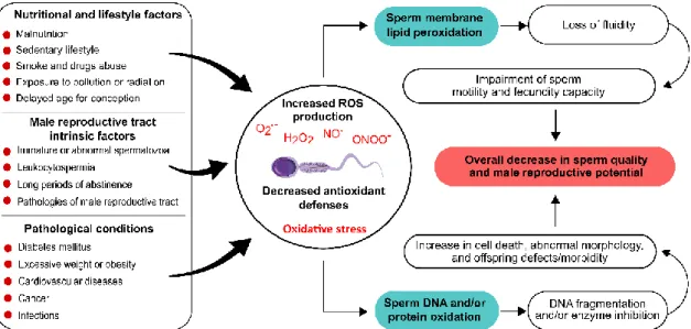
Figure 1.6 Oxidative stress-promoting factors in spermatozoa and the respective outcomes for male reproductive function. ... 34
Figure 1.7 Chemical structures of L-theanine and caffeine. ... 39
Figure 1.8 Chemical structures of the main catechins present in white tea. ... 40
Figure 3.1 Effect of white tea extract in metabolites consumption/production by cultured rat Sertoli cells. ... 60
Figure 3.2 Effect of white tea extract in mRNA and protein levels of glucose transporter 1, glucose transporter 3, phosphofructokinase 1, lactate dehydrogenase and monocarboxylate transporter 4 ... 60
Figure 3.3 Effect of white tea extract in cultured rat Sertoli cells intracellular lactate dehydrogenase activity ... 61
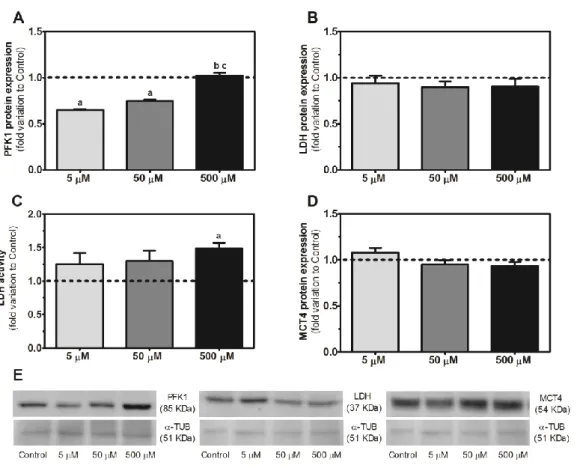
Figure 3.4 Effect of caffeine (5, 50 and 500 μM) on glucose consumption and glucose transporters protein expression in human Sertoli cells ... 73
Figure 3.5 Effect of caffeine (5, 50 and 500 μM) in the expression of phosphofructokinase 1, lactate dehydrogenase and monocarboxylate transporter 4, as well as lactate dehydrogenase activity in human Sertoli cells ... 74
Figure 3.6 Effect of caffeine (5, 50 and 500 μM) in the production of lactate and alanine, as well as the lactate/alanine ratio in human Sertoli cells ... 75
Figure 3.7 Effect of caffeine (5, 50 and 500 μM) in the ferric reducing antioxidant power value, carbonyl groups formation, and lipid peroxidation in human Sertoli cells ... 76
Figure 3.8 Effect of epigallocatechin gallate (5 and 50 µM) in the proliferation of human Sertoli cells ... 88
xx
Figure 3.9 Effect of epigallocatechin gallate (5 and 50 µM) in glucose metabolism of human
Sertoli cells ... 89
Figure 3.10 Effect of epigallocatechin gallate (5 and 50 μM) in pyruvate and lactate metabolism
of human Sertoli cells ... 90
Figure 3.11 Effect of epigallocatechin gallate (5 and 50 μM) in mitochondrial membrane
potential and extracellular oxygen consumption of human Sertoli cells ... 92
Figure 3.12 Effect of epigallocatechin gallate (5 and 50 μM) in oxidative damage levels of
human Sertoli cells ... 93
Figure 3.13 Effect of L-theanine (5 and 50 µM) in human Sertoli cells survival ... 105 Figure 3.14 Effect of L-theanine (5 and 50 µM) in glucose metabolism of human Sertoli cells
... 106
Figure 3.15 Effect of L-theanine (5 and 50 μM) in mitochondrial function of human Sertoli cells
... 107
Figure 4.1 Representative proton nuclear magnetic resonance spectrum of white tea extract
showing the phytocomponents peak assignments ... 117
Figure 4.2 Microscopic image of rat spermatozoa after eosin/nigrosin staining: viable
spermatozoon (left) and non-viable spermatozoon (right). ... 120
Figure 4.3 Ferric reducing antioxidant power of the epididymal spermatozoa storage media,
control medium and media supplemented with freeze-dried white tea or green tea aqueous extracts to a final concentration of 0.5 or 1 mg/mL ... 122
Figure 4.4 Ferric reducing antioxidant power of the epididymal spermatozoa stored in control
medium and media supplemented with freeze-dried white tea or green tea aqueous extracts to a final concentration of 0.5 or 1 mg/mL, during the 24, 48 and 72 h of the experiment ... 123
Figure 4.5 Sperm thiobarbituric acid reactive substances produced in epididymal spermatozoa
stored in control medium and media supplemented with freeze-dried white tea or green tea aqueous extracts to a final concentration of 0.5 or 1 mg/mL, during the 24, 48 and 72 h of the experiment ... 124
Figure 4.6 Spermatozoa viability at collection time and during the 3-day storage in control
medium and media supplemented with freeze-dried white tea or green tea aqueous extracts to a final concentration of 0.5 or 1 mg/mL ... 125
xxi combination of the three compounds ... 136
Figure 4.8 Spermatozoa oxidative profile at the end of incubation in control medium and media
supplemented with caffeine, epigallocatechin gallate, L-theanine or the combination of the three compounds ... 137
Figure 5.1 Effect of white tea consumption in testicular glucose transport of prediabetic rats..
... 148
Figure 5.2 Effect of white tea consumption in testicular glycolytic profile of prediabetic rats..
... 149
Figure 5.3 Effect of white tea consumption in testicular content of lactate and alanine in
prediabetic rats ... 150
Figure 5.4 Effect of white tea consumption in epididymal glucose transport in prediabetic rats.
... 152
Figure 5.5 Effect of white tea consumption in epididymal glycolytic profile in prediabetic rats..
... 152
Figure 5.6 Effect of white tea consumption in the epididymal content of lactate and alanine in
xxiii
Table 3.1 Oligonucleotides and cycling conditions for amplification of glucose transporters 1
and 3, phosphofructokinase 1, lactate dehydrogenase, monocarboxylate transporter 4 and β-2-microglobulin ... 57
Table 3.2 Phytochemical profile of white tea extract in percentage of weight determined for
each compound relative to the total weight of white tea extract. ... 58
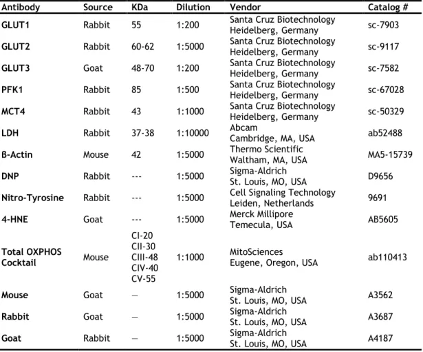
Table 3.3 List of the primary and secondary antibodies used in this study. ... 86 Table 3.4 Metabolites consumption/production and lactate/alanine ratio in human Sertoli cells
from the control group and groups exposed to 5 or 50 µM of epigallocatechin gallate. ... 91
Table 3.5 Protein expression levels of mitochondrial complexes in human Sertoli cells from the
control group and groups exposed to 5 or 50 µM of epigallocatechin gallate. ... 92
Table 3.6 Protein expression levels of mitochondrial complexes in human Sertoli cells from the
control group and groups exposed to 5 or 50 µM of L-theanine. ... 107
Table 3.7 Oxidative damage levels, evaluated by carbonyl groups, nitration and lipid
peroxidation, of human Sertoli cells from the control group and groups exposed to 5 or 50 µM of L-theanine. ... 108
Table 4.1 Phytochemical profile of white tea and green tea extracts determined by proton
nuclear magnetic resonance spectroscopy ... 121
Table 5.1 List of the primary and secondary antibodies used in this study. ... 146 Table 5.2 White tea phytochemical characterization as determined by proton nuclear magnetic
resonance. ... 147
Table 5.3 Effect of white tea consumption on blood glucose levels in prediabetic rats, at the
end of the treatment (non-fasting glucose) and after performing the glucose and insulin tolerance tests. ... 148
Table 5.4 Relationships between lactate content, lactate dehydrogenase protein levels and
activity, monocarboxylate transporter 4 protein levels, alanine content and alanine transaminase protein levels evaluated by Pearson’s correlation coefficient (r) in testis from the control, prediabetic rats drinking water or white tea. ... 151
Table 5.5 Relationships between lactate content, lactate dehydrogenase protein levels and
xxiv
transaminase protein levels evaluated by Pearson’s correlation coefficient (r) in epididymis from the control, prediabetic rats drinking water or white tea ... 155
Table 5.6 Effect of white tea consumption in epididymal sperm quality (motility, viability,
xxv
1H-NMR Proton nuclear magnetic resonance
4-HNE 4-hydroxynonenal
ABP Androgen binding protein
ADP Adenosine diphosphate
AI Artificial insemination
ALT Alanine transaminase
AMH Anti-Müllerian hormone
AMP Adenosine monophosphate
AP Alkaline phosphatase
ART Assisted reproductive technology
AT Annealing temperature
ATP Adenosine triphosphate
ATP5A ATP synthase alpha-subunit
B2M β-2-microglobulin
BEB Blood-epididymal barrier
BSA Bovine serum albumin
BTB Blood-testis barrier
cDNA Complementary deoxyribonucleic acid
CPS Counts per second
D2O Deuterium oxide
DM Diabetes mellitus
DMEM Dulbecco’s modified eagle medium
DMSO Dimethyl sulfoxide
DNA Deoxyribonucleic acid
DNP 2,4-dinitrophenol
DNPH 2,4-dinitrophenylhydrazine
dNTPs Deoxynucleotide triphosphates
DTT Dithiothreitol
EC Epicatechin
ECG Epicatechin gallate
EDTA Ethylenediamine tetraacetic acid
EGC Epigallocatechin
EGCG Epigallocatechin gallate
EGF Epidermal growth factor
EGTA Ethylene glycol tetraacetic acid
ES Ectoplasmic specialization
ETC Electron transport chain
F12 Ham's F12 nutrient mixture
FBS Fetal bovine serum
FDA Food and Drug Administration
FGF Fibroblast growth factor
FSH Follicle stimulating hormone
GJs Gap junctions
GLUT1 Glucose transporter 1
GLUT2 Glucose transporter 2
GLUT3 Glucose transporter 3
GLUTs Glucose transporters
GnRH Gonadotropin releasing hormone
GRAS Generally recognized as safe
GTEA Green tea
GTT Glucose tolerance test
H2A Histone 2A
H2B Histone 2B
H3 Histone 3
H4 Histone 4
xxvi
HEPES Hydroxyethyl piperazineethanesulfonic acid
HIV Human immunodeficiency virus
IDF International Diabetes Federation
IGF-I Insulin-like growth factor I
IGT Impaired glucose tolerance
INT 3-(4-Iodophenyl)-2-(4-nitrophenyl)-5-phenyl-2H-tetrazol-3-ium chloride
IR Insulin resistance
ITS Insulin–transferrin–sodium selenite
ITT Insulin tolerance test
IVF In vitro fertilization
JC1 5,5’,6,6’-tetrachloro-1,1’,3,3’-tetraethylbenzimidazolylcarbocyanine iodide
LDH Lactate dehydrogenase
LDL Low density lipoproteins
LH Luteinizing hormone
MARs Matrix attachment regions
MCT4 Monocarboxylate transporter 4
MCTs Monocarboxylate transporters
MDA Malondialdehyde
M-MLV RT Moloney murine leukemia virus reverse transcriptase
M-PER Mammalian protein extraction reagent
mRNA Messenger ribonucleic acid
MTCO1 Mitochondrially encoded cytochrome c oxidase I
MTT 3-(4,5-dimethylthiazol-2-yl)-2,5-diphenyltetrazolium bromide
NAD+ Nicotinamide adenine dinucleotide, oxidized form
NADH Nicotinamide adenine dinucleotide, reduced form
NDUFB8 NADH dehydrogenase (ubiquinone) 1 beta subcomplex subunit 8
ODF Outer dense fibers
OS Oxidative stress
OXPHOS Oxidative phosphorylation
PBS Phosphate-buffered saline
PDH Pyruvate dehydrogenase
PFK1 Phosphofructokinase 1
PMSF Phenylmethylsulfonyl fluoride
PTM Post-translational modifications
PUFAs Polyunsaturated fatty acids
PVDF Polyvinylidene fluoride
qPCR Quantitative polymerase chain reaction
RNA Ribonucleic acid
ROS Reactive oxygen species
RT Room temperature
SCs Sertoli cells
SDHB Succinate dehydrogenase complex, subunit B, iron sulfur
SP Statistical power
SRB Sulforhodamine B
STF Seminiferous tubular fluid
STZ Streptozotocin
T1DM Type 1 diabetes mellitus
T2DM Type 2 diabetes mellitus
TBA Thiobarbituric acid
TBARS Thiobarbituric acid reactive substances
TGF-α Transforming growth factor alpha
TGF-β Transforming growth factor beta
TJs Tight junctions
TPTZ 2,4,6-tripyridyl-s-triazine
TYH Krebs-Ringer bicarbonate
UQCRC2 Ubiquinol-cytochrome c reductase core protein II
VLDL Very low-density lipoproteins
WHO World Health Organization
xxvii
Publications included in this thesis:
Articles:
1 - Martins AD, Alves MG, Bernardino RL, Dias TR, Silva BM, Oliveira PF. (2013) “Effect of white
tea (Camellia sinensis (L.)) extract in the glycolytic profile of Sertoli cell”. European Journal of Nutrition 53(6):1383-1391 (DOI:10.1007/s00394-013-0640-5).
2 - Dias TR, Martins AD, Reis VP, Socorro S, Silva BM, Alves MG, Oliveira PF. (2013) “Glucose
transport and metabolism in Sertoli cell: relevance for male fertility”. Current Chemical Biology 7(3):282-293 (DOI:10.2174/2212796807999131128125510).
3 - Dias TR, Tomás G, Teixeira N, Alves MG, Oliveira PF, Silva BM. (2013) “White tea (Camellia
sinensis (L.): antioxidant properties and beneficial health effects”. International Journal of Food Science, Nutrition and Dietetics 2(2):19-26 (DOI:dx.doi.org/10.19070/2326-3350-130005).
4 - Dias TR, Alves MG, Tomás GD, Socorro S, Silva BM, Oliveira PF. (2014) “White tea as a
promising antioxidant media additive for sperm storage at room temperature: a comparative study with green tea”. Journal of Agricultural and Food Chemistry 62(3): 608–617 (DOI:10.1021/jf4049462).
5 - Dias TR, Alves MG, Silva BM, Oliveira PF. (2014) “Sperm glucose transport and metabolism
in diabetic individuals”. Molecular and Cellular Endocrinology 396(1-2):37-45 (DOI:10.1016/j.mce.2014.08.005).
6 - Dias TR, Alves MG, Oliveira PF, Silva BM. (2014) “Natural products as modulators of
spermatogenesis: the search for a male contraceptive”. Current Molecular Pharmacology 7(2):154-166 (DOI:10.2174/1874467208666150126155912).
7 - Alves MG, Dias TR, Silva BM, Oliveira PF. (2014) “Metabolic cooperation in testis as a
pharmacological target: from disease to contraception”. Current Molecular Pharmacology 7(2):83-95 (DOI:10.2174/1874467208666150126153830).
8 - Dias TR, Alves MG, Bernardino RL, Martins AD, Moreira AC, Silva J, Barros A, Sousa M, Silva
BM, Oliveira PF. (2015) “Dose-dependent effects of caffeine in human Sertoli cells metabolism and oxidative profile”. Toxicology 328:12-20 (DOI:10.1016/j.tox.2014.12.003).
9 - Dias TR, Alves MG, Casal S, Silva BM, Oliveira PF. (2016) “Single and synergistic effect of
major tea components caffeine, epigallocatechin gallate and L-theanine in rat sperm viability”. Food & Function 7(3):1301-1305 (DOI:10.1039/c5fo01611h).
10 - Dias TR, Alves MG, Rato L, Casal S, Silva BM, Oliveira PF. (2016) “White tea intake prevents
prediabetes-induced metabolic dysfunctions in testis and epididymis preserving sperm quality” The Journal of Nutritional Biochemistry 37:83-93 (DOI:10.1016/j.jnutbio.2016.07.018).
xxviii
11 - Dias TR, Bernardino RL, Meneses MJ, Sousa M, Sá R, Alves MG, Silva BM, Oliveira PF. (2016)
“Emerging potential of natural products as an alternative strategy to pharmacological agents used against metabolic disorders”. Current Drug Metabolism 17(6):582-597 (DOI:10.2174/1389200217666160229113629).
12 - Dias TR, Alves MG, Silva J, Barros A, Sousa M, Casal S, Silva BM, Oliveira PF. (2017)
“Implications of epigallocatechin gallate in cultured human Sertoli cells glycolytic and oxidative profile” Toxicology In Vitro 41:214-222 (DOI:10.1016/j.tiv.2017.03.006).
13 - Dias TR, Alves MG, Casal S, Oliveira PF, Silva BM. (2017) “Promising potential of dietary
(poly)phenolic compounds in the prevention and treatment of diabetes mellitus” Current Medicinal Chemistry 24(4):334-354 (DOI:10.2174/0929867323666160905150419).
14 - Dias TR, Alves MG, Oliveira PF, Silva BM. (2017) “Bioactive Substances from Medicinal
Plants for Metabolic Disorders” Current Medicinal Chemistry 24(4):332-333 (DOI:10.2174/092986732404170302223350).
15 - Dias TR, Bernardino RL, Alves MG, Silva J, Barros A, Sousa M, Casal S, Silva BM, Oliveira
PF. (2019) “L-theanine promotes cultured human Sertoli cells proliferation and modulates glucose metabolism” European Journal of Nutrition 58(7):2961-2970 (DOI:10.1007/s00394-019-01999-2).
Book Chapters:
16 – Dias TR, Alves MG, Neuhaus-Oliveira A, Socorro S, Silva BM, Oliveira PF. (2013)
“Implications of Diabetes on sperm glucose uptake and metabolism” In Glucose Uptake: Regulation, Signaling Pathways and Health Implications, Nova Science Publishers, Inc, New York, USA, pp. 141-168 (ISBN: 978-1-62618-671-2).
17 - Dias TR, Bernardino RL. (2017) “Biochemical events occurring in the epididymis” In: Alves
MG, Oliveira PF (eds) Biochemistry of Andrology, vol 1. Andrology: Current and Future Developments. Bentham Science Publishers, Sharjah, UAE, pp. 178-206 (DOI: 10.2174/97816810850051170101), (ISBN:978-1-68108-501-2).
18 - Dias TR. (2017) “Functional and biochemical aspects of spermatozoa” In: Alves MG, Oliveira
PF (eds) Biochemistry of Andrology, vol 1. Andrology: Current and Future Developments.
Bentham Science Publishers, Sharjah, UAE, pp. 230-256 (DOI:
10.2174/97816810850051170101), (ISBN:978-1-68108-501-2).
19 - Dias TR, Alves MG, Silva BM, Oliveira PF. (2018) “Nutritional factors and male
reproduction” In: Bernard Jégou and Michael K. Skinner (Eds), Encyclopedia of Reproduction, 2nd edition, volume 1, Elsevier, Waltham, MA, USA, pp. 458-464
(DOI:10.1016/B978-0-12-801238-3.64616-0), (ISBN:978-0-12-801238-3).
20 - Dias TR, Carrageta DF, Alves MG, Oliveira PF, Silva BM. (2018) “White tea” In: Seyed Nabavi
and Ana Silva (eds), Nonvitamin and Nonmineral Nutritional Supplements, 1st edition, Academic Press, Elsevier, pp. 437-445 (DOI: 10.1016/B978-0-12-812491-8.00058-8), (ISBN:9780128124918).
1
Chapter 1
Introduction
This chapter includes the following sections:
• Male reproductive biology and physiology
• Diabetes mellitus and male fertility
3
Male reproductive biology and physiology
This section was adapted from the following publications:
- Dias TR, Martins AD, Reis VP, Socorro S, Silva BM, Alves MG, Oliveira PF. (2013) “Glucose
transport and metabolism in Sertoli cell: relevance for male fertility”. Current Chemical Biology 7(3):282-293 (DOI:10.2174/2212796807999131128125510).
- Dias TR, Alves MG, Oliveira PF, Silva BM. (2014) “Natural products as modulators of
spermatogenesis: the search for a male contraceptive”. Current Molecular Pharmacology 7(2):154-166 (DOI:10.2174/1874467208666150126155912).
- Alves MG, Dias TR, Silva BM, Oliveira PF. (2014) “Metabolic cooperation in testis as a pharmacological target: from disease to contraception”. Current Molecular Pharmacology 7(2):83-95 (DOI:10.2174/1874467208666150126153830).
Dias TR, Bernardino RL. (2017) “Biochemical events occurring in the epididymis” In:
Alves MG, Oliveira PF (eds) Biochemistry of Andrology, vol 1. Andrology: Current and Future Developments. Bentham Science Publishers, Sharjah, UAE, pp. 178-206 (DOI: 10.2174/97816810850051170101), (ISBN:978-1-68108-501-2).
- Dias TR. (2017) “Functional and biochemical aspects of spermatozoa” In: Alves MG,
Oliveira PF (eds) Biochemistry of Andrology, vol 1. Andrology: Current and Future Developments. Bentham Science Publishers, Sharjah, UAE, pp. 230-256
Effect of white tea on the reproductive function of diabetic or prediabetic individuals
4
Spermatogenesis
In mammals, about 80% of the testicular mass consists of highly coiled seminiferous tubules [1, 2], while the remaining 20% are Leydig cells and other interstitial components [1]. Spermatogenesis takes place in the seminiferous tubules, of which the main structural elements are Sertoli cells (SCs) [3]. Spermatogenesis refers to the development of mature spermatozoa with half the number of chromosomes (haploid), from the most immature germ cell in the testis, spermatogonium (diploid) [4, 5]. This complex process is divided in three major phases: mitotic, meiotic and spermiogenesis [6]. Firstly, undifferentiated spermatogonia, located along the basement membrane of the seminiferous tubules can be of two types: with dark (Ad) or
pale (Ap) nuclei [7]. Ad cells do not present active mitosis, thus constituting the reserve of
spermatogonial stem cells that allow the maintenance of spermatogenesis from puberty until death. This explains why despite aging in males is associated with a decline in semen volume, sperm motility and normal morphology, sperm concentration is not altered with aging [8]. On the other hand, Ap cells are the spermatogonial steam cells that differentiate into type B
spermatogonia, which then migrate closer to the tubule lumen and differentiate into slightly larger cells called primary spermatocytes (diploid) [7]. In the second phase, primary spermatocytes move into the adluminal compartment of the seminiferous tubules and divide into two secondary spermatocytes (haploid) by meiosis I. Then, each secondary spermatocyte undergo meiosis II resulting in two haploid round spermatids [9]. Finally, spermiogenesis involves the transformation of round spermatids into elongated flagellar spermatids and culminates with the release of spermatozoa (spermiation) into the lumen of the seminiferous tubules [9, 10]. During spermiogenesis, spermatids suffer a number of morphological changes including acrosome formation, nuclear condensation, development of the flagellum, and cytoplasm reorganization, which eventually result in the generation of spermatozoa [9]. Those sequential alterations in the spermatids differentiation are very helpful in the identification of the stages of the spermatogenic cycle [11]. In most mammals, each spermatogenic cycle takes 9-12 days. In the mouse takes about 8.6 days (XII stages) and in the rat 12.9 days (XIV stages) [11]. However, the human spermatogenic cycle is longer, lasting 16 days (VI stages) and four cycles are needed to complete spermatogenesis (more than 70 days) [12].
Spermatogenesis is under a strict hormonal control driven by the communication between the hypothalamus–pituitary axis and the gonad itself (Figure 1.1) [13]. The master regulator of this process is the gonadotropin releasing hormone (GnRH) [14], which triggers the release of both luteinizing hormone (LH) and follicle-stimulating hormone (FSH) from the anterior pituitary (Figure 1.1) [15]. LH binds to receptors on the surface of Leydig cells stimulating the production of testosterone, a steroid hormone that diffuses into the seminiferous tubules. Within the seminiferous tubules, only SCs possess receptors for testosterone and FSH [14]. FSH stimulates SCs division and differentiation, as well as the production of androgen-binding protein (ABP), facilitating the passage of testosterone through the blood-testis barrier (BTB). Thus,
5 testosterone and FSH act synergistically on SCs leading to the secretion of paracrine agents that are essential for spermatogenesis [13]. SCs convert testosterone into estrogen and the circulating levels of these two hormones mediate the feedback inhibition of GnRH through steroid receptors present in the hypothalamic neurons and in the pituitary [16]. SCs also secrete the hormones inhibin and activin that also regulate GnRH and LH/FSH release.
Figure 1.1 Spermatogenesis and its hormonal control. Spermatogonia divides by mitosis originating primary spermatocytes, which transform into secondary spermatocytes after the first meiotic division. Then, meiosis II give rise to round spermatids, which subsequently differentiate into elongated spermatids. The process culminates with the release of spermatozoa into the lumen of the seminiferous tubule (spermiation). Spermatogenesis is mainly regulated by the gonadotropin releasing hormone (GnRH), synthetized by the hypothalamus, which stimulates the anterior pituitary to produce the luteinizing hormone (LH) and follicle-stimulating hormone (FSH). FSH acts only on the Sertoli cells (SCs), present in the seminiferous tubules, stimulating spermatogenesis. SCs secrete hormones inhibin and activin, which regulate GnRH and LH/FSH release. LH acts only on the Leydig cells inducing testosterone production. SCs convert testosterone into estrogen, which has an inhibitory effect on Leydig cells androgen production.
Effect of white tea on the reproductive function of diabetic or prediabetic individuals
6
Sertoli cells
The SCs are highly polarized epithelial cells that extend upwards from the basement membrane, where the peritubular myoid cells reside, towards the lumen of the seminiferous tubules, surrounding the germ cells (Figure 1.1) [17, 18]. SCs play a central role in the development of a functional testis, and hence in the expression of the male phenotype [19]. Testis determination in mammals occurs through the action of the Y-linked gene Sry. The Sry gene encoded protein acts in the supporting cell lineage of the indifferent gonad, triggering a cascade of events that results in the differentiation of these cells into Sertoli rather than follicle cells [20]. Then, SCs undergo a phase of rapid cell proliferation and differentiation [21] activating other cell lineages within the gonad to follow the testicular pathway [20]. At each division of pre-pubertal SCs, the daughter cells generate specialized micro-domains to sustain the amplification of the mitotic spermatogonia [22]. The anti-Müllerian hormone (AMH) is the first product secreted by SCs in the developing testes [23], ensuring the regression of the Mullerian ducts, which is a crucial step for proper male sexual differentiation [24].
Around the onset of puberty, the final phase of SCs differentiation is marked by the cessation of proliferation and irreversible changes in SCs morphology and physiology [18], heralding the switch from an immature, proliferative state to a mature, non-proliferative state [25]. In this terminal differentiation phase, SCs exit from the cell cycle and originate the BTB [21], marking the progressive entry of SCs into adulthood. Besides, the lumen of the tubules is formed and SCs acquire the typical adult characteristics: the nucleus, usually situated in the basal portion of the cell, enlarges and becomes tripartite; the nucleolus becomes more prominent [25]; the cytoplasm becomes voluminous and include cytoplasmic components with a polar distribution; the organelles and inclusions appear in higher amounts in the basal and lower trunk of the cell, whereas mitochondria and smooth endoplasmic reticulum are more abundant in the apical portion [26]. SCs also contain a relatively low proportion of rough endoplasmic reticulum, an elaborate cytoskeleton, lipids and lysosomes [18]. SCs differentiation is also accompanied by the expression of many gene products that are not present in immature cells [27-32]. Nevertheless, there is an interesting feature about adult SCs proliferation. Although it has been reported that SCs cease to divide and proliferate at a certain phase of their adulthood, being that time different between species [25], this affirmation revealed not to be an absolute true. When adult SCs were transplanted into impaired or defective testis, were found to be proliferative, capable of populating the testis and even restoring spermatogenesis [33, 34]. Moreover, several in vitro studies have shown that this cellular type has a proliferative potential when cultured in favorable conditions [35, 36]. In fact, an in vitro model of adult SCs retain many specific characteristics of their derived tissues, being a great model for in vitro toxicology studies [37].
The BTB is one of the tightest blood-tissue barriers in mammals [2]. It is formed by coexisting specialized junctions between adjacent SCs, including tight junctions (TJs), basal ectoplasmic
7 specialization (ES), gap junctions (GJs) and desmosome-like junctions [38-42], such that nothing larger than 1000 dalton can pass from the outside to the inside of the tubule [14, 43]. The BTB divides the seminiferous epithelium into the basal compartment (containing spermatogonia and early spermatocytes) and adluminal (or apical) compartment (containing spermatocytes in different stages of differentiation, spermatids and spermatozoa) [2, 43]. This barrier consists in three components: i) an anatomical/physical barrier to restrict the entry of molecules into the adluminal compartment; ii) an immunological barrier that limits the movement of immune cells and regulates the level of cytokines in the seminiferous epithelium; and iii) a physiological barrier that is highly dynamic to encounter the needs of germ cells [1, 44]. These components create a special microenvironment responsible for the proper development of spermatogenesis. Particularly, BTB allows the control of the ionic composition and pH of the seminiferous tubular fluid (STF) [35, 45, 46]. Thus, BTB dysfunction may lead to the arrest of germ cells differentiation [47, 48].
Sertoli-germ cells cooperation
Germ cells development is a highly organized process that is mainly under the control of SCs [49]. It has been established that each SC can only support a limited number of germ cells (30 to 50) [50], and approximately 75% of developing germ cells undergo spontaneous degeneration [51]. This will determine the daily sperm production, a factor that will impact male fertilizing potential [25, 52]. Mature SCs acquire a characteristic spatial arrangement that provides them the unique capability to interact either morphologically and/or chemically with the different generations of germ cells, with peritubular myoid cells and with the steroid producing Leydig cells. The SCs base is in contact with spermatogonia, while their lateral surfaces send processes around spermatocytes and early spermatids; their apical portion is intimately associated to elongating and elongated spermatids and faces the tubule lumen where spermatozoa are released (Figure 1.1) [18]. There is a well-timed movement of developing germ cells across the BTB consisting of intermittent phases of junctional complexes disassembly and reassembly, which allow the passage of germ cells while maintaining BTB integrity [53]. In the beginning of meiosis, germ cells located outside the barrier pass through the TJs and, once beyond the BTB, they are dependent on SCs supplies [43]. If the passage across BTB is accelerated, this will induce a premature detachment of germ cells from the epithelium and the spermatozoa produced are unable to fertilize the egg due to their immaturity. Likewise, if the process is hampered and germ cells become retained in the epithelium, they will be removed by SCs via phagocytosis. In either case, subfertility or infertility may occur [53], highlighting the importance of a proper SCs function.
In 1865, Enrico Sertoli gave his name to SCs and defined them as “nurse cells” [54]. With this concept, he meant that SCs provide nutrients, regulatory factors, functional glycoproteins and peptides for the development and differentiation of germ cells [55-58]. Particularly, SCs
Effect of white tea on the reproductive function of diabetic or prediabetic individuals
8
provide factors required to fuel germ cells metabolism (lactate, transferrin, ceruloplasmin, ABP) [59, 60]; growth regulatory factors (stem cell factor, transforming growth factors alpha and beta - TGF-α and TGF-β); insulin-like growth factor-I (IGF-I); fibroblast growth factor (FGF); and epidermal growth factor (EGF) [60]. SCs have the ability to metabolize various substrates, but preferentially use glucose [61, 62], even though this monosaccharide (hexose) is present at very low levels in STF due to its rapid metabolism [62]. Glucose transport through SCs cytoplasmic membrane is mediated by glucose transporters (GLUTs), particularly GLUT1, GLUT2 and GLUT3 (Figure 1.2) [63]. Glucose enters the glycolytic pathway and is decomposed to pyruvate, in a rate-limiting process catalyzed by the enzyme phosphofructokinase 1 (PFK1) [64]. This cascade of reactions yields not only two molecules of pyruvate, but also two molecules of adenosine triphosphate (ATP) and two electron-carrying molecules of nicotinamide adenine dinucleotide reduced (NADH) [62, 65]. Under low-oxygen conditions, glycolysis end-products follow into the production of lactic acid, by lactate dehydrogenase (LDH) action [66]. LDH is responsible for the reversible conversion of pyruvate into lactate, with the concomitant oxidation/reduction of NADH to its oxidized form NAD+ [67]. After lactate production, it is
crucial that this product becomes available for the developing germ cells, as it is their preferred substrate for ATP production [60, 68-70]. This event is mediated by active membrane monocarboxylate transporters (MCTs), especially MCT4, that are responsible for lactate transport through the plasma membrane of SCs [63, 71-73] (Figure 1.2). The importance of lactate for normal spermatogenesis is highlighted in a report showing that spermatogenesis in adult cryptorchid testis is improved by intratesticular infusion of lactate [74]. It is well known that spermatogonia may utilize glucose as the major energy substrate [75], but spermatocytes and spermatids suffer a rapid decline in their ATP content in glucose-supplemented media, thus, they require lactate/pyruvate for the maintenance of their ATP concentrations [76, 77]. Interestingly, spermatozoa prefer glucose/fructose as energy source [78]. Nevertheless, an although the regulatory molecular mechanisms by which SCs preferentially export lactate, pyruvate or glucose at each stage of spermatogenesis remain largely unknown [79], it is widely accepted that glucose and its metabolites play a pivotal role at many stages of germ cells development [80].
Pyruvate is an important regulatory point of SCs metabolism as it can follow two other pathways besides lactate production: it can be converted to alanine in a reversible reaction catalyzed by alanine transaminase (ALT) [81], or it can enter the mitochondria to originate acetyl-CoA by the action of pyruvate dehydrogenase (PDH) [82]. Alanine plays a key role in the maintenance of cellular redox status and glucose homeostasis [9]. In fact, the interconversion lactate – pyruvate - alanine is NADH-dependent and the NADH/NAD+ ratio can be estimated by the ratio
lactate/alanine, which is often used as a measure of the cellular redox state [83]. On the other hand, acetyl-CoA can enter the Krebs cycle, where it is converted to citrate [84], or it can be exported to the cytosol and form acetate [85], which can be used for fatty acids and cholesterol synthesis [86]. When there is a Krebs cycle truncation, citrate can also be transported to the
9 cytosol or to extracellular compartment [84]. Under physiological conditions, glucose metabolism is strictly controlled and is essential for a normal spermatogenesis [10]. However, the dysregulation of glucose metabolism may lead to male reproductive disorders [11, 12].
Figure 1.2 Sertoli cells (SCs) metabolic cooperation with developing germ cells. In SCs, glucose from the interstitial space is taken up through high-affinity glucose transporters, GLUT1, GLUT2 and GLUT3, which are present in the plasma membrane. In physiological conditions, most of glucose is converted to pyruvate through a rate-limiting process catalyzed by the enzyme phosphofructokinase 1 (PFK1). Pyruvate can be converted into lactate, alanine or acetyl-coA by the action of lactate dehydrogenase (LDH), alanine transaminase (ALT) or pyruvate dehydrogenase (PDH), respectively. Acetyl-CoA enters the mitochondrion to be used in the Krebs cycle, while lactate is exported to the intratubular fluid by specific monocarboxylate transporters (MCT4). This substrate is then taken up by germ cells to be used as metabolic fuel for adenosine triphosphate (ATP) production.
Spermatozoa ultrastructure
Spermatozoa present a unique and complex morphology. In general, they comprise a head, a midpiece and a tail region (Figure 1.3), commonly known as flagellum [15]. The spermatozoon is smaller than most cells in the body, but its size does not reflect its fundamental function of generating a new human being. The size, shape of the head, length and relative amount of the different components of the flagellum is species-specific [87]. For instance, rodent spermatozoa have hook-shaped heads, while the head of human spermatozoa is pear-shaped. The head of human spermatozoa has a median length of 4.4 µm and width of about 3 µm [88]. The nucleus occupies most of the human sperm head area and contains a haploid set of condensed, genetically inactive chromosomes [7]. The apical half of the nucleus is covered by the acrosome (Figure 1.3), which represents about 48% of the sperm head surface [88]. This
Effect of white tea on the reproductive function of diabetic or prediabetic individuals
10
acrosomal cap is a membrane-enclosed cytoplasmic vesicle originating from the Golgi apparatus during spermatozoa formation [89]. It contains several monosaccharides (e.g. galactose, mannose, fructose) [90] and hydrolytic enzymes with a preponderant role in the penetration of the spermatozoon into the oocyte membranes [15]. The part of the nucleus that is not overlaid by the acrosome cap constitutes the postacrosomal region. The sperm head also contains a small amount of cytoplasm and several cytoskeletal structures, including the dense perinuclear layer that is made of basic proteins (e.g. calicin and cylicin I and II) associated with calmodulin and actin filaments [91]. On the base of the sperm head there is a small structure called connecting piece (or neck) that connects the head to the midpiece. The connecting piece harbors the proximal centriole and the empty vault. The proximal centriole is composed of nine microtubule triplets and has a vital role on the orchestration of cell division in the embryo, while the empty vault is originated after the degradation of the distal centriole during spermiogenesis [92]. The connecting piece provides a basal anchor to the axoneme and is covered by several redundant nuclear envelopes [93]. It was also proposed to be responsible for flagellum beat initiation and alternating directions of bends propagating down the beating flagellum [94].
The midpiece has a cylindrical shape and has about 5-9 µm of length and half the width of the sperm head (Figure 1.3) [95]. Its structure consists of numerous mitochondria spirally arranged around the outer dense fibers (ODF) and the central axial filament - axoneme [87]. The mitochondrial sheath is responsible for the production of ATP, which is the energy supply needed for tail motility during the migration upon the female reproductive tract. The axoneme is essentially a long, specialized cilium formed by a core of microtubules, surrounded by ODF extending from the connecting piece to the principal piece. It has a characteristic “9+2” structure, i.e., two central singlet microtubules encircled by nine outer doublet microtubules (A- and B-tubules). Radial links connect the central microtubule pair to each surrounding microtubule doublet, and nexin bridges connect adjacent doublets [96]. Outer and inner dyneins are observed as projecting “arms” that slide along each outer doublet microtubule. This active sliding has been associated with spermatozoa flagellar movement [97]. A cytoplasmic droplet is frequently found at the midpiece or at the junction of the midpiece with the principal piece in human mature spermatozoa. This tiny droplet-like structure is usually retained after the removal of spermatids cytoplasm by SCs at the end of spermiogenesis, and is suggested to play a key role in sperm volume adaptation [98]. At the distal part of the mitochondrial sheath of sperm midpiece is a traverse septin-based ring called annulus (Figure 1.3) that is the hallmark of the separation between the midpiece and the principal piece [99].
11 Figure 1.3 Human spermatozoon structure. It is constituted by the head, connecting piece, midpiece, principal piece and end piece. The head is mainly composed by the nucleus that is enveloped by the acrosome. The connecting piece contains the proximal centriole surrounded by the nuclear redundant envelopes. The midpiece holds the mitochondrial sheath, which is the site of energy production. These several mitochondria are organized around the outer dense fibers and the axoneme. The axoneme extends from the basis of the head (connecting piece) until the end of the sperm flagellum. Separating the midpiece and the beginning of the tail is a ring-like small structure called annulus. The tail comprises the principal piece that is the lengthiest part of the spermatozoon, and the end piece, the narrowest part. Besides the axoneme, the principal piece also contains the outer dense fibers surrounded by a fibrous sheath. The end piece is mainly composed by the axoneme. The spermatozoon is enveloped by a plasma membrane.
The sperm tail is the only functional flagellum in humans and it can be divided into principal piece and end piece. The principal piece constitutes most of the tail, having an average length of 40-45 μm [95]. It is constituted by the axoneme surrounded by a sheath of supportive fibers composed of two longitudinal columns that run parallel to ODF (Figure 1.3). The main function of principal piece is to propel the spermatozoon towards the oocyte, changing both the amplitude and frequency of the whip like movement of the tail to facilitate sperm hyperactivation and egg penetration.