Universidade de Lisboa
Faculdade de Medicina Dentária da Universidade de Lisboa
Effects of different ways of
polymerization of an universal adhesive
in shear bond strength to dentin
Mariana Luís Batista Guttiérrez
Dissertação
Mestrado Integrado em Medicina Dentária
Universidade de Lisboa
Faculdade de Medicina Dentária da Universidade de Lisboa
Effects of different ways of
polymerization of an universal adhesive
in shear bond strength to dentin
Mariana Luís Batista Guttiérrez
Dissertação orientada por:
Prof.ª Doutora Sofia Arantes e Oliveira Prof.ª Doutora Ana Filipa Chasqueira
Mestrado Integrado em Medicina Dentária 2018
i
Agradecimentos
Quando olho para trás, é com enorme orgulho que vejo todo o percurso que percorri. Nem sempre foi fácil, no entanto tudo culmina neste momento. Nestes cinco anos que passaram, mais depressa do que gostaria de admitir, aprendi e cresci imenso, mas o que me deixa mais espantada foi o descobrir de uma paixão, uma paixão que não sabia que tinha…
Não vejo isto como o fim, mas sim como o início de uma nova e excitante aventura, que encaro com os braços bem abertos.
No entanto, para poder estar aqui tive uma grande ajuda por parte de algumas pessoas muito especiais, por isso quero agradecer:
À minha orientadora, Professora Doutora Sofia Arantes e Oliveira, por toda a sua paciência, sabedoria e amabilidade. Eu sei que não foi um ano fácil, mas conseguiu encontrar sempre forma de me ajudar e garantir que eu tinha tudo o que precisava para a concretização deste projeto. Muito obrigada por tudo, sem si nunca teria conseguido concluir esta dissertação.
À minha coorientadora, Professora Doutora Filipa Chasqueira, por me ter apoiado durante todo este processo, nunca vou esquecer toda a sua simpatia e disponibilidade para me ajudar sempre que precisei.
Aos meus colegas Joana Luís, Margarida Martins e Nuno Prudêncio, por serem os melhores parceiros de laboratório que alguém poderia querer. Um especial agradecimento à Daniela Abreu por toda a sua paciência e boa vontade.
Aos meus amigos Ana Correia, Beatriz Filipe, Bruno Carvalho, Catarina Miao, Mariana Gomes, Rita Tomás e Tiago Ferreira por me aturarem e por me relembrarem constantemente que o mais importante na vida são as pessoas que amamos.
Ao Tiago Policarpo, por todo o apoio e carinho. Muito obrigada por nunca desistires de mim e pela prontidão em auxiliar-me sempre que preciso, incluindo na concretização deste trabalho.
ii Às pessoas mais importantes da minha vida, os meus pais, por todo o apoio e motivação. Muito obrigada por todo o incentivo e esforço para que eu seja uma pessoa melhor. Sem a vossa persistência nunca teria chegado onde cheguei, muito obrigada por não deixarem que me falte nada e principalmente por todo o amor que sempre me deram.
Muito obrigada do fundo do coração!
iii
Resumo
Objetivos: O objetivo deste estudo in vitro é avaliar a influência nos valores de
resistência adesiva à dentina do modo de polimerização de um sistema adesivo universal, aplicado tanto pela estratégia self-etch (SE) como etch-and rinse (ER). Pretende-se assim perceber se a polimerização do adesivo e da primeira camada de compósito em simultâneo é um método viável na prática clínica, uma vez que existe uma contínua necessidade de simplificação dos procedimentos clínicos. Por outro lado, verifica-se a criação de uma camada de resina inibida pelo oxigénio que pode corresponder a toda a extensão da impregnação do adesivo no substrato dentinário, principalmente com a utilização de adesivos do tipo SE que formam camadas híbridas pouco espessas.
Materiais e métodos: 20 molares saudáveis (sem cárie ou restaurações) foram
selecionados, seccionados longitudinalmente e o esmalte interproximal foi removido, de forma a originar duas fatias por dente, dando origem a 40 espécimes. Estes, por sua vez foram divididos equitativamente em 4 grupos:
Grupo SE independente- utilização do sistema adesivo universal pelo modo SE e polimerização independente do adesivo e da primeira camada de compósito; Grupo SE copolimerização – utilização do sistema adesivo universal pelo modo SE e polimerização simultânea com a primeira camada de compósito;
Grupo ER independente- utilização do sistema adesivo universal pelo modo ER e polimerização independente do adesivo e primeira camada de compósito;
Grupo ER copolimerização – utilização do sistema adesivo universal pelo modo ER e polimerização simultânea com a primeira camada de compósito.
Os espécimes foram preparados numa placa de Watanabe para testes de resistência ao cizalhamento e o adesivo utilizado no presente estudo foi o Scotchbond Universal®, que para a estratégia SE foi aplicado sobre o substrato dentinário durante 20 s e de seguida foi seco com jato de ar por 5 s, no grupo SE independente foi logo polimerizado durante 10 s e só depois se procedeu a colocação e polimerização de uma camada de compósito com aproximadamente 2 mm, enquanto que no grupo SE copolimerização procedeu-se à aplicação imediata do compósito e só depois é que se polimerizou o conjunto compósito e adesivo durante 20 s. Nos grupos com aplicação do adesivo segundo uma estratégia ER, o primeiro passo foi a aplicação de ácido fosfórico a 37% no
iv substrato dentinário ao longo de 15 s, acompanhado posteriormente por uma lavagem com jato de água durante mais 15 s. De seguida, o substrato dentinário condicionado foi seco 5 s e o adesivo aplicado ativamente na zona durante 20 s. O adesivo foi então seco com jato de ar por 5 s. No grupo ER independente procedeu-se à polimerização do adesivo durante 10 s e ulterior aplicação da primeira camada de compósito, enquanto que no grupo ER copolimerização só após a aplicação da primeira camada de compósito é que o conjunto foi sujeito a polimerização por 20 s.
Após a conclusão da preparação, os espécimes foram armazenados numa estufa durante as 24 h seguintes.
Passado esse tempo, os espécimes foram testados quanto à resistência adesiva à dentina, e para tal, recorreu-se a uma máquina de testes universais que apresenta um braço inferior fixo e um braço superior móvel. Esta, através da aplicação de forças na zona da adesão, testa os espécimes até à falha, calculando o valor de tensão aplicada nesse momento.
Os valores de resistência adesiva foram obtidos a partir da razão entre a carga medida no momento da falha e a área de secção da interface adesiva, previamente estabelecida.
Após a conclusão do ensaio de resistência adesiva, a interface foi observada a fim de se classificar o tipo de falha de união ocorrida. O tipo de falha foi classificado como adesiva (falha na interface resina/dentina), mista (falha na interface resina/dentina, com inclusão de algum dos substratos vizinhos- resina ou dentina) ou coesiva (falha exclusivamente na dentina ou na resina restauradora) e para tal, os espécimes foram observados recorrendo a um microscópio ótico com uma ampliação de 20 X.
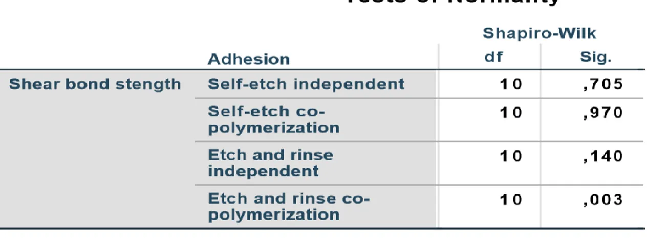
A análise estatística foi realizada no programa SPSS, os testes Shapiro-Wilk e Levene foram realizados para averiguar a normalidade da distribuição de valores e homogeneidade da variância. Após a confirmação destes pressupostos na maioria dos dados obtidos, foi realizado o teste ANOVA de uma dimensão que permitiu efetuar comparações múltiplas.
Resultados: Os 40 espécimes obtidos a partir dos 20 dentes (2 fatias por dente)
foram distribuídos de forma aleatória e de modo a ficarem 10 espécimes por grupo, cada espécime foi testado quanto à resistência adesiva. No grupo dos
v espécimes que foram preparados com polimerização independente e o adesivo foi utilizado segundo uma estratégia SE, o valor máximo atingido foi 29.2 MPa e o mínimo 13.1 MPa, tendo alcançado um valor médio de 21.1 MPa. Os espécimes deste grupo foram os que obtiveram os valores de resistência adesiva significativamente mais elevados comparativamente com todos os outros grupos em estudo (p<0,05). No grupo em que adesivo foi utilizado segundo um modo SE e se procedeu à copolimerização deste com a primeira camada de compósito, o valor máximo foi de 13.8 MPa e o mínimo de 3.3 MPa, tendo sido o valor médio de 8.6 MPa. No grupo da polimerização independente e o adesivo segundo uma estratégia ER, o valor máximo foi de 22.9 MPa e o mínimo 1.9 MPa, e assim o valor médio obtido foi de 14.0 MPa. Por fim, no grupo em que se testou a polimerização simultânea do adesivo aplicado segundo uma estratégia ER e primeira camada de compósito, o valor máximo atingido foi de 28.1 MPa e o mínimo de 2.4 MPa, sendo que a o valor médio foi de 9.5 MPa.
À medida que os espécimes foram testados, o modo de falha foi avaliado através da visualização ao microscópio ótico. No grupo A, 20% das falhas foram adesivas, enquanto que 70% apresentou falha mista, ou seja, a maioria das falhas ocorreu na interface resina/dentina, mas incluiu substrato vizinho (resina ou dentina). Apenas 10% dos espécimes testados deste grupo possuíram falha coesiva. Por outro lado, no grupo SE copolimerização 100% das falhas foram do tipo adesivo, ou seja, a falha ocorreu na interface resina/dentina sem a inclusão de qualquer outro substrato. No grupo ER independente, 80% das falhas ocorridas foram adesivas, 10% mistas e 10% coesivas, isto é, só numa pequena percentagem de espécimes é que a falha ocorreu exclusivamente na dentina ou na resina. Quanto ao grupo ER copolimerização, verificou-se que 90% das falhas eram de origem adesiva enquanto que só 10% foram de origem mista.
Conclusão: A dificuldade em estabilizar a primeira camada de compósito sobre
o adesivo não polimerizado, encontrada neste estudo, refletiu-se na falha entre o compósito e adesivo, principalmente nos grupos da polimerização simultânea, em que havia a formação de uma camada de adesivo muito fluída na superfície dentinária. Após a análise dos resultados, foi possível concluir que a polimerização independente do adesivo aplicado segundo uma estratégia SE apresentou a melhor adesão à dentina, o que se refletiu nos valores mais elevados
vi de resistência adesiva dos espécimes deste grupo. Os restantes grupos não apresentaram diferenças estatisticamente significativas entre si.
Mais estudos devem ser realizados nesta área de forma a testar metodologias que não comprometam a adesão logo no início, como por exemplo a utilização de compósito tipo flow em vez do compósito utilizado neste estudo.
Também foi possível concluir que existe a necessidade de se desenvolverem mais estudos relativamente à profundidade da camada inibida pelo oxigénio nos diferentes tipos de adesivo, e para tal, achamos que seria interessante analisar diferentes tipos de sistemas adesivos com espectroscopia de RAMAN.
Palavras-chave: camada inibida pelo oxigénio; self-etch; etch-and-rinse;
vii
Abstract
Objectives: The aim of this in vitro study was to understand the influence of the
curing mode of an universal adhesive system, applied according to etch-and-rinse (ER) and self-etch (SE) strategies, in the shear bond strength to dentin.
Materials and methods: 20 healthy teeth were selected and sectioned in a way
that originated 40 specimens. These were randomly distributed in 4 groups: SE independent- pre-curing of the adhesive used with a SE strategy; SE co-polymerization- co-polymerization of the composite and the adhesive used with a SE strategy; ER independent- pre-curing of the adhesive used with an ER strategy; ER co-polymerization- co-polymerization of the composite and the adhesive used with an ER strategy.
After 24 h incubation period, the shear bond strength tests were performed. Normality and homogeneity of the data were evaluated by Shapiro-Wilk test and Levene test, respectively, and a one-way ANOVA was used.
Results: Specimens from group SE independent yielded the higher shear bond
strength values comparing to the values obtained with the specimens from the other groups (p<0,05). Shear bond strength values from specimens of Groups SE co-polymerization, ER independent and ER co-polymerization where not significantly different.
The most common failure mode in specimens from group SE independent was mixed, while adhesive failure was prevalent among the other groups.
Conclusion: The separate polymerization of the universal adhesive with a SE
approach and first layer of composite lead to the best bond strength values to dentin.
Key-words: oxygen-inhibited layer; self-etch; etch-and rinse; pre-curing;
ix
Table of contents
Agradecimentos ... i Resumo ... iii Abstract ... vii List of figures ... xi List of tables ... xi Abbreviations ... xiii Symbols ... xiii Units ... xiii 1. Introduction ... 1 2. Objectives ... 53. Materials and methods ... 6
3.1 Experimental design ... 6
3.2 Materials ... 7
3.3 Specimens preparation ... 8
3.4 Shear bond strength protocol ... 10
3.5 Statistical analysis ... 11
4. Results ... 12
4.1 Shear bond strength values analysis ... 12
4.2 Mode of failure analysis ... 14
5. Discussion ... 15
6. Conclusion ... 19
7. Bibliography ... 20 Attachment A- Tables ... I
xi
List of figures
Fig. 1 – Representative scheme of experimental design………...…7
Fig. 2 – Total Etch®………..……….………8
Fig. 3 – Scotchbond Universal®………...……..……….……8
Fig. 4 – Tetric EvoCeram®……...…….………...………..…...…8
Fig. 5 – Watanabe device with the specimen after the adhesive and first layer of composite application...……….…...………10
Fig. 6 – Watanabe device settled in universal test machine...……….11
Fig. 7 – Graphic representing average shear bond strength values for each group….…14 Fig. 8 – Graphic representing % of the type of failure in each group…...……...……15
List of tables
Table 1 – Materials used in the study, with composition and way of application...……….8Table 2 – Shear bond strength values mean, standard deviation, minimum and maximum………..………...……14
Table A.1 - Shapiro-Wilk test to evaluate the normality of values distribution………...…I Table A.2 - Levene Statistic test to evaluate the homogeneity of variance………...I Table A.3 - ANOVA test of one dimension for the analysis of shear bond strength values from the experimental groups……….………...…II Table A.4- Post-hoc test Student-Newman- Keuls to identify statistically significant differences in the experimental groups……….…………III
xiii
Abbreviations
GPDM - glycerophosphoric acid dimethacrylate Hap - hydroxyapatite
ER - etch-and-rinse
HEMA - 2- Hydroxyethyl methacrylate
SE - self-etch
10-MDP - methacryloyloxydecyl dihydrogen phosphate OIL - oxygen-inhibited layer
SPSS - statistic package for social sciences
Symbols
% - percentage pH - hydrogen potential n - sample size p - probability of significanceUnits
MPa - megapascal - unit of pressure µm - micrometres - unit of measurement
h - hours - unit of time
ºC - Celsius degrees – unit of temperature s - seconds - unit of time
nm - nanometres- unit of measurement mm - millimetres - unit of measurement
mW/cm2 - microwatt per square centimetre - unit of radiation intensity
kN - kilonewtons - unit of measurement force
1
1. Introduction
“Extension for prevention”, was proposed by G. V. Black in 1917 as the best surgical approach (Black, 1917). Although this theory proved very efficient and necessary for a long time, due mainly to a poor performance of the dental materials, fortunately, nowadays “minimally-invasive dentistry” (M. V. Cardoso et al., 2011) concept prevails. The pioneer studies in this area were conducted by Kramer and McLean in 1952 (Kramer & Mclean, 1952) who studied the interaction between glycerophosphoric acid dimethacrylate (GPDM) and dentin (Van Meerbeek, Perdigão, Lambrechts, & Vanherle, 1998). However only in 1955, with the introduction of 85% phosphoric acid conditioning to “render the tooth surface more receptive to adhesion” by M. Buonocore (Buonocore, 1955) the technique of enamel acid-etching was created (Van Meerbeek et al., 1998).
Nowadays, it is very common to use enamel etchants with 30-40% phosphoric acid, which are able to produce shear bond strengths of resin composite to enamel of around 20 MPa (Swift, 1998). It is established that enamel adhesion is reliable due to its high percentage of hydroxyapatite (Hap) (Asmussen & Uno, 1992), however in dentine, a strong adhesion is very difficult to obtain. This is due to the hydrophilic and organic nature of dentine (David H. Pashley, 1992) originated by the water and collagen matrix present in its constitution (David H. Pashley, 1996).
Therefore, adhesive technology is constantly being improved. It started with “bonding agents” and developed to “adhesive systems” with improved application. The latest development were the “universal”, “all” or “multi”- purpose adhesive systems which claim to be able to adhere to enamel, dentine, amalgam, metal and porcelain (Van Meerbeek et al., 1998).
Etch-and-rinse (ER) systems can be applied in two or three steps. In these systems, the first step is the etching, followed by the rising procedure, for the complete removal of smear layer and smear plugs in dentine (M. V. Cardoso et al., 2011) and increase surface area and energy in enamel (Muñoz et al., 2013). The acid-etching with 37% phosphoric acid promotes a complete demineralization of 5-8 µm of the intertubular dentin matrix (Tarle, Marović, & Pandurić, 2012), which reveals a microporous network of collagen (Van Meerbeek et al., 2003). This acid is capable of etching enamel and dentine, and also to eliminate residual bacteria due to the low pH (Tarle et al., 2012). The second step is the application of the primer with hydrophilic monomers such as 2-hydroxyethyl
2 methacrylate (HEMA) dissolved in a solvent (acetone, ethanol or water)(M. V. Cardoso et al., 2011). The solvent lowers the solution’s viscosity, allowing HEMA monomers to impregnate the collagen network (Nakajima, Okuda, Pereira, Tagami, & Pashley, 2002). Finally, the application of the adhesive leads to the penetration of hydrophobic monomers into interfibrillar spaces and dentine tubules (M. V. Cardoso et al., 2011). The adhesive is polymerized after application. This process described above is for three-steps ER systems. In two steps systems, the acid- etching is also used, yet the second step is the application of a single product with hydrophilic and hydrophobic monomers, that means that the second bottle has primer and adhesive mixed together (Van Landuyt et al., 2007). The critical step in these systems is after etchant removal and before primer application (Van Meerbeek et al., 2003), if too much water is left in the dentin, the monomer will be diluted (Tay & Pashley, 2003), however, if we over-dry the dentin, the collagen fibers will collapse, and the adhesive is unable to penetrate the interfibrillar spaces (Kanca, 1992).
Self-etch (SE) systems use a hydrophilic acidic primer monomer, which incorporates the smear layer by demineralization and infiltration at the same time (Kenshima, Francci, Reis, Loguercio, & Filho, 2006). These functional monomers have a higher pH comparing to phosphoric acid etchants (Giannini et al., 2015), and according to the pH, these adhesives can be classified as: “strong” (pH<1), “intermediately strong” (pH≈1.5), “mild” (pH≈2) and “ultra-mild” (pH≥2.5). In two steps SE systems, priming and bonding are separated (Frankenberger & Tay, 2005), after the solvent evaporation, the bonding agent is applied to seal the dentin (Tay & Pashley, 2003) The one-step systems contains acidic functional monomers, hydrophilic and hydrophobic monomers, water and organic solvents in one solution (Frankenberger & Tay, 2005). These systems are preferred for cases with difficult moisture control, because, except for those with a “strong” pH, all systems provide an impregnation by the resin monomers in all extension of demineralized dentine, however, the surface area accomplished is not as large as the one with ER systems (Muñoz et al., 2013).
The simplified adhesive systems, whether they are ER or SE, form a permeable membrane that will allow the creation of nano-leakage areas (Sezinando et al., 2015), therefore, and in an attempt to achieve the best adhesion possible, considering the cavity prepared (Hanabusa et al., 2012), a more versatile adhesive system, the “Universal”, “Multi-purpose” or “Multi-mode” adhesives, was created (Muñoz et al., 2013). These are
3 able to be used by ER strategy, SE strategy or enamel selective etching, which propose the use of ER strategy in enamel and SE in dentine surface (Sezinando, 2014). “Universal” adhesives contain a monomer designated 10-methacryloyloxydecyl dihydrogen phosphate (10-MDP), that can interact ionically with the calcium present in HAp and form a calcium salt, through “nanolayering” (Perdigão & Swift, 2015; Sezinando, 2014).
Regardless of the choice, all adhesive systems intend to replace the removed minerals from dental tissue with resin monomers that will became micromechanically interconnected in the porosities after polymerization (M. V. Cardoso et al., 2011). In dentin, a hybrid layer (formed by dentin and adhesive (N. Nakabayashi, Kojima, & Masuhara, 1982)) is created and forms an acid-resistant cloak with the intention of sealing the dentin (N. Nakabayashi, Nakamura, & Yasuda, 1991). The hybrid layer is then protected by a thin layer of adhesive that will allow the connection to composite (Gateva & Kabaktchieva, 2012). This structure allows retention of the restoration and diminishes microleakage around the adhesive interface (Skupien et al., 2010)
It has been shown that morphological differences appear in bond structures originated by ER and SE systems, however the most outstanding is the hybrid layer thickness (Albaladejo, Osorio, Toledano, & Ferrari, 2010). According to Pashley, the hybrid layer should have, at least,0 0,5 µm of depth in the interdiffusion region to be considered a success (David H. Pashley & Tay, 2001). However, the hybrid layer created after the application of the adhesive system depends of several factors, the first variable is the choice between a SE and an ER system (Albaladejo et al., 2010; M. V. Cardoso et al., 2011), hybrid layer thickness might also be inflected by the smear layer’s density (Gateva & Kabaktchieva, 2012).
The hybrid layer achieved by SE has morphological differences depending on the adhesive components (Grégoire & Millas, 2005). One of the differences in SE systems composition is the pH, mild SE adhesives can create thin hybrid layers of 0.4 to 0.5 µm while intermediately strong adhesives achieved a thickness around 1.2 to 2.2 µm. The systems classified as strong were able to promote hybrid layers of 2.5 to 5 µm (Skupien et al., 2010), however the pH value is not a conditioning factor for the adhesive’s efficacy (Grégoire & Millas, 2005). Overall, the values of hybrid layer thickness obtained with these adhesives can be 0.7 µm (Kenshima et al., 2006) to 9 µm (Grégoire & Millas, 2005).
4 In ER systems the hybrid layer thickness varies with etching time (Gateva & Kabaktchieva, 2012) and with different conditioners, because they do not promote the same morphological changes in dentin (Skupien et al., 2010), the number of steps may also produce some differences in the new layer (M. V. Cardoso et al., 2011). For these systems, hybrid layer thickness can vary from 2.44 µm (Skupien et al., 2010) to 7.36 µm (Gateva & Kabaktchieva, 2012).
When the adhesive is applied, regardless of the system used, the oxygen present in the air will create a surface zone where the polymerization is inhibited, known as oxygen-inhibited layer (OIL) (Rueggeberg & Margeson, 1990). The monomers present in the composite resin are able to form interpenetrating systems with the unpolymerized monomers present in this OIL at the adhesive layer, and this mechanism is responsible for the adhesion between composite and adhesive.
The OIL’s thickness is related with the viscosity (which depends the monomer composition) and the mode of activation (Finger, Lee, & Podszun, 1996; Rueggeberg & Margeson, 1990; Ruyter, 1981). The layer is mostly composed by monomers and oligomers that could not originate polymers, and has a consistency similar to a liquid (Liebenberg, 2004).
The thickness of this layer is not constant, there are some studies that concluded that a 15µm in depth can be achieved (Van Meerbeek et al., 1998), while certain adhesives had an OIL of 0.7 µm ± 0.9 µm (Finger et al., 1996).
If the inhibited layer has a certain thickness, the initiator will be consumed, which lowers the degree of conversion and mechanical strength (Kim et al., 2005). Incomplete polymerization allows the formation of gaps, allowing water to infiltrate the space and cause a much quicker degradation (Nunes, Ceballos, Osorio, & Toledano, 2005).
This means that, depending on the adhesive’s constitution, mode of application, time of curing and form of evaluation, the thickness of OIL is variable and there is a possibility that this layer is as thick as the hybrid layer and the layer of adhesive applied in the tooth surface. Furthermore, the pre-curing of the adhesive creates an extra clinical task, which can influence the quality of the adhesion (Eick, Gwinnett, Pashley, & Robinson, 1997).
Therefore, emerges the question: “Is it necessary to separately cure the adhesive before resin application?”
5
2. Objectives
This in vitro study has the aim of:
1. Understanding the influence of the separate or simultaneous curing mode of an universal adhesive system, applied according to SE strategy, in the shear bond strength to dentin, at 24 hours.
H0: The curing mode of a SE adhesive system does not influence the shear bond strength to dentin.
H1: The curing mode of a SE adhesive system does influence the shear bond strength to dentin.
2. Understanding the influence of the separate or simultaneous curing mode of an universal adhesive system, applied according to ER strategy, in the shear bond strength to dentin, at 24 hours.
H0: The curing mode of an ER adhesive system does not influence the shear bond strength to dentin.
H1: The curing mode of an ER adhesive system does influence the shear bond strength to dentin.
6
3. Materials and methods
3.1 Experimental design
For the concretization of the study, the collection of human extracted teeth was necessary, it was accomplished without the identification of the donors and approved by the ethical committee of Faculdade de Medicina Dentária da Universidade de Lisboa. From all the teeth collected, 20 molars were selected, without any cavity or filling, and stored in a 1% Chloramine solution, at 4ºC, for maximum period of 6 months.
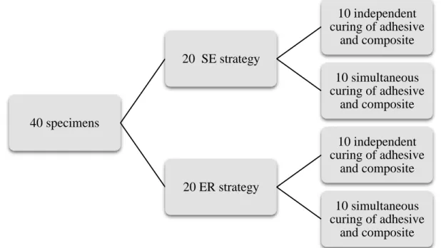
From the 20 molars a total of 40 specimens were obtained, that were randomly divided in 4 different groups.
SE independent: Adhesion with SE strategy and independent curing of the
adhesive and first layer of composite
SE co-polymerization: Adhesion with SE strategy and simultaneous curing of
the adhesive and first layer of composite
ER independent: Adhesion with ER strategy and independent curing of the
adhesive and first layer of composite
ER co-polymerization: Adhesion with ER strategy and simultaneous curing of
the adhesive and first layer of composite
Fig. 1- Representative scheme of experimental design
40 specimens 20 SE strategy 10 independent curing of adhesive and composite 10 simultaneous curing of adhesive and composite 20 ER strategy 10 independent curing of adhesive and composite 10 simultaneous curing of adhesive and composite
7
3.2 Materials
Table 1- Materials used in the study, with composition and way of application
Materials Composition Mode of use
Total- Etch® Ivoclar Vivadent, Schaan, Liechtenstein Lot: V10559 Shelf life: 2018/09
37% phosphoric acid gel pH<1
Etch and Rinse
1. Apply to dentin to condition for 15 s.
2. Wash it with water for 15 s. 3. Dry the surface with soft air jet,
for 5 s. Scotchbond Universal® 3M, Neuss, Germany Lot: 3184625 Shelf life: 2019/06 MDP Phosphate Monomer; Dimethacrylate resins; HEMA; Vitrebond™ Copolymer; Filler; Ethanol; Water; Initiators; Silane
Etch and Rinse and Self-Etch
1. Apply actively for 20 s to dentin. 2. Dry with soft air jet for 5 s and
confirm visually if the dentin surface is bright and without movement.
Independent curing: 1. Curing for 10 s Simultaneous curing:
1. Apply a 2 mm layer of composite 2. Curing for 20 s Tetric EvoCeram® Ivoclar Vivadent, Schaan, Liechtenstein Lot: V17575 Shelf life: 2020/04 Dimethacrylates (17–18% weight);
Barium glass, ytterbium trifluoride, mixed oxide and prepolymer (82–83% weight);
Additives, catalysts, stabilizers and pigments (< 1.0% weight);
Particle size: between 40 nm and 3,000 nm;
Mean particle size: 550 nm
Independent curing:
1. After the adhesive’s curing apply a 2 mm layer of composite 2. Polymerize for 20 s
Simultaneous curing:
1. After applying the adhesive, but before its polymerization, apply a 2 mm layer of composite.
2. Polymerize for 20 s
8
3.3 Specimens preparation
A longitudinal section was made in each molar and the interproximal enamel was removed with a diamond saw (IsoMet TM Diamond Wafering Blades- Buehler Ltd, Lake Bluff, IL, USA) in a cutting machine (Isomet 1000 - Buehler Ltd, Lake Bluff, IL, USA), with the intent of obtain 2 slices of dentin per tooth.
The dentin surface, to be used as substrate, was polished with a silicon carbide sandpaper with a granulometry of 320 (Buehler - Struers, Copenhagen, Denmark), with water, for 5 s, for simulation of the smear layer (Oliveira et al., 2003). The slices were then arbitrarily divided in 4 experimental groups (n=10).
Before the adhesive procedure, the specimens were adapted in the first part of the Watanabe’s device, with the assistance of a polyether adhesive pellicle (Glossy White – Xerox, Connecticut, USA). A hole, with 3 mm diameter, was made in each pellicle to standardize the adhesion area (ISO/TS 11405/2003).
At that point, the Scotchbond Universal® (3M, Neuss, Germany) was applied, with two different strategies: SE and ER.
9
3.3.1. Application protocol of the adhesive system according to a SE strategy
The adhesive was applied actively on the dentine’s substrate for about 20 s.
The adhesive was dried with a soft air jet for 5 s and it was visually confirmed that the surface was bright but there was no adhesive movement.
3.3.2. Application protocol of the adhesive system according to an ER strategy
The 37% phosphoric acid gel was applied over the dentine substrate for about 15 s.
The sample was washed with a water jet for 15 s.
The dentin’s conditionate surface was dried with a soft air jet, for 5 s.
The adhesive was applied actively on the dentine’s substrate for about 20 s.
The adhesive was dried with a soft air jet for 5 s and it was visually confirmed that the surface was bright but there was no adhesive movement.
In 10 specimens of each adhesive strategy, the adhesive system was cured previously for 10 s and a 2 mm layer of Tetric EvoCeram® (Ivoclar Vivadent, Liechtenstein) composite was applied, and polymerized for 20 s.
In the other 10 specimens of each adhesive strategy the adhesive and first layer of composite were polymerized at the same time, for 20 s, following the Fig 1.
The curing unit used was Bluephase LED curing light (Ivoclar Vivadent, Schaan, Liechtenstein), with an intensity of 940 mW/cm2.
The dentin specimen was kept in place by the incorporation of a layer of type IV gypsum (Gilstone – BK Giulini Corp Ludwigshafen, Germany), in the second part of the Watanabe’s device.
Then, the specimens were stored in an incubator for 24 h, before shear bond strength tests.
10
Fig. 5 – Watanabe device with the specimen after the adhesive and first layer of
composite application
3.4 Shear bond strength protocol
Shear bond strength tests were made using an universal mechanical test machine (Instron, model 4502– Instron LTD, Bucks, England), with an inferior fixed arm and a mobile superior arm. This machine, through the application of forces in the adhesion area, test the specimens until the breakpoint, calculating the tension applied in the moment.
The Watanabe devices were adapted to the universal test machine through claws appropriated for the purpose and in a way that the adhesion interface will be aligned with the force application axis. The essay was made with a charging cell of 1 kN and at a speed of 1 mm/min (ISO 11405/2003).
Shear bond values were obtained from the ratio between the strength at the failure moment and the section area of the adhesive’s interface, previously established.
11
Fig. 6- Watanabe device settled in universal test machine
Following the conclusion of shear bond strength tests, the break interface was observed, so the type of union failure was determined. The union failure was classified as adhesive (failure occurred in resin/dentin interface), mixed (failure occurred in resin/dentin interface and included some of the surrounding substrate – resin or dentin) or cohesive (failure occurred exclusively in dentin or in resin)(Luque-Martinez et al., 2014). For this evaluation, the specimens were observed with the help of a stereomicroscope (Meiji Techno Co., model EMZ-8TR, Saitama, Japan).
3.5 Statistical analysis
Means and standard deviation values of shear bond strength for each group, and comparisons between groups were made using the SPSS program (Statistic Package for Social Sciences; IBM SPSS statistics, version 24.0).
The normality of the distribution was determined with the use of Shapiro-Wilk test and the homogeneity of the variance was confirmed using Levene test.
Once the normality and homogeneity in the generality of the parameters was guaranteed, the parametric test ANOVA with one dimension allowed different comparisons between the experimental groups.
The post-hoc test Student-Newman-Keuls was also applied in this analysis to identify statistically significant differences in the diverse groups and the level of statistical meaningfulness was established at 5%.
12
4. Results
4.1 Shear bond strength values analysis
The mean shear bond strength values obtained in each group, along with the standard deviation are displayed in Table 2.
In the group with independent polymerization of the adhesive with a SE approach and the first layer of composite (SE independent), the maximum and minimum shear bond strength values were 29.2 MPa and 13.1 MPa, respectively (Table 2).
In the group where the co-polymerization of the SE approach and first layer of composite (SE co-polymerization) was tested, the maximum shear bond strength was 13.8 MPa and the minimum 3.3 MPa (Table 2).
The results for the ER method, in the independent polymerization (ER independent), achieved a maximum of 22.9 MPa and a minimum of 1.9 MPa (Table 2).
Regarding the group that tested the co-polymerization of the adhesive with an ER strategy simultaneously with the first layer of composite (ER co-polymerization), 28.1 MPa was the maximum value, while the minimum was 2.4 MPa (Table 2).
The specimens from SE independent group yielded higher shear bond strength values than the ones in SE co-polymerization group (p<0,05). They also obtained higher shear bond strength results than the specimens in the groups with independent and co-polymerization ER strategy (p<0,05) (Fig. 7).
There were no statistically significant differences in shear bond strength results between independent polymerization and co-polymerization strategies in the specimens from ER groups, even though a slight tendency to higher shear bond strength values was observed however, the shear bond strength results from specimens in the independent group were slightly higher than co-polymerization group (Fig. 7).
Comparing all groups, SE independent was the technique that achieved the strongest shear bond values, while the group ER co-polymerization was, overall, the group with the worst results.
13
Table 2 – Shear bond strength values mean, standard deviation, minimum and maximum
Fig. 7 – Graphic representing average shear bond strength values (in MPa) for each group Experimental groups Shear bond strength values
Mean Std. deviation Minimum Maximum
SE independent 21.1 5.4 13.1 29.2 SE co-polymerization 8.6 3.3 3.3 13.8 ER independent 14.0 7.1 1.9 22.9 ER co-polymerization 9.5 7,1 2,4 28,1
14
4.2 Mode of failure analysis
Regarding the failure mode, in SE independent group 20% of the failures were adhesive, 70% mixed and 10% cohesive.
In SE co-polymerized group, all specimens had an adhesive failure.
In ER independent, 80% of the failures were adhesive, 10% mixed and 10% cohesive.
Finally, in ER co-polymerized, 90% of the failures were adhesive and 10% mixed. All this data is exposed in Fig. 8.
Fig. 8 – Graphic representing % of the type of failure in each group
0,00 10,00 20,00 30,00 40,00 50,00 60,00 70,00 80,00 90,00 100,00
SE Independent SE Co-polymerized ER Independent ER Co-polymerized
T y p e o f u n io n f ailu re (%) Experimenal groups
Type of union failure after shear bond strength
15
5. Discussion
Traditionally, the adhesive is polymerized after application, and before the addiction of composite’s first layer. However, as shown in literature, there is the formation of an oxygen-inhibited layer (OIL), that is the mechanism of adhesion between adhesive and composite, by the creation of a network between the uncured monomers of the adhesive and the monomers from the composite resin layer (Kim et al., 2005). This inhibited layer can measure as much as the hybrid layer, especially in SE systems, which achieve a lower depth of demineralization, and consequently a shorter adhesive layer (Santini & Miletic, 2008).
Regardless the system of choice, every adhesive needs to be polymerized, although, there are always new adhesives being produce with the intent of reduce clinical steps, saving more time during the appointments, and reducing the risks of mistakes during the application of the adhesive.
Since the adhesive layer is not completely polymerized due to oxygen inhibition, anyway, and in the daily clinic it is essential to save time and diminish the margin for error, the aim of this study was to explore if co-curing the adhesive and first layer of composite produces any improvement or changes in shear bond strength.
For this, an universal adhesive was applied according to a SE and ER approach. In half of the specimens, the adhesive was pre-cured, and the rest was prepared with co-polymerization of the adhesive and first layer of composite.
Shear bond tests were selected to evaluate the bond strength to dentin using two different strategies for polymerization. The values obtained after testing the samples are achieved by dividing the maximum applied force by the bonded cross-sectional area (Versluis, Tantbirojn, & Douglas, 1997). This method has some advantages, for example, specimens preparation is easy (Sudsangiam & van Noort, 1998) and also, it is considered to create a stress condition more similar to the clinical situation than other tests (P. E. C. Cardoso, Braga, & Carrilho, 1998). However, in some studies, the rising of cohesive failures with new adhesives, which are supposed to have improved bond strength, raised some questions regarding the legitimacy of the measurements (Placido et al., 2007). These results can be explained with the fact that a premature failure may occur due to the stress concentration in the substrate instead of the failure in the interface itself (Della Bona & van Noort, 1995).
16 According to some authors, a smaller bonding surface leads to higher bond strength results (Scherrer, Cesar, & Swain, 2010) which can be related to the fact that smaller specimens do not contain as many defects as larger ones (Michael F. Burrow, Thomas, Swain, & Tyas, 2004; D H Pashley, Sano, Ciucchi, Yoshiyama, & Carvalho, 1995). One can never forget that strength values may vary with material properties, loading configuration (Placido et al., 2007), specimen preparation (Michael F. Burrow et al., 2004; Placido et al., 2007) and storage (Michael F. Burrow et al., 2004).
Shear bond tests to dentin performed at 24 h, showed mean values of 21.1±5.4 MPa, for the specimens in pre-cured SE group, in contrast, the specimens in the group where SE adhesive was co-polymerized with the composite achieved a bond strength of 8.6±3.3 MPa. In the ER groups, bonding to dentin was 14.0±7.1 MPa in the specimens with the separate polymerization, while for the specimens from the group with simultaneous curing of adhesive and composite, the values were 9.5±7.1 MPa.
From the analysis of shear bond strength results, it is possible to conclude that the SE approach with independent polymerization had the higher bond strength to dentin, and was the only to present a mean result higher than 20 MPa, which is the starting value for a restoration to be considered a success (M.F. Burrow, Tagami, Negishi, Nikaido, & Hosoda, 1994). Therefore, the null hypothesis of the first objective is rejected.
There were no statistical differences between the other three groups, though the samples in the ER group with independent polymerization showed a tendency to slightly higher values when compared to the ones ER group with co-polymerization. In the light of these results, the null hypothesis of the second objective must be accepted.
The mode of failure that occurred more often in the specimens from the SE independent group was mixed, this means that the failure occurred in resin and dentin interface, but also included the surrounding substrate, however, in the specimens from the other three groups, the main mode of failure was adhesive, that means the failure occurred in the adhesive layer between resin and dentin.
The adhesive failures are often associated with a less resistant interface reflecting lower shear bond values. In mixed failures, typically there is a stronger bonding to dentin (Bouillaguet et al., 2001). Since in 2 groups the adhesive was not cured before applying the resin composite the interaction between both materials could be affected revealing lower shear bond strength values and an adhesive failure mode.
17 Overall, the values of the shear bond tests, performed in this study, exposed a weak bond strength to dentin. There are many critical steps during the application of the adhesive and first layer of composite. In this study, the protocol was created and followed to allow a homogeneous preparation of the specimens. However, the application of the first layer of composite resin, especially in the specimens from the co-polymerization groups, where the adhesive was fluid, led to a lack of attachment between composite and adhesive. The non-cured adhesive layer was very fluid at the surface, making it difficult to stabilize the composite before polymerization and allowing the adhesive to move away from the dentinal surface. This probably led to an interface without a thick adhesive layer, in the specimens of co-polymerization groups, which according to Bouillaguet, is essential to achieve higher bond strength values (Bouillaguet et al., 2001). This is one of the possible explanations for the low values obtained in the co-polymerization groups. McCabe and Rubsy (McCabe & Rusby, 1994) also developed a shear bond strength study, testing two adhesive systems, ATR Bond (Coltene Whaledent, Altstätten Switzerland) and Syntac (Vivadent, Schaan,Liechtenstein). In one group they were pre-cured prior to the composite application, while in the other group they were polymerized simultaneously with the composite. The mean values for the pre-cured groups were 13.20±8.25 MPa for ATR Bond and 6.91±4.62 MPa for Syntac, on the other hand, the co-polymerized groups only achieved mean bond strengths of 4.98±5.24 MPa and 2.67±2.66 MPa, respectively.
Another study conducted a few years later by Chapman et al. (Chapman, Burgess, Holst, Sadan, & Blatz, 2007), tested three self-etching bonding agents in shear bond strength to dentin and enamel. In half the specimens the adhesives were pre-cured, and in the remaining specimens, adhesive and composite were cured at the same time. Their results allowed them to conclude that there were no significant differences in bonding to enamel, however, shear bond strength to dentin decreased significantly in the co-cured groups. They tested the following adhesives: Adper Prompt-L-Pop (3M Espe, Neuss, Germany), Clearfil SE Bond (Kuraray, Tokyo, Japan) and Xeno III (Dentsply Caulk, Pennsylvania, USA), for the pre-cured groups, the dentin shear bond strength results were, respectively, 8.9±3.2 MPa, 15.9±3.2 MPa and 12.9±5.2 MPa, in contrast, the groups where the polymerization was simultaneous, the values achieved were 5.4±1.6 MPa, 6.3±2.5 MPa and 4.0±3.1 MPa.
18 The results of the present study are in accordance with the results displayed by McCabe (McCabe & Rusby, 1994) and Chapman (Chapman et al., 2007).
It is well established that, while being submitted to polymerization, there is a contraction in resin composites (Sakaguchi, Peters, Nelson, Douglas, & Poort, 1992), this may affect the position of the unpolymerized adhesive in dentin orifices (Chapman et al., 2007). According to Chappell et al. (Chappell, Cobb, Spencer, & Eick, 1994) the resin tags create an essential network with the lateral branches of dentin tubules, which may allow the construction of a stronger bond between the adhesive and dentin. However, this bond is weakened by the stress originated with the shrinkage of composite (Chapman et al., 2007; McCabe & Rusby, 1994). Polymerization shrinkage can cause real damage to bonding to dentin, especially when the direction of the contraction is opposite to that of the dentin surface (N. Nakabayashi et al., 1991). This could be one of the factors that justify the low results in the groups where the adhesive was not polymerized before composite application.
Another explanation is proposed by McCabe (McCabe & Rusby, 1994) and reflects about the fact that not enough light intensity can achieve the adhesive. This is due to the 2 mm composite layer, that minimizes the cure efficiency of the adhesive when compared to cases where the adhesive is exposed directly to the light. This theory is supported by studies, that prove the hardness of the composite activated by visible light, decreases from the surface to the depth (Matsumoto et al., 1986; Watts, Amer, & Combe, 1987).
All three theories can justify the low results obtained in our study, however the lack of interaction between composite and adhesive layer felt by the operator probably overshadowed the other two explanations. The assessment of the effect of the co-polymerization between composite and the adhesive in SE mode should be further studied with a protocol that does not compromise the adhesion between composite and dentin, for example the use of flow instead of regular composite may guarantee that the adhesive remains intact on the dentine surface before polymerization.
19
6. Conclusion
Despite the limitations of this in vitro study, we may conclude that the separate polymerization of the first layer of composite and an universal adhesive with a SE approach increases the bond strength to dentin.
From the analysis of the literature and the results of the present study, it should be helpful to conduct further studies with a protocol that does not compromise the adhesion between composite and dentin, for example the use of flow instead of regular composite. Other study that could be interesting to develop in the future, is to evaluate the depth of OIL in the adhesive with RAMAN spectroscopy.
20
7. Bibliography
Albaladejo, A., Osorio, R., Toledano, M., & Ferrari, M. (2010). Hybrid layers of etch-and-rinse versus self-etching adhesive systems. Medicina Oral, Patologia Oral y Cirugia Bucal, 15(1), 112–118. https://doi.org/10.4317/medoral.15.e112
Asmussen, E., & Uno, S. (1992). Adhesion of restorative resin to dentin: chemical and physicochemical aspects. Oper Dent, (5), 68–74.
Black, G. V. (1917). A work in operative dentistry in two volumes. Chicago: Medico-Dental Publishing.
Bouillaguet, S., Gysi, P., Wataha, J. C., Ciucchi, B., Cattani, M., Godin, C., & Meyer, J. M. (2001). Bond strength of composite to dentin using conventional, one-step, and self-etching adhesive systems. Journal of Dentistry, 29(1), 55–61.
https://doi.org/10.1016/S0300-5712(00)00049-X
Buonocore, M. G. (1955). A Simple Method of Increasing the Adhesion of Acrylic Filling Materials to Enamel Surfaces. J Dent Res., 34(6), 849–53.
Burrow, M. F., Tagami, J., Negishi, T., Nikaido, T., & Hosoda, H. (1994). Early Tensile Bond Strengths of Several Enamel and Dentin Bonding Systems. Journal of Dental Research, 73(2), 522–528. https://doi.org/10.1177/00220345940730020701
Burrow, M. F., Thomas, D., Swain, M. V., & Tyas, M. J. (2004). Analysis of tensile bond strengths using Weibull statistics. Biomaterials, 25(20), 5031–5035. https://doi.org/10.1016/j.biomaterials.2004.01.060
Cardoso, P. E. C., Braga, R. R., & Carrilho, M. R. O. (1998). Evaluation of micro-tensile, shear and tensile tests determining the bond strength of three adhesive systems. Dental Materials, 14(6), 394–398. https://doi.org/10.1016/S0300-5712(99)00012-3
Cardoso, M. V., De Almeida Neves, A., Mine, A., Coutinho, E., Van Landuyt, K., De Munck, J., & Van Meerbeek, B. (2011). Current aspects on bonding effectiveness and stability in adhesive dentistry. Australian Dental Journal, 56(SUPPL. 1), 31– 44. https://doi.org/10.1111/j.1834-7819.2011.01294.x
Chapman, L., Burgess, J., Holst, S., Sadan, A., & Blatz, M. (2007). Precuring of self-etching bonding agents and its effect on bond strenght of resin composite to dentin and enamel. Quintessence Int 38, 8, 637–641.
21 anastomosis: A potential factor in adhesive bonding? The Journal of Prosthetic Dentistry, 72(2), 183–188. https://doi.org/10.1016/0022-3913(94)90078-7 Della Bona, A., & van Noort, R. (1995). Shear vs . Tensile Bond Strength of Resin
Shear vs . Tensile Bond Strength of Resin Composite Bonded to Ceramic. J Dent Res., 74(September), 1591–1596. https://doi.org/10.1177/00220345950740091401 Eick, J. D., Gwinnett, A. J., Pashley, D. H., & Robinson, S. J. (1997). Current concepts
on adhesion to dentin, 8(3), 306–335.
Finger, W. J., Lee, K.-S., & Podszun, W. (1996). Monomers with low oxygen inhibition as enamel/dentin adhesives. Dental Materials, 12(4), 256–261.
https://doi.org/10.1016/S0109-5641(96)80032-7
Frankenberger, R., & Tay, F. R. (2005). Self-etch vs etch-and-rinse adhesives: Effect of thermo-mechanical fatigue loading on marginal quality of bonded resin composite restorations. Dental Materials, 21(5), 397–412.
https://doi.org/10.1016/j.dental.2004.07.005
Gateva, N., & Kabaktchieva, R. (2012). Hybrid Layer Thickness in Primary and
Permanent Teeth – a Comparison Between Total Etch Adhesives. Journal of IMAB - Annual Proceeding (Scientific Papers), 18, 2(2012), 191–199.
https://doi.org/10.5272/jimab.2012182.191
Giannini, M., Makishi, P., Almeida Ayres, A. P., Moreira Vermelho, P., Marin Fronza, B., Nikaido, T., & Tagami, J. (2015). S e l f - E t c h Ad h e s i v e S y s t e m s : A Literature Review. Brazilian Dental Journal, 26(1), 3–10.
https://doi.org/10.1590/0103-6440201302442
Grégoire, G., & Millas, A. (2005). Microscopic Evaluation of Dentin Interface Obtained with 10 Contemporary Self-etching Systems: Correlation with Their pH G.
Operative Dentistry, 30(4), 481–491.
Hanabusa, M., Mine, A., Kuboki, T., Momoi, Y., Van Ende, A., Van Meerbeek, B., & De Munck, J. (2012). Bonding effectiveness of a new “multi-mode” adhesive to enamel and dentine. Journal of Dentistry, 40(6), 475–484.
https://doi.org/10.1016/j.jdent.2012.02.012
Kanca, J. (1992). Effect of resin primer solvents and surface wetness on resin composite bond strength to dentin. Am J Dent, 5(4), 213–215.
Kenshima, S., Francci, C., Reis, A., Loguercio, A. D., & Filho, L. E. R. (2006). Conditioning effect on dentin, resin tags and hybrid layer of different acidity
self-22 etch adhesives applied to thick and thin smear layer. Journal of Dentistry, 34(10), 775–783. https://doi.org/10.1016/j.jdent.2006.03.001
Kim, J.-S., Choi, Y.-H., Cho, B.-H., Son, H.-H., Lee, I.-B., Um, C.-M., & Chang-Keun, K. (2005). Effect of Light-Cure Time of Adhesive Resin on the Thickness of the Oxygen-Inhibited Layer and the Microtensile Bond Strength to Dentin. Wiley InterScience. https://doi.org/10.1002/jbm.b.30463
Kramer, I. R. H., & Mclean, J. W. (1952). Alterations in the staining reactions of dentine resulting from a constituent of a new self-polymerizing resin. British Dental Journal, 93, 150–153.
Liebenberg, W. (2004). OXYGEN-INHIBITED LAYER IN ADHESION
DENTISTRY. Journal of Esthetic and Restorative Dentistry, 16(5), 316–323. https://doi.org/10.1111/j.1708-8240.2006.00002.x
Luque-Martinez, I. V., Perdigão, J., Muñoz, M. A., Sezinando, A., Reis, A., & Loguercio, A. D. (2014). Effects of solvent evaporation time on immediate adhesive properties of universal adhesives to dentin. Dental Materials, 30(10), 1126–1135. https://doi.org/10.1016/j.dental.2014.07.002
Matsumoto, H., Gres, J. E., Marker, V. A., Okabe, T., Ferracane, J. L., & Harvey, G. A. (1986). Depth of cure of visible light-cured resin: Clinical simulation. The Journal of Prosthetic Dentistry, 55(5), 574–578.
McCabe, J. F., & Rusby, S. (1994). Dentine bonding-the effect of pre-curing the bonding resin. British Dental Journal, 176, 333–336. Retrieved from http://dx.doi.org/10.1038/sj.bdj.4808447
Muñoz, M. A., Luque, I., Hass, V., Reis, A., Loguercio, A. D., & Bombarda, N. H. C. (2013). Immediate bonding properties of universal adhesives to dentine. Journal of Dentistry, 41(5), 404–411. https://doi.org/10.1016/j.jdent.2013.03.001
Nakabayashi, N., Kojima, K., & Masuhara, E. (1982). The promotion of adhesion by the infiltration of monomers into tooth substrates, 16, 265–273.
Nakabayashi, N., Nakamura, M., & Yasuda, N. (1991). Hybrid Layer as a Dentin‐
Bonding Mechanism. Journal of Esthetic and Restorative Dentistry, 3(4), 133–138. Retrieved from http://doi.org/10.1111/j.1708-8240.1991.tb00985.x
Nakajima, M., Okuda, M., Pereira, P. N., Tagami, J., & Pashley, D. H. (2002). Dimensional changes and ultimate tensile strengths of wet decalcified dentin applied with one-bottle adhesives. Dent Mater, 18(8), 603–8.
23 Nunes, T. G., Ceballos, L., Osorio, R., & Toledano, M. (2005). Spatially resolved
photopolymerization kinetics and oxygen inhibition in dental adhesives. Biomaterials, 26(14), 1809–1817.
https://doi.org/10.1016/j.biomaterials.2004.06.012
Oliveira, S. S. A., Pugach, M. K., Hilton, J. F., Watanabe, L. G., Marshall, S. J., & Marshall, G. W. (2003). The influence of the dentin smear layer on adhesion: A self-etching primer vs. a total-etch system. Dental Materials, 19(8), 758–767. https://doi.org/10.1016/S0109-5641(03)00023-X
Pashley, D. H. (1992). The effects of acid etching on the pulp dentin complex. Oper Dent., 17(6), 229–242.
Pashley, D. H. (1996). Dynamics of the pulpchdentin complex. Crit Rev Oral Biol Med., 7(2), 104–133.
Pashley, D. H., Sano, H., Ciucchi, B., Yoshiyama, M., & Carvalho, R. M. (1995). Adhesion testing of dentin bonding agents: a review. Dental Materials : Official Publication of the Academy of Dental Materials, 11(2), 117–125.
https://doi.org/10.1016/0109-5641(95)80046-8
Pashley, D. H., & Tay, F. R. (2001). Aggressiveness of contemporary self-etching adhe- sives. Part II: etching effects on ungrounded enamel. Dent Mater, 17, 430–444. Perdigão, J., & Swift, E. J. (2015). Universal Adhesives. Journal of Esthetic and
Restorative Dentistry, 27(6), 331–334. https://doi.org/10.1111/jerd.12185
Placido, E., Meira, J. B. C., Lima, R. G., Muench, A., Souza, R. M. de, & Ballester, R. Y. (2007). Shear versus micro-shear bond strength test: A finite element stress analysis. Dental Materials, 23(9), 1086–1092.
https://doi.org/10.1016/j.dental.2006.10.002
Rueggeberg, F. A., & Margeson, D. H. (1990). The Effect of Oxygen Inhibition on an Unfilled/Filled Composite System. Journal of Dental Research, 69(10), 1652– 1658. https://doi.org/10.1177/00220345900690100501
Ruyter, I. E. (1981). Unpolymerized surface layers on sealants. Acta Odontologica Scandinavica, 39(1), 27–32. https://doi.org/10.3109/00016358109162255 Sakaguchi, R. L., Peters, M. C. R. B., Nelson, S. R., Douglas, W. H., & Poort, H. W.
(1992). Effects of polymerization contraction in composite restorations. Journal of Dentistry, 20(3), 178–182. https://doi.org/10.1016/0300-5712(92)90133-W
24 demineralization , adhesive penetration , and degree of conversion of three dentine bonding systems. Eur J Oral Sci, 177–183.
Scherrer, S. S., Cesar, P. F., & Swain, M. V. (2010). Direct comparison of the bond strength results of the different test methods: A critical literature review. Dental Materials, 26(2). https://doi.org/10.1016/j.dental.2009.12.002
Sezinando, A. (2014). Looking for the ideal adhesive - A review. Revista Portuguesa de Estomatologia, Medicina Dentaria e Cirurgia Maxilofacial, 55(4), 194–206. https://doi.org/10.1016/j.rpemd.2014.07.004
Sezinando, A., Luque-Martinez, I., Muñoz, M. A., Reis, A., Loguercio, A. D., &
Perdigão, J. (2015). Influence of a hydrophobic resin coating on the immediate and 6-month dentin bonding of three universal adhesives. Dental Materials, 31(10), e236–e246. https://doi.org/10.1016/j.dental.2015.07.002
Skupien, J. A., Susin, A. H., Angst, P. D., Anesi, R., Machado, P., Bortolotto, T., & Krejci, I. (2010). Micromorphological effects and the thickness of the hybrid layer - a comparison of current adhesive systems. J Adhes Dent, 12(6), 435–442.
https://doi.org/10.3290/j.jad.a18242
Sudsangiam, S., & van Noort, R. (1998). Do dentin bond strength tests serve a useful purpose? The Journal of Adhesive Dentistry, 1(1), 57–67. Retrieved from
http://europepmc.org/abstract/MED/11725686%5Cnhttp://www.ncbi.nlm.nih.gov/ pubmed/11725686
Swift, J. (1998). Bonding systems for restorative materials-a comprehensive review. American Academy of Pediatric Dentistry, 20(2), 80–84.
https://doi.org/10.1016/j.jdent.2013.03.001
Tarle, Z., Marović, D., & Pandurić, V. (2012). Contemporary concepts on composite materials. Rad 514 Medical Sciences, 38(August), 23–38.
Tay, F. R., & Pashley, D. H. (2003). Have dentin adhesives become too hydrophilic? Journal (Canadian Dental Association), 69(11), 726–731.
https://doi.org/10.1016/S0109-5641(03)00110-6
Van Landuyt, K., Snauwaert, J., De Munck, J., Peumans, M., Yoshida, Y., & Poitevin, A. (2007). Systematic review of the chemical composition of contemporary dental adhesives. Biomaterials, 28(26), 3757–85.
Van Meerbeek, B., De Munck, J., Yoshida, Y., Inoue, S., Vargas, M., Vijay, P., Vanherle, G. (2003). Adhesion to enamel and dentin: current status and future
25 challenges. Operative Dentistry-University of Washington-, 28(3), 215–235.
Van Meerbeek, B., Perdigão, J., Lambrechts, P., & Vanherle, G. (1998). The clinical performance of adhesives. Journal of Dentistry, 26(1), 1–20.
https://doi.org/10.1016/S0300-5712(96)00070-X
Versluis, A., Tantbirojn, D., & Douglas, W. H. (1997). Why do shear bond tests pull out dentin? Journal of Dental Research, 76(6), 1298–1307.
https://doi.org/10.1177/00220345970760061001
Watts, D. C., Amer, O. M., & Combe, E. C. (1987). Surface hardness development in light-cured composites. Dental Materials, 3(5), 265–269.
I
Attachment A- Tables
Table A.1- Shapiro-Wilk test to evaluate the normality of values distribution
II
Table A.3- ANOVA test of one dimension for the analysis of shear bond strength values
III
Table A.4- Post-hoc test Student-Newman- Keuls to identify statistically significant