UNIVERSIDADE DE LISBOA
Faculdade de Farmácia
Research Institute for Medicines
(iMed.ULisboa)
Neuron Glia Biology in Health and Disease Group
Endothelial progenitor cells and associated
pathways in multiple myeloma
Maria Margarida Batista Tenreiro
Dissertação de Mestrado para obtenção do grau de Mestre em Ciências
Biofarmacêuticas
Orientadora: Prof.ª Doutora Maria Alexandra Brito
Co-orientadora: Prof.ª Doutora Maria Leonor Correia
The studies presented on this master thesis were performed in the research group “Neuron Glia Biology in Health & Disease”, from Research Institute for Medicines (iMed.ULisboa), Faculty of Pharmacy, Universidade de Lisboa, under the supervision of Maria Alexandra Brito, Ph.D. and Maria Leonor Correia, Ph.D.
This study was supported by Fundação para a Ciência e Tecnologia (FCT – UID/DTP/04138/2013), Portugal.
Outputs ensuing from the present thesis:
Publications in international scientific periodicals with referees
Tenreiro MM, Correia ML, Brito MA. Endothelial progenitor cells in multiple myeloma neovascularization: a brick to the wall (In preparation).
Publications in scientific meetings abstract book
Tenreiro MM, Malhó R, Silveira M, Correia ML, Brito MA (2016): “Assessment of endothelial progenitor cells in bone marrow smears by multiple-labelling immunofluorescence analysis: a novel tool to monitor angiogenesis”. 50ª Reunião da Sociedade Anatómica Portuguesa (SAP) e 3ª Reunião Científica da Associação Anatómica Portuguesa (AAP). Escola de Ciências da Saúde, Universidade do Minho, Braga, April 30th, 2016, Portugal.
Tenreiro MM, Malhó R, Silveira M, Correia ML, Brito MA (2016): “A novel tool to monitor angiogenesis in bone marrow smears: Detection and quantificiation of endothelial progenitor cells by multiple-labeling immunofluorescence analysis”. 21st Congress of the European Hematology Association. Copenhagen, June 9-12th, 2016, Denmark.
Posters
Tenreiro MM, Malhó R, Silveira M, Correia ML, Brito MA (2016): “Endothelial progenitor cells in multiple myeloma”. Reunião Anual da Sociedade Portuguesa de Hematologia. Hotel Solverde, Espinho, November 17-19th, 2016, Portugal (Accepted).
Tenreiro MM, Malhó R, Silveira M, Correia ML, Brito MA (2016): “Células endoteliais progenitoras: um possível biomarcador do mieloma múltiplo”. XX Jornadas Científicas de Análises Clínicas e de Genética Humana. Faculty of Pharmacy, Universidade do Porto, October 7-8th, 2016, Portugal.
Tenreiro MM, Malhó R, Silveira M, Correia ML, Brito MA (2016): “Assessment of endothelial progenitor cells in bone marrow smears by multiple-labelling immunofluorescence analysis: a novel tool to monitor angiogenesis”. 50ª Reunião da Sociedade Anatómica Portuguesa (SAP) e 3ª Reunião Científica da Associação Anatómica Portuguesa (AAP). Escola de Ciências da Saúde, Universidade do Minho, Braga, April 30th, 2016, Portugal.
Abstract
Multiple myeloma (MM) is characterized by the clonal expansion of plasma cells (PCs) in the bone marrow (BM) that leads to bone destruction, anaemia and renal failure. Although there are several therapeutic options nowadays, there is still no effective cure and the standard survival up to 4 years. The evolution from the asymptomatic stage of monoclonal gammopathy of undetermined significance (MGUS) to MM and the progression of the disease itself are related with cellular and molecular alterations in the BM microenvironment, namely, the development of the vasculature. In postnatal vasculogenesis, there is stimulation of the recruitment of BM progenitors known as endothelial progenitor cells (EPCs) to the tumour vasculature, which will incorporate newly-forming blood vessels and differentiate into endothelial cells. The mobilization of EPCs is tightly controlled by cells and molecules in the BM microenvironment. With this retrospective study, we intended to evaluate the potential of EPCs as biomarkers for MM progression and response to therapy, while assessing their relationship with PCs and the signalling receptors C-X-C motif chemokine receptor (CXCR)4 and platelet-derived growth factor receptor (PDGFR)-β in sequentially collected BM smears from MM patients in different disease stages. Thus, we aimed to: 1) develop a method to quantify EPCs in BM smears from MM patients, 2) establish the temporal evolution of EPCs levels and verify their relationship with PCs, and 3) evaluate the content of CXCR4+ and PDGFR-β+ cells and their connection with EPCs. We examined the percentage of EPCs with multiple immunofluorescence, and with single immunofluorescence the percentage of CXCR4+ and PDGFR-β+ cells, from sequentially collected BM smears from followed MM patients in two main groups: i) patients that evolved from MGUS to MM, and ii) patients with MM that received treatment. Our results show that MM patients with higher BM PCs at diagnosis had significantly higher levels of BM EPCs in MGUS in comparison to the patients with lower MM PCs. On the other hand, MM patients who entered remission after treatment displayed lower initial levels of EPCs than MM patients who did not achieve remission. Both CXCR4+ and PDGFR-β+ cells levels were correlated with EPCs levels throughout the analysed stages, which suggests that these receptors may be involved in EPC-related molecular processes such as recruitment to the tumour location and proliferation in MM. Taken together, our findings highlight for the first time in sequential archived BM samples that EPCs can constitute a biomarker for an aggravated progression from MGUS to MM and a worse response to therapy. Moreover, we also underline the need to study the mechanisms related to EPC-mediated vasculogenesis in MM.
Keywords: endothelial progenitor cells, monoclonal gammopathy of undetermined
Resumo
O mieloma múltiplo (MM) é caracterizado pela expansão clonal de plasmócitos na medula óssea (MO) que leva à destruição do osso, anemia e insuficiência renal. Embora existam atualmente várias opções terapêuticas, não existe uma cura efetiva e o tempo médio de vida após diagnóstico é de 3 a 4 anos. A evolução do estado assintomático de gamopatia monoclonal de significado indeterminado (GMSI) para MM e a progressão da doença em si estão relacionadas com alterações celulares e moleculares dentro do microambiente da MO, nomeadamente com o desenvolvimento da vasculatura. Na vasculogénese pós-natal existe estimulação do recrutamento de progenitores da MO conhecidos como células endoteliais progenitoras (CEPs) para a vasculatura do tumor. As CEPs incorporam os vasos sanguíneos recém-formados e diferenciam-se em células endoteliais. A mobilização das CEPs é altamente controlada pelas células e moléculas no microambiente da MO. Este estudo retrospetivo teve como objetivo principal avaliar o potencial das CEPs como biomarcadores para a progressão de MM e resposta à terapia, assim como determinar a relação das CEPs com os plasmócitos e recetores de sinalização CXCR4 e recetor β do fator de crescimento derivado de plaquetas (PDGFR-β) em amostras colhidas sequencialmente de esfregaços de MO de pacientes com MM em várias fases da doença. Assim sendo, os objetivos específicos deste trabalho foram: 1) desenvolver um método de quantificação de CEPs em esfregaços de MO de pacientes com MM, 2) estabelecer a evolução temporal dos níveis de CEPs e verificar a sua relação com os plasmócitos, 3) avaliar o conteúdo de células positivas para os recetores CXCR4 e PDGFR-β e a sua ligação aos plasmócitos. Examinámos a percentagem de CEPs recorrendo a tripla imunofluorescência e, com imunofluorescência única, a percentagem de células CXCR4+ e PDGFR-β+, a partir de esfregaços de MO recolhidos sequencialmente de doentes com MM seguidos em dois grupos principais: i) doentes que evoluíram de GMSI para MM, e ii) doentes com MM que receberam tratamento. Os nossos resultados mostram que os doentes com MM com conteúdo de plasmócitos na MO mais elevados no momento do diagnóstico apresentaram níveis significativamente mais elevados de CEPs na MO em GMSI em comparação com os pacientes com conteúdo menor de plasmócitos em MM. Por outro lado, os doentes com MM que entraram em remissão após o tratamento apresentaram níveis iniciais mais baixos de CEPs do que os doentes com MM que não atingiram remissão. Ambos os níveis das células CXCR4+ e PDGFR-β+ foram correlacionados com os níveis de CEPs ao longo das fases de doença analisadas, sugerindo que estes recetores podem estar envolvidos em processos moleculares relacionados com as CEPs tais como recrutamento para localização do
tumor e proliferação em MM. Deste modo, os nossos resultados destacam pela primeira vez em amostras de MO arquivadas sequencialmente que as CEPs podem constituir um biomarcador para uma progressão agravada de GMSI para MM e uma pior resposta à terapia. Além disso, também destacamos a necessidade de estudar os mecanismos relacionados com a vasculogénese mediada por CEPs em MM.
Palavras-chave: células endoteliais progenitoras, gamopatia monoclonal de significado
Acknowledgements/Agradecimentos
O meu primeiro agradecimento é dirigido à Professora Doutora Maria Alexandra Brito, a orientadora da minha tese. Gostaria de lhe agradecer por toda a orientação que me deu ao longo deste projeto, por tudo o que me ensinou e por me ter feito evoluir tanto ao longo deste ano de tese.
Gostaria de agradecer à minha co-orientadora, a Professora Doutora Maria Leonor Correia, por todos os conselhos, disponibilidade, e ajuda que me deu ao longo deste trabalho. Obrigada também por todo o conhecimento que me foi transmitindo ao longo deste ano.
Queria também agradecer à Doutora Margarida Silveira por me ter cedido os esfregaços de medula óssea, sem os quais o meu trabalho não teria sido possível.
Agradeço ao Professor Doutor Rui Malhó da Faculdade de Ciências da Universidade de Lisboa (FCUL) por me ter permitido usar os vários microscópios da FCUL e por me dado liberdade para os usar por períodos de tempo tão prolongados. Agradeço também toda a orientação que me deu ao longo das minhas marcações de fluorescência. Ao Telmo Nunes, também da FCUL, gostaria de deixar um obrigado pela simpatia e boa disposição, e por tudo o que me ensinou acerca de fluorescência e microscópios.
Aos meus amigos de Lisboa, Joana, Ricardo, Ana Isabel, Mickael, Zé e Rita, as primeiras pessoas a quem pude chamar de amigas após entrar no mundo académico. Sei que ao longo dos meus 2 anos de mestrado cada vez tive menos disponibilidade para estar com vocês, mas mesmo assim, cada reunião que tivemos nunca me soube a diferente. Desde as conversas mais parvas às mais interessantes e estimulantes, de todas as aventuras que vivemos e todos os momentos que passámos, obrigada por serem o grupo com quem pude partilhar dos melhores anos da minha vida.
À Sara e à Tânia, as minhas colegas de mestrado, queria-vos agradecer por terem escolhido este mestrado comigo. Sinto que tive imensa sorte por serem minhas colegas e, consequentemente, amigas. Valorizo imenso a confiança que fomos construindo ao longo destes 2 anos. A ti, Tânia, tenho que agradecer por estares sempre disponível para me ouvir e ajudar. Obrigada por seres tão fofinha. Só espero ter feito por ti tanto quanto fizeste por mim. Sara, a ti agradeço por seres a pessoa única que és, por compreenderes e participares nas minhas brincadeiras que sempre alegravam o meu dia. No fundo, obrigada às duas por tornarem este ano melhor, quer nos momentos bons ou maus. Sempre que precisarem, têm aqui uma amiga para a vida.
À minha Alicia fofinha, obrigada por continuares a ser a minha melhor amiga mesmo em Paris. O facto de estarmos a quilómetros de distância e mesmo assim seres uma constante na minha vida e poder contar contigo faz-me sentir uma sortuda por ter cruzado caminhos contigo. Não consigo agradecer-te o suficiente por todo o apoio que me deste ao longo deste ano, por todos os conselhos e por me fazeres sempre sentir compreendida. Obrigada pela amizade incondicional, do fundo do coração.
Ao Francisco, obrigada por toda a paciência, compreensão e encorajamento nos momentos menos bons. Por toda a força e apoio que me deste, por tentares sempre por um sorriso na minha cara e por todo o amor diário. Sobretudo, obrigada por me fazeres levar as coisas menos a sério.
À minha querida família, só tenho que agradecer tudo o que me deram. Aos meus gatos Riscas e Preta, porque à sua maneira, foram fazendo cada dia mais bonito. À minha avó Maria, quero agradecer por ser a pessoa mais querida que eu conheço e que me faz sempre sorrir. Ao meu irmão Diogo, obrigada por teres o dom de me fazer rir em qualquer altura. Para os meus pais, obrigada por me tornarem na pessoa que sou hoje. Ao meu pai, obrigada por tentares sempre estimular e ajudar-me, por teres sempre incentivado a seguir e lutar pelos meus objetivos e por contribuíres para a realização dos meus sonhos. À minha querida mãe, obrigada por seres a melhor mãe que podia pedir. Obrigada por todo o carinho, todo o amor, toda a paciência e compreensão não só ao longo destes 2 anos, mas de toda a minha vida.
i
Index
Figure Index ... ii
Table Index ... iii
Abbreviations ... iv
1. Introduction ... 1
1.1. The bone marrow and its microenvironment ... 1
1.2. Formation of blood vessels ... 4
1.3. Endothelial progenitor cells ... 5
1.3.1. Characteristic markers ... 6
1.4. Multiple myeloma ... 10
1.4.1. Compromised bone marrow microenvironment ... 13
1.4.2. Malignant angiogenesis ... 14
1.4.3. Endothelial progenitor cells-induced vasculogenesis ... 15
2. Goals ... 23
3. Materials and Methods ... 24
3.1. Chemicals ... 24
3.2. Patients ... 24
3.3. Development of a multiple immunofluorescence method for assessment of endothelial progenitor cells levels in bone marrow smears ... 29
3.4. Single immunofluorescence of CXCR4 and PDGFR-β ... 31
3.5. Confocal immunofluorescence and bright-field microscopy ... 31
3.6. Statistics ... 32
4. Results ... 33
4.1. Identification of endothelial progenitor cells in bone marrow smears by multiple labelling immunofluorescence analysis ... 33
4.2. Endothelial progenitor cells levels according to MM progression and response to therapy ... 33
4.3. Alterations in CXCR4+ cells percentage from MGUS to MM and from MM to cREM ……….36
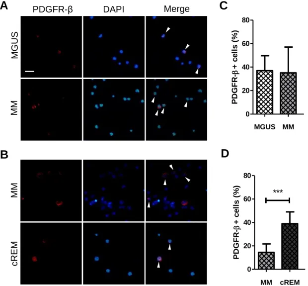
4.4. Alterations in PDGFR-β+ cells percentage from MGUS to MM and from MM to cREM ……….37
4.5. Correlation between plasma cells, endothelial progenitor cells, CXCR4+ and PDGFR-β+ cells levels from MGUS to MM and from MM to cREM ... 39
5. Discussion ... 42
6. Conclusions and future perspectives ... 47
7. References ... 49
ii
Figure Index
Introduction
Figure 1: Schematic representation of the bone marrow (BM) microenvironment and
respective niches………..……….…..…3
Figure 2: Schematic representation of the origin and differentiation of endothelial
progenitor cells (EPCs) and haematopoietic stem cells (HSCs) populations and their respective characteristic markers………...……….…………..10
Figure 3: Stages of multiple myeloma progression and respective diagnosis
parameters………..…...11
Figure 4: Schematic representation of the proposed mechanisms for endothelial
progenitor cells (EPCs) mobilization in the bone marrow (BM) and neovascularization in multiple myeloma (MM)………,………..………….….………….17-18
Materials and Methods
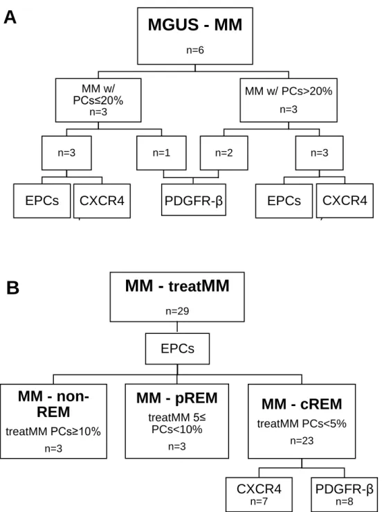
Figure 5: Schematic representation of the organization of the groups of
patients.……….………..………...26
Figure 6: Schematic representation of the optimized multiple immunofluorescence
method for endothelial progenitor cells (EPCs) detection in bone marrow (BM) smears…….………..…………..………...30
Results
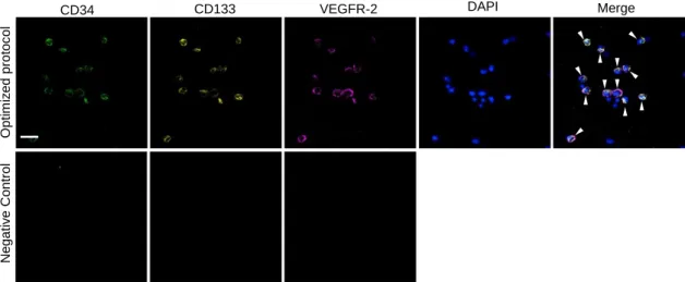
Figure 7: Identification of endothelial progenitor cells (EPCs) in bone marrow (BM)
smears by triple-labelling immunofluorescence analysis of cluster of differentiation (CD)34, CD133 and vascular endothelial growth factor receptor (VEGFR)-2, with nuclei visualization by 4',6-diamidino-2-phenylindole (DAPI)……….33
Figure 8: Plasma cells (PCs) and endothelial progenitor cells (EPCs) in the bone marrow
(BM) of patients with monoclonal gammopathy of undetermined significance (MGUS), multiple myeloma (MM) and patients after treatment……….…………35-36
Figure 9: Bone marrow (BM) C-X-C motif chemokine receptor (CXCR)4+ cells from monoclonal gammopathy of undetermined significance (MGUS), multiple myeloma (MM) and complete remission (cREM) patients………..………37
Figure 10: Bone marrow (BM) platelet-derived growth factor receptor (PDGFR)-β+ cells from monoclonal gammopathy of undetermined significance (MGUS), multiple myeloma (MM) and complete remission (cREM) patients………..…………...38
iii
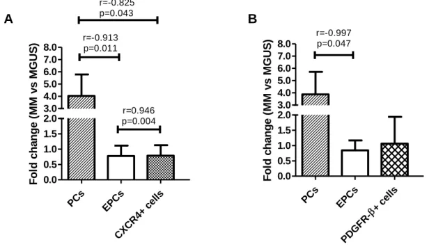
Figure 11: Changes in the levels of plasma cells (PCs), endothelial progenitor cells
(EPCs), C-X-C motif chemokine receptor (CXCR)4+ cells and platelet-derived growth factor receptor (PDGFR)-β+ cells in multiple myeloma (MM) versus monoclonal gammopathy of undetermined significance (MGUS) and correlation coeficients between the studied parameters……….40
Figure 12: Changes in the levels of plasma cells (PCs), endothelial progenitor cells
(EPCs), C-X-C motif chemokine receptor (CXCR)4+ cells and platelet-derived growth factor receptor (PDGFR)-β+ cells in complete remission (cREM) versus multiple myeloma (MM) and correlation coefficients between the studied parameters.…...41
Table Index
Table 1: Markers expressed by putative endothelial progenitor cells and/or whose
expression has been deemed characteristic of putative endothelial progenitor cells……….………..……….7-8
Table 2: Parameters experimentally analysed by immunofluorescence in each group of
patients...27
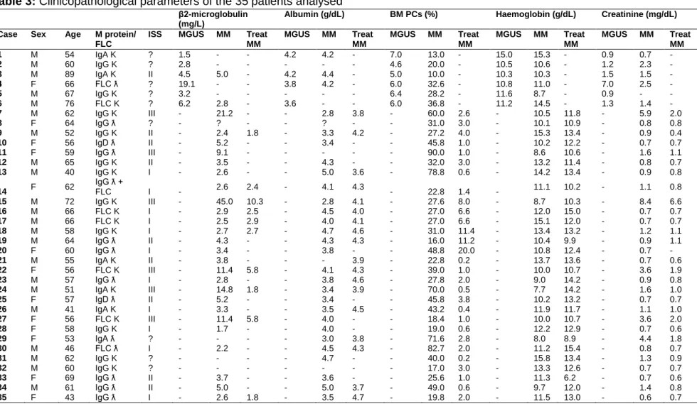
Table 3: Clinicopathological parameters of the 35 patients analysed……...…………..28 Table 4: Summary of the antibodies and experimental conditions used for multiple
immunofluorescence analysis.………..………...…..……….………...31
Supplementary Table 1: Experimental conditions tested during protocol optimization of
multiple labelling of CD34, CD133 and VEGFR-2………...………..61-62
Supplementary Table 2: Tested combinations of the antibodies CD34, CD133 and
iv
Abbreviations
bFGF, basic fibroblast growth factor BS, blocking solution
BSA, bovine serum albumin BM, bone marrow
BMSC, bone marrow stromal cell CD, cluster of differentiation
cEPC, circulating endothelial progenitor
cell
cREM, complete remission CSF1, colony-stimulating factor 1 CSF1R, colony-stimulating factor 1
receptor
CXCL12, C-X-C motif chemokine ligand
12
CXCR4, C-X-C motif chemokine
receptor 4
DAPI, 4',6-diamidino-2-phenylindole EC, endothelial cell
ECM, extracellular matrix ELAM-1, endothelial-leukocyte
adhesion molecule 1
EPC, endothelial progenitor cell E-selectin, endothelial selectin F, female
FLC, free light chain Flk-1, fetal liver kinase 1 GS, goat serum
HSC, haematopoietic stem cell
ICAM, intercellular adhesion molecule Ig, immunoglobulin
IGF, insulin-like growth factor IL, interleukin
IPO, Instituto Português de Oncologia
ISS, international staging system KDR, kinase insert domain receptor LCA, leukocyte common antigen LECAM-2, leukocyte-endothelial cell
adhesion molecule 2
LFA-1, lymphocyte-function-associated
antigen
M, male
Mc, monoclonal
MCAM, melanoma cell adhesion
molecule
M-CSFR, macrophage
colony-stimulating factor receptor
MGUS, monoclonal gammopathy of
undetermined significance
MM, multiple myeloma
MMP, matrix metalloproteinase MNC, mononuclear cell
M protein, monoclonal protein MSC, mesenchymal stem cell MVD, microvascular density non-REM, not in remission OB, osteoblast
OC, osteoclast ON, overnight PB, peripheral blood
PBS, phosphate buffered saline PC, plasma cell
Pc, polyclonal
PDGF, platelet-derived growth factor PDGFR-β, platelet-derived growth
factor receptor β
PECAM-1, platelet endothelial cell
v
pREM, partial remission
RANKL, receptor activator of nuclear
factor-kappa B ligand
REM, remission
ROS, reactive oxygen species RS, rabbit serum
RT, room temperature
SCFR, stem cell growth factor receptor SDF-1α, stromal cell-derived factor 1α SMM, smoldering multiple myeloma TGF, transforming growth factor TLR, toll-like receptor
treatMM, treated multiple myeloma USA, united states of america VCAM-1, vascular cell adhesion
molecule 1
VE-cadherin, vascular endothelial
cadherin
VEGF, vascular endothelial growth
factor
VEGFR-2, vascular endothelial growth
factor receptor 2
VLA-4, very-late-antigen 4 vWF, von Willebrand factor %, percentage
?, inconclusive information -, unavailable information #, catalogue reference
1
1. Introduction
The bone marrow (BM) constitutes the primary site for haematopoiesis, a process through which blood cells are produced and the amount of each cell population that goes into circulation is regulated (Anthony and Link 2014). The BM is mainly composed by haematopoietic stem cells (HSCs) and non-haematopoietic stromal cells. While the HSC population originates all the blood cell types, the non-haematopoietic cells give rise to a variety of cells important to the BM maintenance (Wickramasinghe et al. 2011). The BM is characterized by a peculiar microenvironment that provides proper growth factors and conditions for the development of blood cells. In order to maintain an adequate haematopoiesis, the BM is dependent on a highly efficient blood supply (Iversen 1997). Several pathologies can affect the BM microenvironment, including multiple myeloma (MM), which is one of the most common hematologic malignancies. It is predominant in the elderly population and remains an incurable disease (Rajkumar 2014). While the first documented case of MM goes back to 1844, nowadays the annual incidence in the United States population is 4.3 per 100000 (Kyle and Rajkumar 2008). The disease is characterized by an increase in antibody producing plasma cells (PCs) in the BM, as well as overstimulation of blood vessel formation (Giuliani et al. 2011). In this disorder, endothelial progenitors may be recruited to the tumour surroundings, stimulating the vasculature development through vasculogenesis (Caiado and Dias 2012). Since studies about MM vasculogenesis are lacking, we focused on the importance of the alterations in the BM microenvironment and the formation of blood vessels while taking a special interest on endothelial progenitors.
1.1. The bone marrow and its microenvironment
The BM is found amongst bone trabeculae of spongious bone and in the diaphysis of long bones. This bone compartment constitutes the main site of haematopoiesis, in which all blood cells are originated from a common pluripotent stem cell. Haematopoiesis can be divided in initial cell proliferation, commitment to a cell lineage, and cell differentiation, where biochemical, functional, and structural changes give rise to a specific cell type (Wickramasinghe et al. 2011). The BM microenvironment, or stroma, is mainly composed by both HSCs and non-haematopoietic or stromal cells. HSCs have unlimited self-renewal and are able to differentiate in all the blood cell types from the myeloid and lymphoid lineages (Krause 2002). The rich BM microenvironment is composed by several cell populations. Facing the bone matrix, there are endosteal osteoblasts (OBs) and osteoclasts (OCs) that are responsible for bone formation and resorption, respectively. The substantial blood vessels are lined by endothelial cells
2
(ECs). Adipocytes constitute the cells with the largest dimensions and their amount is inversely related with the BM cellularity. The population of BM stromal cells (BMSCs) englobes OBs, ECs, mesenchymal stem cells (MSCs), among others. MSCs are non-haematopoietic stromal cells that only constitute about <0.01% of the BM. These stromal cells can differentiate into multilineages, being able to originate OBs, chondrocytes and adipocytes (Wickramasinghe et al. 2011). The use of the definition of BMSCs and MSCs amongst the literature is not consensual, as some authors choose to refer to both as the same cell population, even though they constitute different cell types (Nemeth and Mezey 2015). All mentioned cells are connected through a network of extracellular matrix (ECM) (Klamer and Voermans 2014, Romano et al. 2014), containing soluble factors like cytokines and where important cell-cell interactions and cell-ECM interactions take place (Krause 2002), as illustrated in Figure 1. Inside the BM, distinct microenvironments can be found. These microenvironments are called niches and their function is to regulate the outcome of the cells they harbour (Balderman and Calvi 2014). These niches constitute the endosteal/osteoblastic niche and the vascular/arteriolar niche (Guerrouahen et al. 2011). The endosteal niche is located in the inner part of the bone cavity, near the endosteum, the layer that internally lines the bone (Bydlowski et al. 2013). To confirm the behaviour of HSCs in the BM, Xie et al. transplanted HSCs and observed that HSCs home to the BM endosteum, through its vasculature. There, HSCs are maintained, but can also undergo proliferation upon BM damage (Xie et al. 2009). As endosteum OBs are the main component of the niche, they contribute to HSCs quiescent state by down-regulating HSCs proliferation and differentiation (Nilsson et al. 2005) and by inducing bone adhesion, through angiopoietin-1 secretion (Arai et al. 2004). Moreover, MSCs act as a supportive niche for HSCs by participating in their homing and maintenance (Battiwalla and Hematti 2009, Mendez-Ferrer et al. 2010). Interestingly, HSCs that are more mature and have a higher proliferation level disperse from the endosteal niche (Lo Celso et al. 2009). During stress situations, OCs cleave C-X-C motif chemokine ligand (CXCL)12/stromal cell-derived factor 1, which stimulates HSCs mobilization (Kollet et al. 2006). Then, HSCs migrate from the endosteum and reach a vascular region where they start proliferation. This region constitutes the vascular/arteriolar niche, in which ECs are a strong component (Guerrouahen et al. 2011). It has been considered that this niche is divided in quiescent arteriolar niche and cycling sinusoidal niche (Klamer and Voermans 2014). The BM has a unique blood supply that differs from the remaining circulatory system. The main blood source constitutes the nutrient artery, which enters through the cortex (bone periphery) leading to the sinusoids. These are highly permeable capillaries formed by ECs with a discontinuous basement membrane (Iversen 1997, Marenzana and Arnett 2013).
3
Figure 1: Schematic representation of the bone marrow (BM) microenvironment and
respective niches. Close to the endosteum, the endosteal or osteoblastic niche is characterized by the presence of bone osteoblasts and osteoclasts. Osteoblasts induce haematopoietic stem cells homing into this location and contribute to their quiescent state. The location of stem cells is secured through C-X-C motif chemokine ligand (CXCL)12 and CXCL12-abundant reticular cells, near both the endosteum and the vasculature. The BM vasculature plays a key role in the proliferation and differentiation of stem cells. The source of the BM vasculature is named the nutrient artery which gives origin to sinusoids, capillaries that lack a continuous endothelial lining. The arteriolar niche has haematopoietic stem cells in an intermediate self-renewing state, while the sinusoidal niche is thought to contribute more significantly to both haematopoietic and mesenchymal stem cells proliferation and differentiation into myeloid and lymphoid cells, and osteoblasts and adipocytes, respectively.
CXCL12
CXCL12-abundant reticular cells Osteoblast
Osteoclast Haematopoietic stem cell
Mesenchymal stem cell
Endothelial cell
Adipocyte
Myeloid lineage cell Lymphoid lineage cell
Endosteal/osteoblastic Niche Bone
Quiescence
Stem cells proliferation and differentiation Stem cells quiescent
state Sinusoid Proliferation Self-renewal Quiescence Differentiation Extracellular matrix Differentiation Homing
Arteriolar Niche Sinusoidal Niche
Vascular/Arteriolar Niche
Proliferation
Nutrient artery
4
The arteriolar niche is thought to be closer to the endosteum, where it contributes to the quiescent state of HSCs, while the sinusoidal niche may be spread through the BM and contributes more significantly to haematopoietic stem and progenitor cells proliferation and differentiation (Guerrouahen et al. 2011, Klamer and Voermans 2014). The arterial blood vessels microenvironment has a characteristic low reactive oxygen species (ROS) level, while the highly permeable sinusoids have higher ROS levels, which promote HSCs to migrate towards the higher ROS state and differentiate (Itkin et al. 2016). In the arteriolar quiescent niche, MSCs also remain in their quiescent state (Kunisaki et al. 2013). In addition, the proximity of HSCs to the vasculature is maintained mainly by the C-X-C motif chemokine ligand (CXCL)12 and C-X-C motif chemokine receptor (CXCR)4 interaction, as CXCL12-abundant reticular cells are found near sinusoidal ECs or close to the endosteum. CXCL12 is thus essential to HSCs niche-homing (Sugiyama et al. 2006).
1.2. Formation of blood vessels
Blood vessels are constituted by a monolayer of ECs that form a vascular wall, which is surrounded by the basement membrane. Embedded in this membrane, are mural cells such as smooth muscle cells in larger vessels and pericytes at pre-capillary arterioles, capillaries, and post-capillary venules (Sá-Pereira et al. 2012). Additionally, the ECM provides a structural framework to the blood vessels (Jacob et al. 2001). In the adult, the vasculature remains mostly in a quiescent state (Charpentier and Conlon 2014). The formation of new blood vessels in the adult constitutes the process of neovascularization. Blood vessels can be formed either by vasculogenesis or angiogenesis. Although vasculogenesis is characteristic of embryonic development, postnatal vasculogenesis has also been demonstrated, in both physiological and pathological conditions (Asahara et al. 1999, Morales-Ruiz and Jiménez 2005). During embryonic development, vasculogenesis is defined as de novo differentiation of mesodermal precursors into precursor ECs, also known as angioblasts or endothelial progenitor cells (EPCs), which will ultimately differentiate into ECs. EPCs proliferate and integrate into the primary capillary plexus, a primate vascular network. This initial network is then completed through angiogenesis (Asahara et al. 1999, Papetti and Herman 2002) that represents the most common process of vascular development. In the adult, physiological angiogenesis is mostly related to the ovarian cycle and wound healing. On the other hand, in pathological events, the absence of angiogenesis is characteristic of cardiac failure, while excessive angiogenesis is found in chronic inflammation and cancer (Griffioen 2012). This process of blood vessel development is present in tumour growth, invasion and metastasis, and is also considered to have a key role in several
5 haematological malignancies (Otjacques et al. 2011). Angiogenesis can occur by sprouting angiogenesis or intussusceptive angiogenesis. Sprouting angiogenesis begins with the degradation of the ECM and basement membrane by proteolytic enzymes. ECs migrate, adhere, and proliferate in order to form a vascular sprout that is stabilized by a basement membrane and the recruitment of mural cells. Intussusceptive angiogenesis is characterized by the division of pre-existing vessels through transcapillary tissue pillars that are stabilized by the invagination of the ECM and pericytes (Morales-Ruiz and Jiménez 2005). The angiogenic process is highly regulated through soluble factors that are mainly responsible for controlling ECs proliferation, migration and endothelium stability, as well as by membrane-bound factors that control cell adhesion and by chemokines that are involved in chemotaxis (Papetti and Herman 2002, Jakob et al. 2006).
1.3. Endothelial progenitor cells
The term EPCs was first reported in 1997 by Asahara et al. (Asahara et al. 1997). Ever since, EPCs are defined as BM derived progenitors with high proliferative ability that have the potential to differentiate into cells of the endothelial lineage (Laurenzana et al. 2015). It was estimated that EPCs represent up to 26% of ECs in recently formed vessels (Murayama et al. 2002). BM-derived EPCs participate in postnatal vasculogenesis, in physiological processes such as tissue growth, and in pathological events like myocardial ischemia, stroke, artherosclerosis and cancer (Caiado and Dias 2012, Moschetta et al. 2014), a topic that will be developed later in the review. Furthermore, these cells contribute to re-endothelialisation in scenarios of tissue injury, which is why EPCs have been considered as an important tool for therapy in tissue repair (Tenreiro et al. 2016). EPCs also aid in maintaining the vasculature through the production of angiogenic factors that stimulate the proliferation, function and survival of ECs. Therefore, they have an indirect but important role in angiogenesis (Laurenzana et al. 2015). It is important to highlight that the notion of angiogenesis is often used when referring to any new blood vessel formation (Kovacic et al. 2008). Thus, EPCs are repeatedly connected with that process even though EPCs are defined as key players in vasculogenesis and not angiogenesis, as stated in the previous section. EPCs are considered to develop from hemangioblasts, which also give origin to HSCs (Moschetta et al. 2014). EPCs are kept in a quiescent BM niche that is thought to have low oxygen tension and an elevated amount of CXCL12 that is responsible for maintaining them (Balaji et al. 2013). In cases of trauma or wound-healing generating hypoxia, EPCs are stimulated to leave this niche, reach the proliferative niche and then are able to go into circulation (Velazquez 2007). Then, EPCs home into their target tissue and are activated.
6
Afterwards, EPCs adhere to the ECs of the vessel and begin transendothelial migration for vascular remodelling. After crossing the endothelial monolayer, EPCs migrate through the basement membrane and ECM, a mechanism that depends on extracellular proteases. When reaching the vessel remodelling site, EPCs differentiate into ECs and/or interact with the ECs. Although the functional activity of EPCs is mostly under investigation, it is considered that their differentiation involves adhesion to the ECM components controlled by integrins, proliferation and survival induced by growth factors, and maturation and acquisition of the endothelial phenotype (Caiado and Dias 2012). The peculiar properties of these cells in the development of blood vessels have attracted special interest and, although there are many studies about them, the knowledge about these cells is far from being complete. In addition, the precise process of origin of EPCs remains to be fully clarified, mainly due to the controversial identification of these cells, as it is portrayed in the next section.
1.3.1. Characteristic markers
Until today, EPCs remain with no uniform definition and no specific cell-surface antigen (Yoder 2012, Tenreiro et al. 2016). For that reason, it is important to underline the developments in this topic. After isolation from peripheral blood (PB), EPCs were firstly identified with the markers cluster of differentiation (CD)34 and vascular endothelial growth factor receptor (VEGFR)-2 (Asahara et al. 1997). Since then, besides being distinguished based on the expression of markers, EPCs are also isolated based on their functional and clonal expansion features (Balaji et al. 2013). EPCs display the capacity to phagocyte low density lipoprotein LDL, and bind to Ulex europaeus lectin-1 (Song et al. 2010). In order to characterize EPCs, researchers usually isolate PB, BM or cord blood mononuclear cells (MNCs), and perform the respective culture in specific cell growth medium in order to outgrow putative EPCs or, alternatively isolate and identify putative EPCs through multiple markers expression (Eggermann et al. 2003, Braunstein et al. 2006, Song et al. 2010, Yang et al. 2011, Amini et al. 2012). Several authors have used and attributed different markers for putative EPCs, as depicted in Table 1.
7
Table 1: Markers expressed by putative endothelial progenitor cells and/or whose
expression has been deemed characteristic of putative endothelial progenitor cells Marker Other
names
Cellular expression
Function Reference CD34 - Transmembrane Adhesion molecule
between EC and haematopoietic stem cells
Cell migration
(Ria et al. 2008, Shi and VandeBerg 2015)
VEGFR-2 Flk-1 KDR CD309
Transmembrane Receptor 2 for VEGF. Involved in the formation of blood vessels (Eggermann et al. 2003, Case et al. 2007, Timmermans et al. 2007, Fadini et al. 2008) CD133 Prominin-1 AC133
Transmembrane Unclear function (Peichev et al. 2000, Shmelkov et al. 2005, Ria et al. 2008)
CD45 LCA Transmembrane T and B cell protein tyrosine phosphatase involved in lymphocyte activation and proliferation (Trowbridge and Thomas 1994, Yoder 2012) CD31 PECAM-1 Transmembrane and soluble Adhesion molecule in EC intercellular junctions and angiogenesis (Peichev et al. 2000, Kalinowska and Losy 2006, Song et al. 2010, Amini et al. 2012) CD146 MCAM Transmembrane and soluble Adhesion molecule in EC adherens junctions (Peichev et al. 2000, Bardin et al. 2001) E-selectin CD62E ELAM-1 LECAM-2 Transmembrane and soluble Adhesion molecule in EC (Peichev et al. 2000, Ley 2003) VE-cadherin Cadherin 5 type 2 CD144 Transmembrane and soluble Adhesion molecule in EC (Peichev et al. 2000, Eggermann et al. 2003, Ria et al. 2008, Vestweber 2008) Tie-2 Tyrosine-protein kinase receptor
Transmembrane Receptor tyrosine kinase, involved in proliferation and differentiation (Schnürch and Risau 1993, Fadini et al. 2008, Ria et al. 2008) c-kit SCFR Tyrosine-protein kinase Kit CD117
Transmembrane Cytokine receptor involved in cell survival, proliferation and differentiation (Nocka et al. 1990, Peichev et al. 2000, Cananzi and De Coppi 2012) CD14 - Transmembrane and soluble
Co-receptor for the TLR, Innate immune response (Eggermann et al. 2003, Lau et al. 2014) CD115 CSF1R M-CSFR
8
CD105 Endoglin Membrane TGF-β receptor complex receptor
(Cheifetz et al. 1992, Eggermann et al. 2003)
vWF - Soluble Platelet adhesion
molecule
(Eggermann et al. 2003, Lenting and Christophe 2015) CXCR4 CD184 Transmembrane Chemokine receptor for
SDF-1α/CXCL12
(Peichev et al. 2000, Kucia et al. 2004)
CD, cluster of differentiation; CSF1, stimulating factor 1; CSF1R, colony-stimulating factor 1 receptor; CXCL12, C motif chemokine ligand 12; CXCR4, C-X-C motif chemokine receptor type 4; EC-X-C, endothelial cell; ELAM-1, endothelial-leukocyte adhesion molecule 1; E-selectin, endothelial selectin; Flk-1, fetal liver kinase 1; KDR, kinase insert domain receptor; LCA, leukocyte common antigen; LECAM-2, leukocyte-endothelial cell adhesion molecule 2; M-CSFR, macrophage colony-stimulating factor receptor; MCAM, melanoma cell adhesion molecule; PECAM-1, platelet endothelial cell adhesion molecule 1; SCFR, stem cell growth factor receptor; SDF-1α, stromal cell-derived factor 1α; TLR, toll-like receptor; TGF, transforming growth factor; VE-cadherin, vascular endothelial cadherin; VEGF, vascular endothelial growth factor; VEGFR-2, vascular endothelial growth factor receptor 2; vWF, von Willebrand factor.
Several cell populations of putative EPCs have been described, which makes it difficult to establish connections between the existing studies. Most studies have focused on isolating putative EPCs from PB and detailed studies exploring EPCs in the BM are rare to find. The types of putative EPCs have been mostly defined based on the maturation state and/or according to cells isolated from MNCs in vitro (Fig. 2), while differentiation in vivo remains elusive. Indeed, it remains a challenge to establish equivalencies between EPCs in vivo and in vitro, since EPCs in culture may gain or lose characteristics that are not present in non-cultured cells. EPCs can be classified based on maturation state, proliferative potential, endothelial markers, morphology and capacity to form blood vessels (Moschetta et al. 2014). Peichev and colleagues observed that CD34+VEGFR-2+ cells also express CD133 and have the capacity to migrate and differentiate into adherent mature ECs. As these cells become mature ECs, they lose CD133 expression (Peichev et al. 2000). Supporting this, Quirici et al. isolated EPCs from BM CD133+ cells that later gave rise to ECs. After 3 weeks of culture, these cells were negative for CD45 and CD14 and positive for several EC markers such as Ulex
europaeus lectin-1, von Willebrand factor (vWF), CD105, endothelial selectin, vascular
cell adhesion molecule 1 and vascular endothelial (VE)-cadherin (Quirici et al. 2001). On the other hand, CD34+cells were isolated from MNCs from human umbilical cord blood and it was reported that CD34+CD45+ cells formed haematopoietic progenitor cells, while CD34+CD45- formed EPCs with endothelial colony forming activity. Thus, the authors of this study stated that true EPCs had no expression of the haematopoietic lineage-specific and common leukocyte antigen CD45 (Case et al. 2007). Contrarily to the previously
9 mentioned study, functional EPCs were attained from CD34+ cells isolated from mouse BM MNCs and these EPCs were positive for CD45 after culture (Yang et al. 2011). Hence, these contradictory reports make for a difficult interpretation, more specifically, of the relevance of the expression of CD45, which is characteristic of haematopoietic cells (Amini et al. 2012). Even so, the combination of CD34, CD133 and VEGFR-2 for the identification of EPCs is generally used (Massa et al. 2005, Caiado et al. 2011, Blix et al. 2015).
Ultimately, when identifying EPCs, it is crucial to bear in mind the methodology used for their isolation and identification, to not intensify the incoherency regarding the terminology and the protocols for isolating putative EPCs. The markers used in the several studies to identify EPCs are not exclusive to EPCs as they can also be expressed by HSCs and, therefore, do not allow a clear and effective identification of these cells (Fadini et al. 2008). This field of investigation deserves further research considering the potential and importance that these cells harbour, the interest in a better knowledge of their characterization, as well as in the development of new techniques for their isolation and differentiation in EPCs with consistent and reliable phenotypes.
10
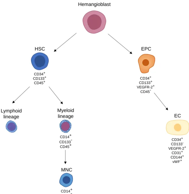
Figure 2: Schematic representation of the origin and differentiation of endothelial
progenitor cells (EPCs) and haematopoietic stem cells (HSCs) populations and their respective characteristic markers. Hemangioblasts can differentiate into HSCs and EPCs. HSCs give rise to cells of the lymphoid and myeloid lineages. In in vitro culture, mononuclear cells (MNCs) can be isolated and differentiated into putative EPCs. True EPCs originated from the hemangioblast express cluster of differentiation (CD)34, vascular endothelial growth factor receptor (VEGFR)-2 and CD133. Ultimately, EPCs give rise to endothelial cells (ECs), which express typical EC markers such as CD34, VEGFR-2, CD31, CD144 and von Willebrand factor (vWF).
1.4. Multiple myeloma
MM represents about 10% of all hematologic malignancies, for which there is still no cure. Patients are usually diagnosed at 65 years old and have a median survival of 3 to 4 years (Rajkumar 2014). MM is characterized by BM accumulation of malignant PCs and high levels of a monoclonal immunoglobulin (Ig), mostly IgG or IgA, or free light chains (FLC) that are detected in blood and/or urine (Bianchi and Anderson 2014). PCs
Lymphoid lineage Myeloid lineage CD14+ CD133 -CD45+ EC CD34+ CD133 -VEGFR-2+ CD31+ CD144+ vWF+ HSC CD34+ CD133+ CD45+ Hemangioblast MNC CD14+ CD45+ CD34+ CD133+ VEGFR-2+ CD45-EPC
11 constitute terminally differentiated lymphoid B cells, which are located in the BM and the medulla of lymph nodes. These cells synthesize Igs that act as antibodies against antigens. When clonal expansion of PCs is altered, it leads to an increased synthesis of a monoclonal Ig that translates as an elevated monoclonal peak in serum electrophoresis. The visual anomalies in PCs include abnormal chromatin network, irregular nuclear outline and cytoplasm colour, and inclusions with different origins connected to irregular trafficking or catabolism related to Ig synthesis by PCs. The diverse morphological characteristics constitute various types of PCs. For instance, immature PCs are named plasmablasts, which are characteristic of patients with higher BM PCs levels and proliferation rate. On the other hand, patients with predominantly mature PCs are usually correlated with a better prognostic. Multiple nuclei can also be observed in PCs, as they are a part of PC burden (Ribourtout and Zandecki 2015). Most importantly, MM is associated with lytic bone disease, due to osteolytic resorption and suppression of bone formation. This leads to the main cause of malaise in MM, namely the excruciating bone pain (Heider et al. 2006).
Clinical staging of malignancies such as MM allows a reliable classification of the disease progression and prediction of patient survival. As a matter of fact, MM belongs to a spectrum of distinct PC disorders, as illustrated in Figure 3. The first manifestation of this disease consists of monoclonal gammopathy of undetermined significance (MGUS), followed by the intermediate smoldering MM (SMM) and, lastly, MM. These stages can be clinically distinguished through the percentage (%) of BM PCs, serum monoclonal protein (M protein) and organ end damage (Kyle 1978, Kyle and Greipp 1980, Kyle et al. 2002, Bianchi and Anderson 2014).
Figure 3: Stages of multiple myeloma progression and respective diagnosis parameters.
BM PCs %, percentage of plasma cells in the bone marrow; MGUS, monoclonal gammopathy of undetermined significance; M protein, monoclonal protein; MM, multiple myeloma; SMM, smoldering multiple myeloma.
MGUS
BM PC % ≤
10
Serum M
protein ≤ 30 g/L
No organ end
damage
SMM
BM PC % ≥
10
Serum M
protein ≥ 30 g/L
No organ end
damage
MM
BM PC % ≥
10
Serum M
protein ≥ 30 g/L
Organ end
damage
12
MGUS is characterized by serum monoclonal Ig of 30 g/L or less and 10% or less of BM PCs, without lytic bone lesions, and no evidence of anaemia, hypercalcaemia and renal insufficiency. It is thought that factors such as genetic changes, several cytokines important in myeloma bone disease and angiogenesis in the BM are responsible for the progression of MGUS to MM, which risk is around 1% per year (Kyle and Rajkumar 2006). MGUS diagnosis is also made through the amount of M protein in serum protein electrophoresis, by immunofixation or an irregularity in a serum FLC assay (Kyle et al. 2004). However, this disorder ends up underdiagnosed, due to the absence of MM symptoms. The asymptomatic stage between MGUS and MM consists of SMM. It was firstly identified in patients with a % of PCs and levels of M protein higher than in MGUS (BM PCs ≥ 10%; M protein level ≥ 30 g/L) that presented no symptoms and remained clinically stable for 5 or more years (Kyle and Greipp 1980). The progression from this state to MM can vary from 2 to 19 years, depending on several factors such as serum M protein levels, presence of IgA isotype, serum FLC ratio, % of BM and PB PCs, cytogenic abnormalities and Bence Jones proteinuria (Gao et al. 2015, Gentile et al. 2015). After the MM malignancy is set up, the symptoms usually include fatigue, bone pain, anaemia, renal failure, hypercalcemia and even peripheral neuropathy and amyloid light chain amyloidosis. The diagnosis is based on BM PCs ≥ 10%, serum M protein ≥ 30 g/L and the presence of end-organ failure (Bianchi and Anderson 2014). MM can be classified according to two staging systems: the Durie-Salmon and the International Staging System (ISS). Since the Durie-Salmon system quantifies the tumour burden but not the bone lesions, the ISS fills that gap by dividing patients into fractions based upon the prognostic factors β2-microglobulin and serum albumin levels (Durie and Salmon 1975, Greipp et al. 2005).
MM is known for its clinical and biological heterogeneity, which is responsible for inconstant response to treatment. Biomarkers can be used to distinguish this heterogeneity and to choose the most adequate treatment option. Biomarkers are defined as a biochemical, cellular/molecular substance or characteristic that is correlated to a regular or pathogenic biological process, or to a response to a certain therapy, and can be measured in an accurate and reproducible way (Naylor 2005). The most commonly used biomarkers for MM diagnosis are PC labelling index, BM-infiltration rate, serum M protein and FCL, and levels of albumin and β2-microglobulin. More uncommonly, are calcium and serum creatinine levels, and cytogenetic abnormalities (Kastritis et al. 2013, Rajshenkhar and Shaji 2015). Even with the high amount of biomarkers for MM, there is a need to complete this list with more sensitive biomarkers in order to deal with the progression from an asymptomatic state to MM, which is still
13 unpredictable due to the characteristic heterogeneity of MM (Landgren and Morgan 2014). Moreover, with increasing MM knowledge and therapy options, new biomarkers can help physicians to identify which patients would benefit from a specific therapeutic option and the response the patient may display.
There is still no therapy for asymptomatic MM or SMM and so it is advisable a close follow-up until MM symptoms evolve (Gentile et al. 2015). Regarding MM, in 1983 the median survival of MM was less than 2 years with only 2.2% of the patients surviving for longer than 10 years with chemotherapy (Kyle 1983). Over the last decade, new therapies have emerged to treat MM, which resulted in a better outcome (Chng et al. 2014). The therapies currently available to attenuate MM symptoms consist of alkylating agents, immunomodulatory drugs such as thalidomide, proteasome inhibitors like bortezomib, immunotherapy with monoclonal antibodies and vaccines, chemotherapy, long-acting steroids and, in the worst cases, autologous stem cell transplant (Bianchi and Anderson 2014, Rajkumar 2014). Nowadays, it is considered that standard risk patients have an overall survival of 6-7 years, whereas high risk patients only seem to survive for 2-3 years even with autologous stem cell transplantation (Rajkumar 2014). Even with the large amount of therapies available, patients do not maintain remission (REM) for long and relapse is expected (Kurtin 2013). Hence, despite the latest evolution in MM treatment, it remains an incurable disease, which demands new therapeutics not only concerning the tumour cells, but also to their surrounding microenvironment.
1.4.1. Compromised bone marrow microenvironment
Since MM PCs accumulate in the BM, its microenvironment plays an important role in MM progression. In fact, BMSCs and MSCs support the development of MM, whether it is by cell-cell interactions or by cytokines secretion. MM MSCs and their progenitors differ from normal MSCs, as they greatly support the growth and survival of MM PCs (Wallace et al. 2001, Corre et al. 2007). MM PCs show higher levels of proliferation when incubated with MM MSCs, namely through expression of translation initiation factors, which contributes highly to the transformation of normal PCs to malignant PCs (Attar-Schneider et al. 2015). MM PCs and MSCs communicate mainly through the CXCR4 signalling pathway (Feng et al. 2010). Not only do BMSCs support the growth of tumour cells, but they also contribute to the MM bone disease, as described below. This hallmark of MM consists in the imbalance between bone formation and degradation. It is connected to chronic pain, bone fractures, spinal cord compressing and, thus, a poor life quality (Walker et al. 2014). Myeloma bone disease is achieved through the inhibition of OBs, which are responsible for bone formation, and the
14
promotion of OCs that are involved in bone resorption. There is an increase in OBs inhibitors from the Wnt pathway, which is essential for OB differentiation, therefore reducing OBs activity. Moreover, the dysregulation of interleukin (IL)-3 and IL-7 expression leads to a disturbance of OBs survival. On the other hand, the OCs regulators tumour necrosis factor-related apoptosis-inducing ligand and receptor activator of nuclear factor-kappa B ligand (RANKL) levels are higher in MM patients (Kristensen et al. 2014, Romano et al. 2014). RANKL is secreted when MM PCs adhere to BMSCs and bind to its receptor RANK in OCs progenitor cells, leading to stimulation of OCs activity, differentiation and bone resorbing. BMSCs also secrete osteoprogesterin, which inhibits OCs differentiation by RANK binding. However, in MM, osteoprogesterin is present in low levels, which leads to a further promotion of OCs differentiation. IL-6 also promotes the action of osteoclastogenic factors and bone degradation (Romano et al. 2014, Walker et al. 2014). Interestingly, Lawson et al. recently reported that, even though most of MM PCs remained circulating in the BM, colonizing MM PCs migrated outwards endocortical bone and remained in the endosteal bone niche. Furthermore, the endosteal niche inhibits MM PCs growth as OBs contribute to suppress MM PCs proliferation and to keep MM PCs in a dormant state. Remarkably, this inhibition is reversible, as OCs activity and bone resorption lead to a reduction in bone surface and release of MM PCs from the endosteal niche, further contributing to tumour development (Lawson et al. 2015).
1.4.2. Malignant angiogenesis
Both angiogenesis and vasculogenesis have been subject of discussion in MM (Giuliani et al. 2011). Tumour angiogenesis differs greatly from physiologic angiogenesis as it is characterized by an increase in ECs proliferation and in the levels of the factors that regulate this process. In tumours, angiogenesis results in an abnormal and aberrant vasculature with heterogeneous ECs, irregular blood flow, instability and elevated permeability, as is the case of MM (Jakob et al. 2006, Giuliani et al. 2011). Early on and until nowadays, it is considered that an “angiogenic switch” occurs from MGUS to MM. In fact, MGUS is considered an avascular phase, whereas the development of blood vessels is characteristic of MM (Vacca et al. 1999, Calcinotto et al. 2015). This switch is caused by the malignant cells and leads to the detachment of pericytes, vessel dilation and sprouting (Giuliani et al. 2011). Interestingly, MGUS BM samples showed the capacity to decrease angiogenesis, while samples from SMM and MM patients stimulated angiogenesis in vitro (Kumar et al. 2004). These results point to a switch in the BM involving pro- and anti-angiogenic factors that participate in paracrine interactions between MM PCs, ECs and BMSCs (Jakob et al. 2006). In fact, assessment of microvascular density (MVD) and expression of angiogenic factors demonstrated that
15 BM angiogenesis is increased in MM (Di Raimondo et al. 2000, Lee et al. 2015). In 1994, it was firstly demonstrated that BM MVD was significantly increased as well as BM angiogenesis in patients with MM in comparison with MGUS (Vacca et al. 1994). Numerous studies have since suggested the prognostic potential of BM MVD, as higher levels correlate with MGUS evolution to MM and shorter progression-free survival. BM MVD was also connected with the level of BM PCs and PC labelling index, as reviewed by Giuliani et al. (Giuliani et al. 2011).
1.4.3. Endothelial progenitor cells-induced vasculogenesis
EPCs have a central role in vasculogenesis, as previously mentioned. These cells have been studied in several haematological malignancies as non-Hodgkin’s lymphoma, leukaemia and MM. Since EPCs have the same mesodermal progenitor as HSCs, which will originate the malignant cells in MM, it was proposed that the origin of EPCs may be from the same malignant clone as haematopoietic cancer cells (Moschetta et al. 2014). The analysis of neovascularization through MVD requires a BM biopsy, which is a very invasive procedure. On the other hand, EPCs can be isolated and evaluated less invasively from PB, so research turned to exploit that method (Zhang et al. 2005). Accordingly, EPCs in the context of MM were firstly mentioned by Zhang et al., in 2005, regarding circulating EPCs (cEPCs) in PB. The study showed that cEPCs levels were higher in MM in comparison to healthy controls and that these levels were correlated with those of M protein and β2-microglobulin, suggesting that EPCs can constitute a biomarker for MM progression (Zhang et al. 2005). Later on, it was confirmed that cEPCs levels in MM were correlated with the evolution of the disease from ISS I to ISS III stage (Bhaskar et al. 2012). A recent study evaluated PB and BM levels of EPCs in healthy individuals and in SMM and MM patients. The control levels were significantly lower in comparison to the disease levels in the two compartments. In PB, the EPCs levels in SMM were slightly lower than in MM, while in the BM, SMM levels were slightly higher than in MM. This study shows that even in early stages of MM, the mobilization and proliferation of EPCs in the BM is substantial in comparison to healthy conditions (Moschetta et al. 2016). BM cells positive for VEGFR-2, a marker used for EPC identification, were also depicted to be higher in MM patients (with Salmon-Durie stage II/III, active disease and BM-infiltration rates of ≥20%) when comparing to MGUS patients (Udi et al. 2011). Regarding treatment, only BM CD34+VEGFR-2+ EPCs levels were significantly higher in MM in comparison to REM (Udi et al. 2011). Thalidomide treatment led to a decline in cEPCs (Zhang et al. 2005). With chemotherapy, cEPCs declined in responders to therapy, while they increased in non-responders (Bhaskar et al. 2012). More recently, concerning the combination of bortezomib with dexamethasone, higher
16
levels of cEPCs were correlated with a later response to this treatment (Wang et al. 2015), which underlines cEPCs potential as a prognostic biomarker. Additionally, in autologous stem cell transplant, higher EPCs levels in PB stem cell autografts were associated with a lower overall survival after the therapy (Blix et al. 2015). Regarding different treatments and their effect on EPCs, the levels of BM EPCs were higher when novel agents were administrated (thalidomine, lenalidomine and bortezomib) in comparison to stem-cell transplant treatment and to stem cell transplant plus novel agents (Udi et al. 2011). There seems to be lack of significant information about the amount of EPCs in the BM regarding different stages of MM, as most studies have focused on PB EPCs. Furthermore, since different responses to therapy are associated with different levels of EPCs, these levels could be used in order to predict the patients response to treatment and which treatment option would be best in a particular scenario. Still, this topic remains much unexplored, as there is an absence of studies regarding BM levels of EPCs and their correlation to the different therapies available.
Mobilization path in the bone marrow
The exact mechanisms related to the path EPCs make in the BM in the malignant scenario of MM are still to be studied. Considering that the tumour site is located within the BM, it is uncertain if EPCs need to intravasate into the vasculature to reach the tumour location, as they do to reach a location distant from the BM, or if EPCs simply migrate to the tumour location within the BM itself, as illustrated in Figure 4A. Even so, several critical phases are involved in vasculogenesis. First, EPCs must leave their BM niche and be recruited to the tumour microenvironment (Fig. 4B-C). Upon reaching the vasculature surrounding the tumour niche, EPCs home to the vasculature by ECs stimulation (Fig. 4D). EPCs adhere to ECs and initiate transendothelial migration (Fig. 4E). Upon integrating the endothelium, EPCs differentiate into mature ECs (Fig. 4F). This process is further completed with the stabilization of blood vessels provided by pericytes (Fig. 4G).
17
Figure 4: Schematic representation of the proposed mechanisms for endothelial
progenitor cells (EPCs) mobilization in the bone marrow (BM) and neovascularization in multiple myeloma (MM). EPCs are formed in the BM, where MM plasma cells (PCs) proliferate. EPCs are mobilized to the tumour location, but the question to if EPCs travel through the blood stream or the BM itself remains unanswered (A). Within these hypotheses, initially, EPCs remain in the quiescent niche near the endosteum, osteoblasts and osteoclasts. EPCs are released from the quiescent niche mainly due to matrix metalloproteinase (MMP)-9 secreted by both osteoclasts and MM PCs, which degrade the extracellular matrix in the niche, facilitating EPCs mobilization (B). EPCs are mobilized to the tumour site attracted by the ligands C-X-C motif chemokine ligand (CXCL)12, vascular endothelial growth factor (VEGF) and platelet-derived growth factor (PDGF)-B secreted by MM PCs that bind to the respective receptors C-X-C motif chemokine receptor (CXCR)4, vascular endothelial growth factor receptor (VEGFR)-2 and PDGF receptor (PDGFR)-β expressed by EPCs (C). Once at the vasculature site, endothelial cells (ECs) induce EPCs homing to the vasculature mediated by the same molecules mentioned in (B). Both CXCL12 and PDGF-B can be produced by ECs, whereas the production of VEGF by these cells can stimulated by CXCL12 secreted by MM PCs (D). Then, EPCs must adhere to the endothelial wall of the vessel being
PDGF-B VEGF Osteoblast Endothelial progenitor cell Multiple myeloma plasma cell Endothelial cell CXCR4 VEGFR-2 PDGFR-β C Bone Bone marrow ? ? MMP-9 CXCL12 Sinusoid A ICAM β2 integrin VCAM-1 ICAM-1 D E Osteoclast Pericyte LFA-1 VLA-4 B Bone marrow Sinusoid Extracellular matrix G Sinusoid F
18
remodelled. This process can be done through integrins lymphocyte-function-associated antigen (LFA)-1 and very-late-antigen (VLA)-4 on EPCs binding to intercellular adhesion molecule (ICAM)-1 and vascular cell adhesion molecule (VCAM)-1, respectively. To finish incorporating the vessel, EPCs transmigrate mainly through β2 integrins binding to ICAM (E). For EPCs to differentiate, these cells must adhere to components of the extracellular matrix, such as fibronectin. Several molecules like VEGF and CXCL12 stimulate EPCs differentiation, which can be produced by MM PCs (F). Lastly, ECs secrete PDGF-B and recruit PDGFR-β+ pericytes to stabilize the vasculature. MM PCs are also capable of recruiting pericytes through the same secretion (G).
EPCs reside in their stem cell niche characterized by high levels of CXCL12 (Caiado and Dias 2012). The main component controlling the detachment of stem cells from their niche seems to be matrix metalloproteinases (MMPs). These are a group of zinc-dependent endopeptidases with the capacity to degrade the ECM, thus contributing to the proliferation, invasion, metastasis, angiogenesis, and progression of cancer (Kessenbrock et al. 2015). More specifically, MMP-9 activation leads to progenitor cells, such as EPCs, leave their quiescent niche and move to a proliferative vascular niche (Heissig et al. 2002). Altered levels of MMP-9 are observed in MM. MMP-9 serum levels in MM patients were higher in Durie-Salmon stages II and III than in stage I (Alexandrakis et al. 2007). In fact, MMP-9 levels were higher when the murine MM PCs 5T33MM cells were co-cultured with BM ECs than with lung ECs (Van Valckenborgh et al. 2002). Interestingly, MMP-9 is secreted by MM PCs, whose upregulation is stimulated by BM ECs through hepatocyte growth factor (Van Valckenborgh et al. 2002, Vande Broek et al. 2004). Moreover, OCs can also secrete MMP-9 that will degrade the ECM and induce a vascular endothelial growth factor (VEGF) discharge (Ribatti et al. 2014). The contribution of MM BMSCs in cell detachment from the niche remains unclear, as it was observed that MM BMSCs secreted only MMP-1 and -2 but not MMP-9, which is the MMP most relevant in this process (Barillé et al. 1997). On the other hand, it seems that MMP-9 is not an indicator of bone disease in MM (Munemasa et al. 2007), which means that it could be relevant in some other event of the disease, such as neovascularization.
The recruitment of EPCs into the target location is a critical step in neovascularization. EPCs are mobilized and home to the location where they will incorporate the blood vessels and differentiate into ECs. A recently published study revealed that BM EPCs are mobilized to the location of MM PCs in the BM, which displacement is driven by the malignant cells (Moschetta et al. 2016). Many factors are involved in EPC mobilization such as CXCL12, VEGF, and also platelet-derived growth factor (PDGF)-B. The chemokine CXCL12 is produced by BMSCs (Nagasawa et al. 1994, de Nigris et al. 2012), BM ECs (Yun and Jo 2003), immature OBs and mesenchymal adipocytes (Lapidot and Kollet 2002). CXCL12 stimulates the motility of