FACULDADE DE MEDICINA
Dissecting the Cellular and Molecular Mechanisms of
IL-7-mediated Leukemia T- cell Survival, Proliferation
and Cell Growth
Daniel Filipe Silva Ribeiro
Orientador: Prof. Doutor João Pedro Taborda Barata
Tese especialmente elaborada para a obtenção do grau de Doutor em Ciências Biomédicas - Especialidade em Biologia Celular e Molecular
Júri:
Presidente: Doutor José Augusto Gamito Melo Cristino, Professor Catedrático e Presidente do
Conselho Científico da Faculdade de Medicina da Universidade de Lisboa, Presidente do Júri
Vogais: Doutor Scott Kenneth Durum, Senior Investigator do Cancer and Inflammation Program,
Director da Cytokines and Immunity Section, Center for Cancer Research - National Cancer Institute;
Doutora Vera Sofia Correia Martins, Investigadora Principal, do Instituto Gulbenkian de Ciência;
Doutor Paulo Jorge Monteiro da Silva Lúcio, Especialista de Reconhecido Mérito e Competência, da Fundação Champalimaud;
Doutor Edgar Rodrigues Almeida Gomes, Especialista de Reconhecido Mérito e
Competência, Investigador, do Instituto de Medicina Molecular da Faculdade de Medicina da Universidade de Lisboa;
Doutora Ana Cristina Gomes Espada de Sousa, Investigadora Principal, e Professora Associada Convidada, com Agregação, da Faculdade de Medicina da Universidade de Lisboa;
Doutor João Pedro Taborda Barata, Professor Associado Convidado, da Faculdade de Medicina da Universidade de Lisboa, (Orientador).
Aluno recipiente da bolsa SFRH/BD/69781/2010 da Fundação para a Ciência e Tecnologia 2016
Preface
This thesis presents data obtained during the research work developed at the Instituto
de Medicina Molecular and at the University Medical Center Utrecht in the period between
January 2011 and May 2016 in the scope of my PhD project and under the supervision of João T. Barata, PhD.
This thesis is organized in 6 chapters, which are preceded by a summary written in Portuguese and by an abstract. Before the description of the results obtained, an introductory review of the subject is provided in chapter 1 and the aims of the work are also detailed at the end of chapter 1. In chapters 2, 3, 4 and 5 the original data obtained during this research project is presented and discussed. A general discussion, which integrates and puts into perspective all the results, is presented in chapter 6.
The data presented in this dissertation is purely the result of my own work and it is clearly acknowledged in the text whenever data or reagents produced by others were utilized. I was financially supported by a scholarship from Programa SFRH, Fundação para a Ciência e Tecnologia, Portugal. This work has not been submitted for any degree at this or any university.
The opinions expressed in this publication are from the exclusive
responsibility of the author.
The printing of this thesis was approved by Conselho Científico da
Faculdade de Medicina de Lisboa on the 20
thof September 2016.
Acknowledgements
Começo por agradecer ao meu orientador, o Prof. João Barata, pela oportunidade e apoio que sempre me deu para desenvolver este trabalho. É alguém que eu admiro e respeito não só como um óptimo cientista, mas também como excelente pessoa e um amigo. São estas facetas que me inspiram e motivam a seguir em frente.
Em pé de igualdade, agradeço aos meus pais, sem os quais nunca teria sido possível chegar até aqui. Sempre acreditaram em mim e me apoiaram em tudo. Penso que todo o apoio (e ralhetes) que me deram deu frutos. Obrigado Mãe. Obrigado Pai. Também não posso esquecer a maninha mais nova, eheh. Obrigado por me aturares Miúda.
Já estou há tanto tempo no lab e já por cá passou tanta gente que que é impossível nomeá-los a todos (UBCA forever :P). Mas alguns são inesquecíveis. Quero agradecer ao grupo que tão bem me recebeu quando cheguei. As noites de jogos, jantares, saídas, Andanças, concertos e o maravilhoso tempo passado com vocês no lab. Já agora também o apoio científico :) Obrigado Nádia Correia (e André), Leila Martins, Ana Gírio, Ana Silva, Bruno Cardoso e Catarina Henriques.
Como nada do que é bom dura para sempre, saem uns e entram outros. Aos que vieram depois (mais velhos ou mais novos) foi sempre espectacular trabalhar, e divertir-me, convosco. Sinto-me em casa. Obrigado Rita Fragoso, Alice Melão, Joana Silva (a nossa), Inês (da Rita), Mariana, Margarida, Vanda, Leonor, Padma Akkapedi P, Cláudia Faria (as corridas), Carlos, Teresa Serafim, Joana Matos, Isabel Alcobia, Rita Silva (da Isabel) e Inês Antunes. Não posso esquecer as minhas orientandas, a Maui, Inês Lopes e Marta Abreu. Ainda há mais… Não posso esquecer a Marta ‘Martinez’, a Joana Silva (da Marta) e Inês Martins. Que bons almoços nos Advogados :P
Aos meus amigos mais próximos tenho de agradecer a vossa amizade de longa data, a paciência, os jantares, os jogos, a ComicCon e o apoio. À Claudia, ao Fábio, à Dora, à Ana, ao Hugo, à Cristina, ao Daniel um grande abraço a todos e um muito obrigado. E às ‘membras’ mais novas da comunidade um beijinho (Jade e Safira).
Por fim, senão isto nunca mais acaba, agradeço a todos os escritores de ficção cientifica e fantasia, pelo escape mental da realidade que me proporcionam. Sim, estou particularmente a pensar em Game of Thrones de George R.R. Martin.
Sumário
A leucemia linfoblástica aguda de células T (LLA-T) constitui um subtipo agressivo de LLA, o cancro pediátrico mais comum. Apesar do grande sucesso obtido com regimes quimio-terapêuticos ajustados ao risco, a sua eficácia está frequentemente associada a efeitos secundários substanciais e os casos que não respondem a terapia ou que recidivam têm muito mau prognóstico. Portanto, são necessárias melhores terapias focadas na eficiência e especificidade contra as células leucémicas. Compreender a biologia e patogénese molecular que contribuem para o desenvolvimento de LLA-T é fundamental para atingir este objectivo. A interleucina-7 (IL-7) e o seu receptor (IL-7R; heterodímero constituído pelas subunidades IL-7Rα/IL7R e γc/IL2RG) são essenciais para o desenvolvimento de células T normais, existindo igualmente evidência de que a sinalização mediada por IL-7 promove leucemia. Ratinhos que sobre-expressam IL-7 desenvolvem linfomas de células B e T, e a expressão aumentada de IL-7Rα, presente em ratinhos AKR/J, promove o desenvolvimento de tumores de células T. Adicionalmente, a IL-7 promove a expansão de LLA-T in vivo e sobrevivência e proliferação celular in vitro. Nós estudámos a existência de mutações activadoras do IL-7R em LLA-T e descobrimos que 9% dos pacientes ao diagnóstico são portadores de mutações somáticas activadoras de IL7R. A maioria das mutações introduz uma cisteína não-emparelhada no exão 6 que promove homodimerização de cadeias IL-7Rα, resultando em sinalização constitutiva exclusivamente dependente de Jak1. Também revelámos que as mutações em IL7R promovem transformação celular e formação de tumores. É importante salientar que a sinalização do IL-7R mutante, e consequente aumento da viabilidade e proliferação celulares, são significativamente limitadas por inibidores da via Jak/STAT5 (Capítulo 2).
No passado, demonstrámos que a IL-7 promove sobrevivência e proliferação de células leucémicas pela activação da via de sinalização PI3K/Akt/mTOR. No entanto, a observação de que a formação de linfomas murinos mediada por IL-7 requer STAT5 e o facto de células LLA-T com mutação no IL-7R serem sensíveis a inibidores da via Jak/STAT5, levou-nos a investigar o papel desta última via em LLA-T. Neste trabalho nós demonstrámos que STAT5 é essencial para o papel da IL-7 na viabilidade, crescimento e proliferação de células de LLA-T. Contudo, verificámos também que o efeito da IL-7 via STAT5 na sobrevivência das células leucémicas é independente da expressão de Bcl-2. Para tentar identificar o mecanismo envolvido, efectuámos análise de sequenciação de nova geração (NGS) que revelou que a cinase PIM1 é um alvo directo de STAT5 no contexto de
IL-7 e é necessário para os efeitos funcionais do eixo de sinalização IL-7-Jak/STAT5. Adicionalmente, os nossos estudos sugerem que a IL-7 diminui a expressão de BCL6 e promove a transcrição de um transcrito alternativo (Capítulo 3).
A autofagia pode mitigar o stresse em células cancerígenas resultante, por exemplo, de proliferação mediada por oncogenes ou de quimioterapia. No entanto, quando persistente, o seu papel protector pode alterar-se para o que é designado de morte mediada por autofagia. Dado que a IL-7 promove activação de mTOR, o principal regulador negativo da autofagia, decidimos estudar se a IL-7 poderia regular autofagia em LLA-T. Os nossos estudos demonstram que a IL-7 regula autofagia em LLA-T de uma forma complexa, que envolve a activação de vias pro- (MEK/Erk) e anti- (PI3K/Akt/mTOR) autofágicas. Dependendo do contexto microambiental, a IL-7 usa uma ‘estratégia flexível’ para alterar a via de sinalização requerida para a sobrevivência. Num microambiente rico em nutrientes (baixa autofagia) a IL-7 inibe autofagia e a sobrevivência celular depende da activação da via PI3K/Akt/mTOR. No entanto, num microambiente pobre em nutrientes a IL-7 passa a aumentar a autofagia e a sobrevivência depende da via MEK/Erk (Capítulo 4).
A 7 mantém o tamanho celular e activação metabólica em células T normais. A IL-7 também promove a expressão de Glut1 e hexocinase II (HK2), ambos envolvidos em glicólise. Em LLA-T, demonstrámos previamente que a IL-7 regula o crescimento celular, uso de glucose e expressão de Glut1. Usando dados de NGS obtidos no Capítulo 2, nós aprofundámos o conhecimento relativo à regulação do metabolismo celular em LLA-T mediado por IL-7. Os nossos resultados sugerem que a IL-7 tem um impacto bastante mais generalizado na regulação de glicólise em células de LLA-T do que antecipado. A análise da expressão génica mostrou que a IL-7 promove a expressão precoce de vários genes da glicólise, incluindo os envolvidos em pontos-chave de regulação glicolítica (Capítulo 5).
Tomados em conjunto, os estudos apresentados nesta tese expandem consideravelmente o nosso conhecimento do papel do eixo de sinalização IL-7/IL-7R em LLA-T. A descoberta de mutações oncogénicas no IL-7R poderá ter importantes implicações terapêuticas em LLA-T. Adicionalmente, nós fornecemos evidências claras de que as vias Jak/STAT5/PIM1 e MEK/Erk poderão constituir novos alvos terapêuticos. Finalmente, desvendámos papéis que a IL-7 tem em importantes processos fisiológicos como autofagia e glicólise, o que não apenas aumenta o entendimento corrente da biologia da IL-7 e da leucemia T mas poderá também contribuir para a criação de novas estratégias terapêuticas em LLA-T.
Palavras-chave (5): IL-7; LLA-T; microambiente; vias de sinalização; alvos terapêuticos
Abstract
T-cell acute lymphoblastic leukemia (T-ALL) constitutes an aggressive subset of ALL, the most frequent childhood malignancy. Although risk-adjusted chemotherapeutic regimens are currently extremely effective, they frequently associate with significant long-term side effects. Moreover, cases that do not respond to therapy or that relapse have dismal prognosis. Thus, better therapies focused on efficacy and specificity against T-ALL cells are necessary. Understanding the biology and molecular pathogenesis of T-cell leukemogenesis is critical to carry out this goal.
Interleukin-7 (IL-7) and its receptor (IL-7R; heterodimer constituted by IL-7Rα/IL7R and γc/IL2RG subunits) are essential for normal T-cell development and there is considerable evidence that IL-7-mediated signaling may promote leukemogenesis. Mice overexpressing IL-7 develop B- and T-cell lymphomas and increased expression of IL-7Rα, present in AKR/J mice, promotes development of T-cell tumors. Furthermore, IL-7 promotes T-ALL expansion in vivo and leukemia cell survival and proliferation in vitro. We assessed whether activating IL-7R mutations could occur in T-ALL. We found that 9% of T-ALL patients harbor somatic gain-of-function IL7R mutations. The majority introduced an unpaired cysteine in exon 6 and promoted IL-7Rα homodimerization, which led to constitutive signaling that relied exclusively on Jak1. We found that IL7R mutations promote cell transformation and tumor formation. Importantly, mutant IL-7R signaling (and consequent increase in viability and proliferation) was targetable with Jak/STAT5 pathway inhibitors (Chapter 2).
Previously, we have shown that IL-7 promotes leukemia cell survival and proliferation
in vitro by activating PI3K/Akt/mTOR signaling pathway. The observation that IL-7-driven
murine lymphomagenesis requires STAT5 and the fact that IL-7R-mutated T-ALL are sensitive to Jak/STAT5 pathway inhibitors, led us to investigate the role of this pathway in T-ALL. Here, we showed that inhibition of STAT5 in T-ALL completely abrogates IL-7-mediated T-ALL cell viability, growth and proliferation. Importantly, we demonstrated that survival mediated by IL-7 via STAT5 was independent from expression of Bcl-2 family members. Next-generation sequencing analysis (NGS) revealed that PIM1 kinase is a direct STAT5 target in the context of IL-7 signaling and that PIM1 is required for IL-7/Jak/STAT5-mediated functional effects. In addition, we provide evidence that IL-7 downregulates the expression of BCL6 and promotes transcription of an alternate transcript (Chapter 3).
Autophagy may mitigate stress, such as that induced by oncogene-driven proliferation or chemotherapy, in cancer cells. However, when persistent, its protective role may shift to what is called autophagic cell death. Since IL-7 promotes activation of mTOR, a master negative regulator of autophagy, we decided to explore whether IL-7 may also regulate T-ALL cell autophagy. We demonstrated that IL-7 modulates autophagy in T-T-ALL cells in a complex manner that involves triggering both pro- (MEK/Erk) and anti- (PI3K/Akt/mTOR) autophagic signaling pathways. Our data suggest that depending on the microenvironmental cues, IL-7 uses a 'flexible strategy' to shift the signaling pathway required for survival. In a nutrient-rich microenvironment (low autophagy) IL-7 inhibits autophagy and survival relies on PI3K/Akt/mTOR, while in nutrient-poor conditions (high autophagy) IL-7 promotes autophagy and survival relies on MEK/Erk pathway activation (Chapter 4).
IL-7 maintains cell size and metabolic activity in normal T-cells. Also, IL-7 promotes expression of Glut1 and hexokinase II (HK2), both involved in glycolysis. In T-ALL, we previously showed that IL-7 mediated cell growth, promoted glucose use and Glut1 expression. Using NGS data obtained in Chapter 2, we extended the knowledge on IL-7-mediated T-ALL cell metabolism. We provide significant evidence that IL-7 is broadly involved in upregulation of glycolysis in T-ALL. Gene expression analysis showed that IL-7 promotes very early expression of several glycolytic genes, including those involved in key stages of glycolysis regulation (Chapter 5).
Taken together, the studies presented in this work significantly expand our understanding of the role of the IL-7/IL-7R signaling axis in T-ALL. The discovery of oncogenic IL7R mutations may have important therapeutic implications in T-ALL. In addition, we provide clear evidence that targeting Jak/STAT5/PIM1 and MEK/Erk pathways in IL-7 signaling constitute new promising therapeutic targets. We also unravel new roles for IL-7 in important physiological processes, such as autophagy and glycolysis, which may help devise new therapeutic strategies in T-cell leukemia.
Keywords (5): IL-7, T-ALL, microenvironment, signaling pathways, therapeutic targets
Abbreviations
1,3BPG 1,3-bisphosphoglycerate
2PG 2-phosphoglycerate
3PG 3-phosphoglycerate
ABL1 Abelson murine leukemia viral oncogene homolog 1
ALDO Aldolase
ALL Acute lymphoblastic leukemia
AML Acute myeloid leukemia
AMPK 5' AMP-activated protein kinase
AQP Aquaporin
Atg Autophagy-related
ATP Adenosine triphosphate
bHLH basic Helix-loop-helix BM Bone marrow CA Carbonic anhydrase CCL C-C motif ligand CCR C-C chemokine receptor CD Cluster of differentiation CDK Cyclin-dependent kinases
CDKN Cyclin-dependent kinase inhibitor
ChIP Chromatin immunoprecipitation
CLP Common lymphoid progenitor
CMP Common myeloid progenitor
CNS Central nervous system
CRLF2 Cytokine receptor-like factor 2
CXCL C-X-C motif ligand
CXCR C-X-C chemokine receptor
DC Dendritic cells
DEG Delayed early gene
Deptor DEP domain-containing mTOR-interacting protein
DHAP Dihydroxyacetone phosphate
DLBCL Diffuse large B-cell lymphoma
Dll Delta-like ligand
DN Double negative
DP Double positive
ECM Extracellular matrix
EGIL European Group for the Immunological Characterization of
Leukemias
eIF Eukaryotic translation initiation factor
EMSA Electrophoretic mobility shift assay
ENO Enolase
ER Endoplasmic reticulum
Erk Extracellular signal–regulated kinase
ETV ETS-related
EZH Enhancer of zeste homolog
F1,6BP Fructose-1,6-bisphosphate
F2,6BP Fructose-2,6-bisphosphate
F6P Fructose-6-phosphate
FBXW F-box/WD repeat-containing protein
FoxO Forkhead-box O
FSC Forward scatter
G6P Glucose-6-phosphate
GABARAP Gamma-aminobutyric acid receptor-associated protein
GADP Glyceraldehyde 3-phosphate
GAPDH Glyceraldehyde 3-phosphate dehydrogenase
GLUT Glucose transporter
GPCR G-protein coupled receptors
GPI Glucose-6-phosphate isomerase
GSEA Geneset enrichment analysis
HCQ Hydroxychloroquine
HK Hexokinase
HOX Homeobox
HSC Hematopoietic stem cell
ICAM Intercellular adhesion molecule
ICN Intracellular Notch
IEG Immediate-early genes
IGF insulin-like growth factor
IGFR insulin-like growth factor receptor
IkB Inhibitor of NF-κB
IKK Inhibitor of NF-κB kinase
IL Interleukin
IL-2RG Interlukin-2 receptor gamma
IL-7 Interleukin-7
IL-7R Interleukin-7 receptor
IL-7Rα Interleukin-7 receptor alpha cahin
Jak Janus kinase
JH2 Jak homology 2
JNK c-Jun N-terminal kinase
KEGG Kyoto Encyclopedia of Genes and Genomes
LDH Lactade dehydrogenase
LFA Lymphocyte function-associated antigen
LIC Leukemia initiatin cell
LMO LIM-domain only
LMPP Lymphoid-primed multipotent progenitor
LRG Late response genes
LYL Lymphoblastic leukemia derived sequence
MAPK Mitogen-activated protein kinase
MAPKK Mitogen-activated protein kinase kinase
MAPKKK Mitogen-activated protein kinase kinase kinase
MEF2C Myocyte-specific enhancer factor 2C
MEK See MAPKK
MFI Mean fluorescence intensity
MHC Major histocompatibility complex
mSIN mammalian stress-activated protein kinase interacting protein
mTOR mammalian(mechanistic) target of rapamycin
mTORC mTOR complex
NAD(H) Nicotinamide adenine dinucleotide
NF1 Neurofibromin 1
NF-κB Nuclear factor kappa B
NGS Next-generation sequencing
OSM Oncostatin M
OXPHOS Oxidative phosphorylation
PAS Periodic-acid Schiff
PDK 3-phosphoinositide dependent protein kinase
PE Phosphatidylethanolamine PEP Phosphoenolpyruvate PFK Phosphofructokinase PFK2-F2,6BPase (PFKFB) Fhosphofructokinase-2-fructose-2,6-bisphosphatase PGK Phosphoglycerate kinase PGM Phosphoglycerate mutase PH Pleckstrin homology PHF PHD finger
PHLPP PH domain and leucine rich repeat protein phosphatase
PI Phosphatidylinositol
PI3K Phosphatidylinositol-3-kinase
PI3P Phosphatidylinositol-3-phosphate
PIAS Protein inhibitors of activated stats
PIP2 Phosphatidylinositol-4,5-bisphosphate
PIP3 Phosphatidylinositol-3,4,5-trisphosphate
PK Pyruvate kinase
PKB (Akt) Protein kinase B
PKC Protein kinase C
PLC Phospholipase C
PP2A Protein phosphatase 2a
PRAS40 Proline-rich AKT substrate 40 kDa
PTEN Phosphatase and tensin homolog
RAG Recombination-activating genes
Raptor Regulatory-associated protein of mTOR
Rictor Rapamycin-insensitive companion of mTOR
RTE Recent thymic emigrants
RTK Receptor tyrosine kinase
RUNX Runt-related transcription factor
S6K p70 ribosomal S6 kinase
SAPK Stress activated protein kinase
SCF Stem cell factor
SGK Serum- and glucocorticoid-induced protein kinase
SHIP Phosphatidylinositol-3,4,5-trisphosphate 5-phosphatase
SOCS Suppressor of cytokine signaling
SP Single positive
SSC Side scatter
STAT Signal transducer and activator of transcription
TCA Tricarboxylic acid
TCR T-cell receptor
TGF Transforming growth factor
TM Transmembrane
TPI Triose-phosphate isomerase
Treg Regulatory T-cell
TSC Tuberous sclerosis complex
TSLP Thymic stromal lymphopoietin
TSLPR Thymic stromal lymphopoietin receptor
TSS Transcription start site
ULK unc-51-like kinase
VCAM Vascular cell adhesion protein
VDAC Voltage-dependent anion channels
VLA Very late antigen
Vps34 Vesicle protein sorting
WBC White blood cells
Wnt Wingless-related integration site
Table of contents
PREFACE ... v ACKNOWLEDGEMENTS ... vii SUMÁRIO ... ix ABSTRACT ... xiii ABBREVIATIONS ... xvTABLE OF CONTENTS ... xix
INDEX OF FIGURES ... xxv
INDEX OF TABLES ... xxix
CHAPTER 1 ... 1
1.1 Hematopoiesis and T-cell development: a brief overview ... 3
Hematopoiesis ... 3
T-cell development ... 3
1.1.2.1 Mouse ... 4
1.1.2.2 Human ... 5
1.2 Acute Lymphoblastic Leukemia (ALL) ... 7
Epidemiology and causes ... 7
Biological characteristics ... 7
Symptoms and treatment ... 9
1.3 T-cell Acute Lymphoblastic Leukemia (T-ALL) ... 10
1.4 Genetic abnormalities in T-ALL ... 10
TCR loci-associated chromosomal translocations ... 11
Cell Cycle regulators ... 11
Signal transduction elements ... 12
1.4.4.1 Alterations in IL-7/IL7R-related signaling mediators ... 13
Other important alterations ... 13
1.5 Microenvironment in T-ALL ... 14
Cell-to-cell contact ... 15
Secreted factors: chemokines ... 15
Secreted factors: cytokines and growth factors ... 16
1.5.3.1 The γ-common chain (γC) family of cytokines ... 16
1.6 The IL-7/IL-7R complex ... 17
The γC subunit ... 17
The IL-7Rα subunit ... 17
Interleukin-7 ... 18
IL-7/IL-7R signaling ... 19
1.7 IL-7-triggered downstream signaling pathways ... 21
PI3K/Akt pathway ... 21
1.7.1.1 Classification of PI3Ks... 21
1.7.1.2 Activation and inactivation of PI3K/Akt pathway ... 22
1.7.1.3 Downstream targets of Akt activation ... 23
mTOR pathway ... 24
Jak/STAT pathway ... 26
MAPK pathway ... 27
1.7.4.1 MEK/Erk pathway ... 27
1.7.4.2 p38MAPK and JNK/SAPK ... 28
1.8 Autophagy and cell metabolism ... 29
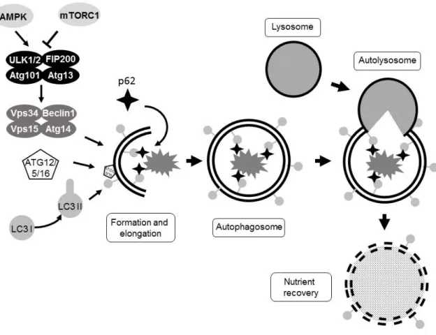
Autophagy ... 29
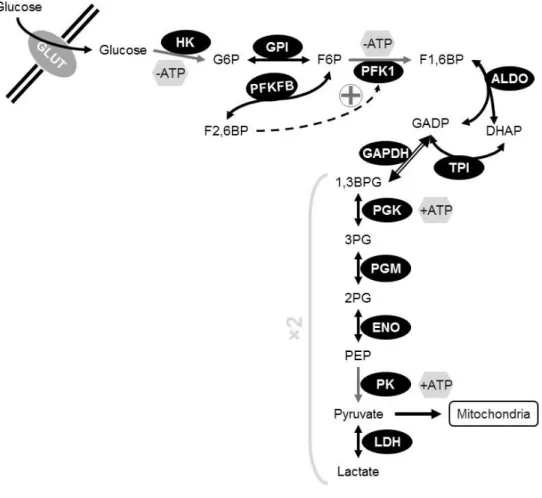
Cell metabolism: a brief overview of aerobic glycolysis ... 32
1.9 Aims ... 34
1.10 References ... 36
CHAPTER 2 ... 57
2.1 Abstract ... 59
2.3 Methods ... 61
2.4 Results ... 67
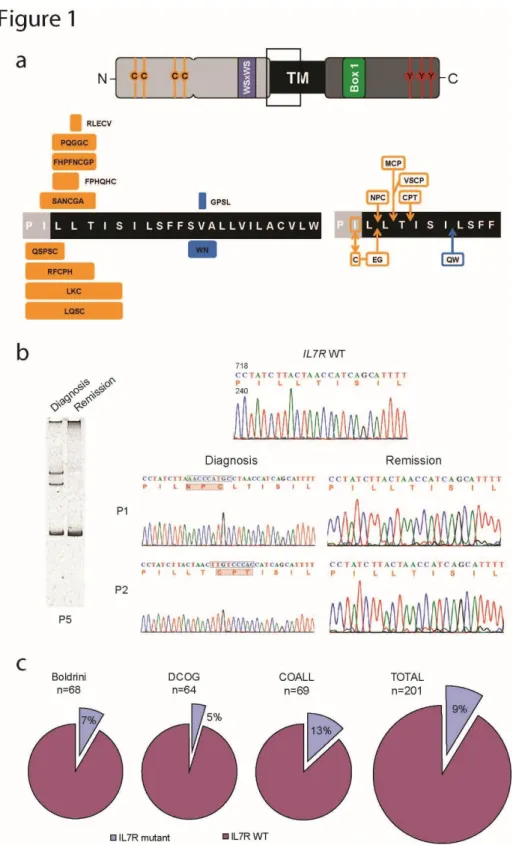
Somatic IL7R mutations in diagnostic pediatric T-ALL patient samples ... 67
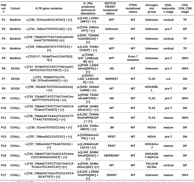
Biological and clinical features associated with IL7R mutations ... 70
IL7R mutations induce constitutive signaling, independently of IL-7, c and JAK3 ... 74
Constitutive signaling from IL7R mutants is associated with homo-dimerization/oligomerization via disulfide bond formation ... 76
IL7R mutations induce cellular transformation in vitro and promote tumor formation in vivo ... 78
Targeting IL7R mutant cells with JAK/STAT pathway pharmacological inhibitors ... 80
2.5 Discussion ... 82 2.6 Acknowledgements ... 85 2.7 Authorship contributions ... 85 2.8 References ... 86 2.9 Supplementary Data ... 89 Supplementary Figures ... 90 Supplementary References ... 108 CHAPTER 3 ... 109 3.1 Abstract ... 111 3.2 Introduction ... 112 3.3 Methods ... 113 3.4 Results ... 117
IL-7 activates the Jak/STAT5 pathway in T-ALL ... 117
STAT5 is mandatory to mediate IL-7 pro-survival, growth and proliferation effects in T-ALL cells ... 118
STAT5-dependent transcriptional network analysis of IL-7-stimulated T-ALL ... 121
IL-7 downregulates BCL6 expression in T-ALL in a STAT5-dependent manner ... 124
IL-7-dependent activation of PIM1 is required for increased survival and proliferation of T-ALL cells ... 125
3.6 References ... 133 CHAPTER 4 ... 137 4.1 Abstract ... 139 4.2 Introduction ... 140 4.3 Methods ... 142 4.4 Results ... 144
4.4.1 IL-7 regulates the expression of key metabolic pathway genes in T-ALL cells ... 144
4.4.2 IL-7 promotes glycolytic flux and early expression of glucose metabolism-related genes in
T-ALL ... 146 4.5 Discussion ... 148 4.6 References ... 151 CHAPTER 5 ... 155 5.1 Abstract ... 157 5.2 Introduction ... 158 5.3 Methods ... 160 5.4 Results ... 162
IL-7 inhibits autophagy in T-ALL in nutrient-rich conditions ... 162 IL-7-dependent activation of PI3K/Akt/mTOR pathway inhibits, whereas MEK/Erk promotes, autophagy in T-ALL cells ... 162 IL-7 relies on MEK/Erk activity and autophagy to promote survival in nutrient-poor conditions ... 167
5.5 Discussion ... 170
5.6 Acknowledgments ... 172
5.7 Authorship Contributions ... 172
5.8 Conflict of Interest Disclosures... 172
CHAPTER 6 ... 175
6.1 IL-7R signaling and leukemogenesis: a new oncogene revealed ... 178
6.2 The Jak/STAT5 pathway: novel mediators of IL-7/IL-7R effector signaling in T-cell ALL ... 179 6.3 IL-7 and T-ALL cell autophagy: a balance between PI3K/Akt/mTOR and MEK/Erk
signaling ... 181 6.4 Cell metabolism in T-ALL: does IL-7/IL-7R signaling play a role? ... 182
6.5 Novel molecular targets with therapeutic potential against leukemia ... 183
Index of figures
Chapter 1
Figure 1. Model overview of human and mouse T-cell development. 6 Figure 2. Schematic representation of major genetic alterations in
T-ALL driving survival and proliferation. 14
Figure 3. The IL-7/IL-7R signaling complex. 19
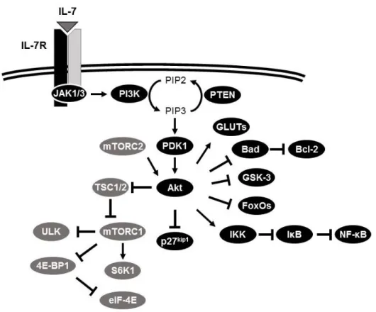
Figure 4. IL-7-mediated activation of PI3K/Akt/mTOR pathway and
potential downstream effectors. 26
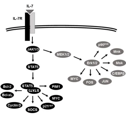
Figure 5. IL-7-mediated activation of JAK/STAT5 and MEK/Erk
pathways and potential downstream effectors. 29
Figure 6. Overview of autophagy. 32
Figure 7. Schematic view of glycolysis. 33
Chapter 2
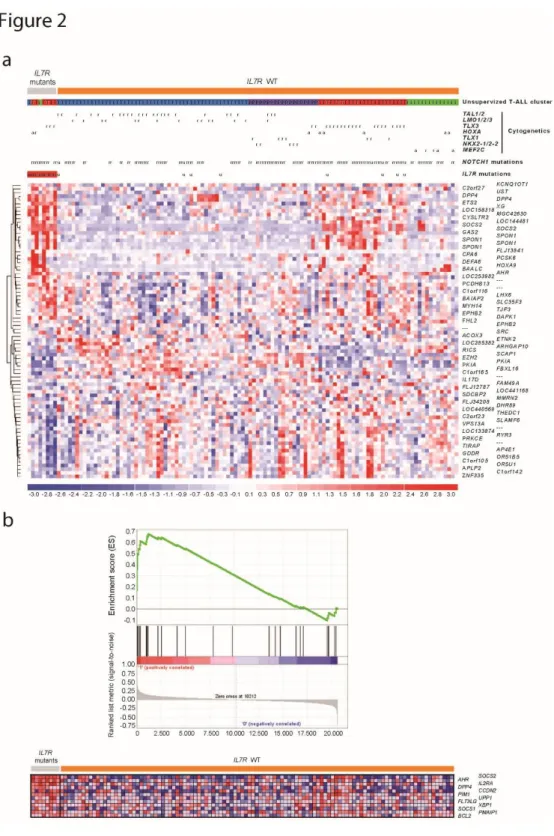
Figure 1. IL7R exon 6 somatic mutations in pediatric T-ALL. 68 Figure 2. Molecular signatures associated with IL7R mutation in
T-ALL. 72
Figure 3. IL7R mutations induce constitutive signaling in a manner that is independent of IL-7, c and JAK3 and relies on disulfide bond
promotion of homodimer formation. 75
Figure 4. IL7R mutations induce cell cycle progression, increase cell
viability, and promote growth factor independence. 77
Figure 5. In vivo tumorigenic effect of IL7R mutations. 79 Figure 6. Targeting IL7R mutants using JAK/STAT pathway inhibitors. 81
Chapter 3
Figure 1. IL-7 induces Jak/STAT5 pathway activation in T-ALL cells. 117 Figure 2. STAT5 knockdown abrogates IL-7-mediated T-ALL cell
viability and cell growth. 118
Figure 3. STAT5 inhibition abrogates IL-7-mediated T-ALL cell
Figure 4. STAT5 inhibition abrogates IL-7-mediated T-ALL upregulation of CD71, and modulation of p27kip1 and Cyclin A
expression, but not Bcl-2 upregulation in T-ALL cells. 121
Figure 5. Cross-analysis of STAT5 ChIP-seq and RNA-seq data on
IL-7-stimulated TAIL7 cells. 122
Figure 6. Quantitative PCR validation of ChIP-seq and RNA-seq data
using the S5i in TAIL7 cells. 123
Figure 7. BCL6 protein is downregulated by IL-7 and is a direct target of STAT5-mediated mRNA downregulation and alternative
transcription. 124
Figure 8. IL-7 upregulates PIM1 via STAT5. 126
Figure 9. PIM1 inhibition abrogates IL-7-mediated T-ALL cell
viability and proliferation. 127
Figure 10. PIM1 inhibition partially abrogates IL-7-mediated Bcl-2
upregulation in T-ALL cells. 128
Chapter 4
Figure 1. IL-7 promotes gene expression of multiple functional pathways, with emphasis on metabolic and sugar-related pathways in
T-ALL. 145
Figure 2. IL-7 increases glucose use and lactate production flux and promotes expression of key glycolysis-related metabolic genes in
T-ALL. 147
Chapter 5
Figure 1. IL-7 inhibits autophagy in T-ALL cells. 164 Figure 2. IL-7 dependent activation of PI3K/Akt/mTOR pathway
inhibits, whereas MEK/Erk pathway promotes, autophagy in T-ALL
cells. 165
Figure 3. Flow cytometric analysis of LC3 shows IL-7-dependent modulation of LC3 turnover by PI3K/Akt/mTOR and MEK/Erk
Figure 4. In serum-poor culture IL-7 promotes T-ALL cell viability by
MEK/Erk-dependent promotion of autophagy. 168
Figure 5. In serum-poor culture inhibition of autophagy abrogates
IL-7-mediated T-ALL cell viability. 169
Chapter 6
Index of tables
Chapter 2
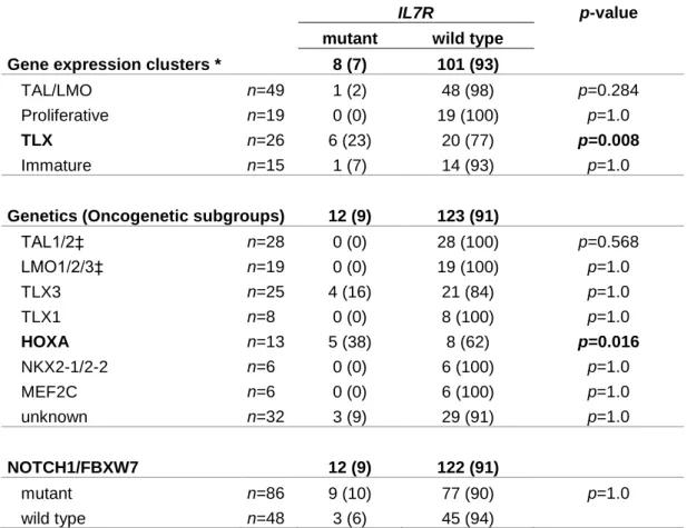
Table 1. Mutational and immunophenotypical characteristics of IL7R
mutant T-ALL patients. 69
Table 2. Association of IL7R mutations with genetic features of
CHAPTER 1
1.1 Hematopoiesis and T-cell development: a brief overview
Hematopoiesis
Hematopoiesis is the process of formation and maturation of blood cellular elements. These include red blood cells (RBCs), white blood cells (WBCs) and platelets. Postnatally and throughout life, hematopoiesis is restricted to the bone marrow (BM) [1]. However, under extreme stress it may occur extramedullary [1-4]. The hematopoietic system is very dynamic, allowing for a fine-tuned and controlled output of newly generated cells under different circumstances. For instance, increased erythropoiesis often occurs in response to hypoxia [5, 6] or malignancy [4].
The challenges posed to the hematopoietic system are met by a hierarchy of stem, progenitor and mature cells, each with a defined role. At the top of the hierarchy sits the hematopoietic stem cell (HSC), a multipotent cell type, capable of self-renewing and giving rise to all hematopoietic cell types [7-9]. During differentiation, two major branches in the hierarchy are established, the myeloid branch and the lymphoid branch [10, 11]. Whereas the later stages of cell maturation are relatively well characterized for each of the various cell types, the early stages of maturation and lineage establishment are less clear. Recent data from murine models suggest the existence of a branching point where the common myeloid progenitor (CMP) gives rise only to cells of the myeloid lineage and the lymphoid-primed multipotent progenitor (LMPP) is capable of giving rise to cells of both the myeloid and lymphoid lineage [11-15].
T-cell development
The preferred model organism to study the hematopoietic development and, in particular, T-cell development is the mouse. To date an extensive list of outs, knock-ins and humanized mouse models have been generated [11] that allow studying both human and murine T-cell development, with their many similarities and important differences.
The thymus is the organ where functional T-cells develop and mature. The thymus is seeded by different populations of precursors coming from the bone marrow which present lymphoid potential. The most studied of these precursors, in mouse and humans, are the common-lymphoid progenitor (CLP) and the LMPP [15-19]. Thymic development is a multi-step process (Figure 1). Under the influence of the thymic microenvironment, the different populations seeding the thymus undergo progressive T-cell lineage restriction,
becoming the most immature thymic cells, early-thymic precursors (ETPs), and culminating with the generation of CD4+ and CD8+ mature T-cells. As the cells progress through the developmental pathway, they acquire T-cell identity and lose the potential to generate other lineages.
The traditional classification of thymic cell populations, or subsets, is based on the expression of the co-receptors CD4 and CD8 [20]. Briefly, thymocytes begin their development as CD4- CD8- (double negative; DN) cells, progress through a CD4+ CD8+ (double positive; DP) stage and then become either mature CD4+ CD8- (single positive CD4; SP4) or mature CD4- CD8+ (single positive CD8; SP8) cells. Additionally, commitment to αβ and γδ T-cell receptor (TCR)-expressing cells occurs at the DN stage. Moreover, particularly for αβ T-cells, key events such as β-selection and positive and negative selection take place. Cells that have not yet rearranged the TCR β-chain may be referred to as pro-T cells while cells that pass β-selection until the DP stage may be referred to as pre-T cells [21, 22]. More detailed subsets have been identified for both mouse and human thymocytes, though they differ in the expression of cell surface markers between the two species (see sub-sections below) (Figure 1).
Although the main focus of this introduction is the classification of developing thymic subsets based on cell surface markers, it is unavoidable to refer that a number of growth factors and signaling pathways play key roles in thymocyte development in mouse and human. The Stem cell factor (SCF) / Kit (CD117) signaling pathway and the interleukin(IL)-7 (IL-interleukin(IL)-7) / IL-interleukin(IL)-7 receptor (IL-interleukin(IL)-7R) mostly sustain proliferation and viability at the early stages of pro-T cells [23-27]. The Wingless-related integration site (Wnt) pathway was also found to be important in sustaining proliferation of DN cells [28]. Importantly, the Notch signaling pathway is the chief element that is mandatory to establish T-cell lineage commitment and identity in both mouse and humans [29, 30].
1.1.2.1 Mouse
In the mouse, the DN1 stage (CD44+ CD25-) constitutes a broad population of cells which contains the earliest ETPs (Lineage/Lin-/low CD117hi CD44+ CD25-) capable of efficiently originating T-lineage progeny, high proliferative potential and B-/myeloid- lineage potential [16, 31, 32]. The DN2 stage (CD117hi IL-7Rα/CD127hi CD44+ CD25+) further restricts fate towards the T-cells, by loss of some myeloid- and total B-lineage potential, and still retain high proliferative potential [16, 33, 34]. In the DN3 stage (CD117 -CD44- CD25+) several important events take place. T-cell lineage commitment is completed
[20, 35]. Cells reduce proliferation and either TCR β-chain rearrangement occurs, committing to αβ-lineage, or TCR γ- and δ-chain rearrangements, committing to γδ-lineage [36]. Within the αβ-lineage, a successful β-chain rearrangement coupled with expression of pre-TCR α-chain (pTα) at the cell surface (pre-TCR) that is signaling productive, allows transition to the next stages [37]. Cells that fail to productively rearrange the β-chain die (β-selection). The TCR signal strength is powerful enough for these cells to enter a pre-TCR-dependent proliferative burst and transition to the DN4 stage (CD117- CD27+ CD44 -CD25-) and then to the DP stage where both CD4 and CD8 co-receptors are expressed [38]. The DP stage comprises ~85% of total thymocytes. In this stage cell undergo a proliferative block and rearrange the TCR α-chain [39]. Thymocytes will continue to mature if they express TCRs with the appropriate characteristics. DP cells interact with antigen presenting cells in the thymus displaying Major Histocompatibility Complex (MHC) molecules to determine their fate. If TCR signals are too weak the developing T cells do not receive enough survival signals and die by neglect. Otherwise, cells will undergo the process of positive selection [40]. Effective interaction with MHC class I will promote development of CD8 SP T-cells and with MHC class II will promote development of CD4 SP T-cells [41]. However, when TCR signals are too strong and self-reactivity may develop, cells are actively killed by negative selection [42], or under particular circumstances, CD4+ cells may develop into regulatory T-cells (Treg) [43].
1.1.2.2 Human
Human thymopoiesis shares many similarities with the murine counterpart. Key events such as T-cell lineage commitment and β-selection during the DN stage, negative and positive selection and CD4/CD8 SP lineage commitment at the DP stage are largely similar. However, DN subset classification based in the murine CD44 vs CD25 expression does not have the same representation in human thymocytes [44]. The early T-cell precursors that seed the human thymus are CD34+ CD38- CD1a- (DN1). Cells then upregulate CD38 (CD34+ CD38+ CD1a-; DN2), followed by CD1a (CD34+ CD38+ CD1a+; DN3) [45, 46]. Analysis of gene expression profiling and TCR gene rearrangements suggests that an overlap between mouse and human DN stages of development can be established. CD34+ CD38- CD1a -resemble mouse DN1/ETP, CD34+ CD38+ CD1a- the mouse late DN1/DN2 and CD34+ CD38+ CD1a+ mouse DN3 [46]. CD1a upregulation is strongly correlated with T-cell lineage commitment [47] and β-selection can occur as early as this stage [46]. Acquisition of CD7
-of CD2 and CD5 expression at the CD34+ CD38+ CD1a- stage [48]. Cells then lose expression of stem cell marker CD34, progressively gain CD4, CD8 and surface CD3 to become DP thymocytes (CD3+ CD4+ CD8+), which, after TCR α-chain rearrangement, undergo positive and negative selection, to become either mature CD4 or mature CD8 SP T-cells [44]. Acquisition of maturity is accompanied by loss of CD38 and CD1a expression [49, 50].
Figure 1. Model overview of human and mouse T-cell development. Precursors migrate from the
bone marrow to the thymus. Thymic T-cell development starts at the double negative (DN) stage. It progresses to the double positive (DP) and later to the single positive stage (SP). Important surface markers are represented for each stage. The β-selection and positive and negative selection events are indicated. Further details in the text.
1.2 Acute Lymphoblastic Leukemia (ALL)
Hematopoiesis is regulated by numerous factors. For instance, T-cell development is under the control of survival, proliferative and differentiation signals, such as those elicited by IL-7, Notch or TCR, and determined by recombination-activating gene (RAG)-mediated DNA double-strand break activity during TCR maturation [44, 51]. Although the process is tightly monitored, developing precursors are at risk of transformation. Malignant transformation of cells of lymphoid origin will result in leukemia or lymphoma.
Epidemiology and causes
ALL is the most common childhood cancer, accounting for 26% of the cases. It is more common in males than in females and more prevalent in white than black children [52, 53]. There is evidence that ALL may develop in utero. Studies in identical twins show leukemias with identical genetic rearrangements [54-56]. Additionally, analysis of neonatal blood spots showed the presence of leukemic genetic lesions before the diagnosis of ALL [57, 58]. Despite this, the peak of incidence is characteristically at ages 2 to 4 [52].
The exact causes for ALL are not clearly known, although some genetic conditions associate with predisposition for leukemia development. ALL has increased risk in genetic disorders such as Down syndrome [59], Fanconi anemia [60], Bloom syndrome [61], neurofibromatosis [62], and ataxia-telangiectasia [63].
There is also evidence that non-genetic factors may increase the risk of ALL development. For example, ionizing radiation exposure (e.g. during medical treatment or atomic disasters) was shown as an important physical factor contributing to increased ALL risk [64-66]. Infectious agents (or abnormal responses against them) have also been postulated to contribute to ALL [67, 68]. Other factors include exposure to environmental pesticides, parental smoking and diet of the mother. Secondary leukemia as consequence of cancer therapy is more associated with the development of acute myeloid leukemia (AML) than ALL [67, 69].
Biological characteristics
ALL is characterized by an abnormal accumulation of immature lymphoid cells, or blasts, arrested in their development and bone marrow involvement superior to 20%. Presence of masses in other organs and peripheral blood involvement may vary [70]. ALL originates from malignant clones of B- or T-cell lineage, and the origin is believed to be in
the BM or thymus [71]. ALL is therefore a heterogeneous cancer with combined morphologic, immunologic, cytogenetic and molecular genetic characteristics [72].
Morphological and cytochemical characteristics per se have limited ALL sub-classification value and usage is mostly applied to distinguish ALL from AML [73]. ALL blast population cells tend to be small, homogeneous, with a central large nucleus, fine chromatin and scant cytoplasm. Cytochemical analysis of myeloperoxidase, acid phosphatase and periodic-acid Schiff (PAS) stainings, help complementing the diagnostics [74].
Immunophenotyping by flow cytometry and cytogenetic analysis of DNA lesions constitute the gold standard of ALL classification and sub-typing. In childhood, around 85% of cases present with a B phenotype and 13-15% present with a T phenotype. In adults, around 75% have a B phenotype and 25% are of T-cell origin [75]. In our studies, we adopted the criteria of the European Group for Immunological Characterization of Leukemias (EGIL) [76], which correlate the immunophenotype at which the leukemia cells are arrested with that of normal developing lymphocyte precursors. In the case of T-cell leukemia, 4 groups are recognized: pro-T or T-I (cytoplasmic CD3+, CD7+), pre-T or T-II (cCD3+ CD7+ CD2+ and/or CD5+), cortical-T or T-III (cCD3+, Cd1a+) and mature-T or T-IV (CD3+, CD1a-). Recently, a novel sub-type was identified, early T-cell precursor ALL (ETP-ALL), which is defined as CD1a-, CD8-, CD5low/- and expressing at least a myeloid or stem cell surface marker [77].
ALL often presents cytogenetic abnormalities involving numeric and structural chromosomal changes. A comprehensive study of cytogenetics showed that cytogenetic features have biological and prognostic significance [78]. ALL can be classified under 5 major modal groups: diploid (46 chromosomes, no evident structural abnormalities; 31-40%), high hyperdiploid (>50 chromosomes; 23-26%), low hyperdiploid (47-50 chromosomes; 10-11%), pseudodiploid (46 chromosomes with structural abnormalities; 18-26%), hypodiploid (<45 chromosomes; 6%). Regarding translocations, ALL may be divided according to the Lund Chromosomal Group as: t(9;22)(q34;q11.2) or a Philadelphia chromosome (Ph+); t(4;11)(q21;q23); t(8;14)(q24;q32) or del(8q); other 14q+ abnormalities; del(6q). The classification recognizes 10 groups, being the remaining related to modal chromosome number [78, 79]. Although these findings are useful for predicting clinical outcome and response to treatment, they are not totally accurate. For example, up to 20% of children with favorable genetic features (TEL-AML1 fusion and hyperdiploidy >50 chromosomes) will eventually relapse, although a third of those with high-risk abnormalities
(the Philadelphia chromosome with BCR-ABL fusion and the t(4;11) with MLL-AF4 fusion) can be cured with chemotherapy alone [80]. This fraction is currently even higher due to the introduction of tyrosine kinase inhibitors such as Imatinib and Dasatinib [81-83]. Additionally, genetic factors intrinsic to the individual (e.g. drug-metabolizing enzyme polymorphisms), rather than those acquired by the leukemic cell, may have an important impact on treatment outcome [84].
Symptoms and treatment
Most clinical symptoms of ALL relate to the collapse of normal hematopoiesis. The common clinical signs include fatigue and lethargy due to developing anemia. Bleeding and excessive bruising occurs due to thrombocytopenia. Neutropenia may lead to predisposition to infections and fever. Thymic masses may lead to shortness of breath and superior vena cava syndrome. Tumor spread to the meninges may result in headaches and central nervous system (CNS) involvement [85].
Survival rates of children with ALL has improved dramatically across the decades. In the 1960s, 5-year survival rate was 10%, whereas currently it reaches up to 90% (85% of event-free survival) [86]. These great improvements were built on top of significant advances such as those observed in the biological characterization of ALL, development of more effective drugs and risk-adjusted multi-agent therapy [86, 87]. In adults, however, treatments have been less successful, classically only achieving ~40% of 5-year event-free survival [75]. Lately, however, the application of intensive chemotherapy pediatric protocols on adult T-ALL patients was able to improve 7-year event free survival to 63% [88]
The current treatment approach for ALL includes 3 main phases. A remission induction phase, an intensification (consolidation) phase and continuation long-term treatment [87]. During the remission-induction phase the main objective is to reduce leukemia burden and restore normal hematopoiesis. Therapy includes administration of glucocorticoids (prednisone or dexamethasone), vincristine and at least another agent (asparaginase or anthracycline). Exceptionally, for high risk ALL, regimens include 4 or more drugs [87]. The intensification (consolidation) treatment happens after restoration of hematopoiesis. This phase will deal with possible drug-resistant leukemic cells and decrease the chance of relapse [86]. Treatment regimens may vary, and have different degrees of success, but often include reinduction therapy, high doses of methotrexate and mercaptopurine, and pulses of vincristine and corticosteroid plus high-dose asparaginase
transplantation, which is especially beneficial to very-high risk patients [93, 94]. During continuation treatment, regimens are adjusted for long-term tolerance. Mercaptopurine and methotrexate are regularly used in this phase [87]. Increased risk of relapse to the CNS associate with factors such as T-cell immunophenotype, hyperleucocytosis, high-risk genetic abnormalities, and presence of leukemic cells in cerebrospinal fluid. These cases require particular attention to CNS-directed treatment [87, 95-97].
Although the success of the treatments is evident, relapses still occur and aggressive treatments frequently impose severe long-term side effects. Side effects include osteoporosis [98], osteonecrosis [99], thrombocytic complications [100], secondary tumors [101], cardiac dysfunction, along with others [102, 103]. Therefore, a continuous effort is required to develop new, less toxic drugs and therapeutic strategies with improved efficacy against leukemic cells and less side effects. To achieve this, it is indispensable to investigate and improve the knowledge of the etiology and biology of leukemia.
1.3 T-cell Acute Lymphoblastic Leukemia (T-ALL)
T-ALL is a subtype of ALL, characterized by the emergence of malignant immature blasts of T-cell origin arrested during development. T-ALL often presents with higher risk factors such as high white blood cell counts (WBC; >50000/μL), older age, mediastinal mass and enlargement of the spleen, liver, and lymph nodes [72]. Historically, T-ALL cases had higher risk and poorer prognosis than B-ALL cases [104-106]. However, improved protocols, based on risk-adjusted chemotherapy, improved the outcome of T-ALL patients to such an extent that currently ALL patients with a T-cell phenotype benefit from better 5-year disease-free survival for children (up to 78%) [86] and adults (65%; 7-5-year survival) [88, 107]. Relapses still occur in approximately 25% of the cases, and T-cell phenotype is associated with poorer prognosis after relapse [86, 108, 109].
1.4 Genetic abnormalities in T-ALL
The malignant transformation of healthy thymocytes into T-cell leukemia is believed to be a progressive, multi-step, process where several cell-autonomous mechanisms accumulate to promote a proliferative and survival advantage and a differentiation block to pre-malignant cells, which associates with abnormal signaling and eventually results in leukemia. Those factors range from point and small mutations, to epigenetic changes and to
large chromosomal alterations. Moreover, the microenvironment often impinges on those alterations by promoting and complementing an already signaling-aberrant cell [110-112].
TCR loci-associated chromosomal translocations
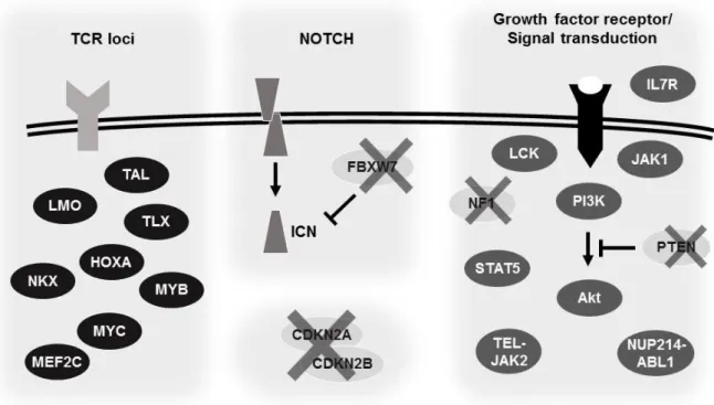
In T-ALL, non-random chromosomal translocations involving the juxtaposition of promoter and enhancer elements of TCR gene loci (TCA@ 14q11, TRB@ 7q34-35, TRG@ 7p15, TRD@ 14q11) and a developmentally important transcription factor are common, found in ~35% of the cases [113] (Figure 2). This leads to unregulated over-expression of the translocated gene and subsequent differentiation blockade [114]. Different families and groups of transcription factors are involved in these abnormalities. Importantly, several groups have classified T-ALL into distinct oncogenetic subgroups that are characterized by rearrangements and aberrant expression of transcription factors and share a similar gene expression profile [115-118]. Major groups include the basic helix-loop-helix (bHLH) family members (SCL/TAL1, TAL2, LYL1 and bHLHB2), the LIM-domain only (LMO) family (LMO1, LMO2, LMO3), several homeobox family members (HOX11/TLX1, HOX11L2/TLX3, NKX2.1, NKX2.2, NKX2.5, HOXA@ cluster) and proto-oncogenes such as TAN1 (truncated from of NOTCH1), MYC, MYB and MEF2C (reviewed in [119]).
Cell Cycle regulators
Cell cycle deregulation is a hallmark of cancer. Deregulation may occur by increased activity/expression of cell cycle promoters, inactivation of cell cycle blockers, or both (Figure 2). The most common occurrence in T-ALL is the deletion of the
CDKN2A/CDKN2B locus on chromosome 9p21, present in more than 70% of cases [120,
121]. These genes code for the inhibitors of cyclin-dependent kinase (CDK) 4 proteins (p16INK4a and p15INK4b, respectively), thus disrupting the cyclin-CDK complexes [122, 123]. Deletion of these genes will lead to phosphorylation and inactivation of the retinoblastoma protein (Rb), thus promoting cell cycle progression [124]. Additionally, the CDKN2A locus, via an alternative reading frame, also codes for p14ARF protein, a negative regulator of HDM2 [125]. Loss of p14ARF will lead to p53 downregulation by allowing HDM2 to promote p53 degradation. Consequently, a decrease in p53 activity, decreases p21Cip1 expression, which does not allow proper DNA repair during cell cycle, leading to accumulation of DNA damage [125, 126].
NOTCH1
NOTCH1 is a gene that has a major role in hematopoiesis. It regulates maintenance of
stem cells [127] and is required for T-cell lineage specification [30, 128].
NOTCH1 is also one of the most frequently altered genes in T-ALL (Figure 2). Its role
in leukemia was first described with the involvement in the translocation t(7;9)(q34;q34.3), but this is a uncommon event (~3% of T-ALL cases) [129]. However, the magnitude of
NOTCH1 importance in cancer was only revealed later on, when it was found that >50% of
T-ALL cases had activating mutations in the gene [130]. Although the mechanisms vary,
NOTCH1 translocations or mutations will result in the accumulation of the intracellular,
activated form of NOTCH (ICN) [131]. In addition, mutations in the F-box/WD repeat-containing protein 7 (FBXW7) gene, found in ~15% of T-ALL patients [132, 133], lead to the stabilization, and consequent increase in activity, of the ICN protein.
Signal transduction elements
During thymopoiesis, both pre-TCR and TCR engagement is required for normal T-cell development. The TCR complex activates a cascade of multiple signaling pathways including the rat sarcoma/ mitogen associated protein kinase (Ras/MAPK), the phosphatidylinositol-3-kinase/ protein kinase B (PI3K/PKB(Akt)) and the phospholipase C γ/ calcineurin (PLCγ/calcineurin) pathways [134, 135]. These pathways are also recruited by pre-TCR signaling transduction.
In T-ALL, multiple signaling components are targets of mutations or translocations (Figure 2). The Src family protein tyrosine kinase lymphocyte-specific protein tyrosine kinase (Lck) is central in TCR signaling [136]. Although rare, ectopic expression of LCK can occur in T-ALL due to the t(1;7)(p34;q34) translocation [137].
The Abelson murine leukemia viral oncogene homolog 1 (ABL1) is a downstream target of Lck [138]. Various ABL1 rearrangements occur in T-ALL. The famous BCR-ABL1 fusion gene, though present, is uncommon [139]. The most common rearrangement is the NUP214-ABL1 (~6%) [140].
The Ras pathway also suffers from mutations. Activating NRAS mutations occur in 4-10% [141]. Additionally, deletion or inactivating mutations in the Neurofibromin 1 (NF1) gene, a negative regulator of the Ras pathway, are found in 3% of the cases [142].
1.4.4.1 Alterations in IL-7/IL7R-related signaling mediators
Altogether, a major fraction of T-ALL cases presents alterations in two major signaling pathways, the PI3K/Akt and the Janus kinase/ signaling transducer and activator of transcription (Jak/STAT) pathways. Both are critical for IL-7R-mediated function in normal thymocytes and T-ALL (discussed later on) [143-145].
The PI3K/Akt pathway is the target of mutations in T-cell leukemia. The most common are in the phosphatase and tensin homolog (PTEN). PTEN is a tumor suppressor and the major negative regulator of the PI3K/Akt pathway [146]. It is mutated in T-ALL (5-30% of the cases) due to non-sense and frameshift mutations and gene deletion occurs in 10% of the cases [147]. Mutations in PI3K family members are uncommon in T-ALL, although gain-of-function mutations in PIK3CA (p110α) and inactivating mutations in
PIK3R1 (p85α) have been reported, each in 5% of the T-ALL cases analyzed. AKT mutations
are even less frequent (around 2% of T-ALLs) [147].
The Jak/STAT pathway is also a target for mutation in T-ALL. The oldest known alteration is the ETV6(TEL)-JAK2 fusion due to the translocation t(9;12)(p24;p13) [148]. Somatic JAK1 gain-of-function occur mostly in adult T-ALL (18% of the cases) and more rarely in pediatric T-ALL (2% of the cases) [149, 150]. JAK3 mutations are found in both adult (12% of the cases) [151] and pediatric (7%-25% of the cases) T-ALL [150, 152]. More recently, STAT5B gain-of-function mutations have been mutations have been reported in 8% of the patients [153, 154].
Other important alterations
Alterations on the MYB locus found in T-ALL, which lead to protein overexpression, include the chromosomal translocation t(6;7)(q23;q32), associated with high expression of proliferation and mitotic genes [155], and duplication of the MYB locus [156].
Mutations in chromatin remodeling genes that ultimately benefit T-cell leukemia progression have been reported to occur in EZH2, SUZ12 [157] and PHF6 [158].
Lastly, inactivating mutations in phosphatases other than PTEN were found recently in PTPN2 [159] and PTPRC (CD45) [160] genes.
Figure 2. Schematic representation of major genetic alterations in T-ALL driving survival and proliferation. Genetic abnormalities are grouped according to their nature. Represented are common
translocations involving TCR loci; NOTCH activating mutations and FBXW7 inactivation; deletion of cell cycle regulators; and mutations in signaling transduction elements and growth factor receptors. Ovals represent either gene families (e.g. NKX) or individual genes (e.g. CDKN2A). The cross over a gene indicates inactivation of the gene or protein. Detailed information is in the text. TCR, T-cell receptor; TAL, T-cell acute lymphoblastic leukemia gene family; LMO, LIM-domain only gene family; TLX, T-cell leukemia homeobox gene family; HOXA, Homeobox A gene cluster; NKX, NK2 homeobox gene family; MEF2C, Myocyte specific enhancer factor 2C; ICN, Intracellular Notch FBXW7, F-box/WD repeat-containing protein 7; CDKN2A/2B, Cyclin-dependent kinase inhibitor 2A/2B; IL7R, Interleukin-7 receptor; JAK, Janus kinase; LCK, Lymphocyte-specific protein tyrosine kinase; PI3K, Phosphatidylinositol-3-kinase family; PTEN, Phosphatase and tensin homolog AKT, also known as protein kinase B (PKB); STAT, Signal transducer and activator of transcription; TEL, also known as ETS variant 6 (ETV6); NUP214, Nucleoporin 214; ABL1, Abelson tyrosine-protein kinase.
1.5 Microenvironment in T-ALL
A tumor is not a homogeneous entity consisting purely of malignant cells entirely self-sufficient. In contrast, it is highly heterogeneous containing both malignant and non-malignant cells of several origins, as well as components such as extracellular matrix and secreted factors. Together, all elements that constitute the tumor microenvironment interact, where the final consequence is to the benefit of the cancer cells [161, 162]. The microenvironment provides cancer cell survival, protects from chemotherapy and may support metastization [112, 162-164]. The studies on the involvement of the
microenvironment in T-ALL have focused mostly in the bone marrow niche, since this is a major site of leukemia burden.
Cell-to-cell contact
Cell adhesion molecules are expressed in T-ALL cells, such as very late antigen 4 / vascular cell adhesion protein 1 (VLA-4/VCAM-1) and lymphocyte function-associated antigen 1 / intercellular adhesion molecule 1 (LFA-1/ICAM-1) [165]. T-ALL cells cultured in BM stroma have increased survival dependent on LFA/ICAM-1 interactions [166]. Interestingly, and in contrast with B-ALL, in vitro survival of T-ALL cells on BM stromal cultures appears to correlate with better patient outcome [167]. As mentioned above, the Notch1 receptor is commonly mutated in T-ALL, originating ligand-independent activation of the pathway. However, there is still room for the canonical ligand-dependent Notch signaling to play a role in T-ALL pathogenesis. Blocking of delta-like ligand 4 (Dll4), a Notch ligand, or of the Notch1/2/3 receptors themselves impairs T-ALL growth in vivo [168] and T-ALL cell escape from dormancy is associated with Notch3-Dll4 interaction in the microenvironment [169]. It is also noteworthy that PTEN deficient T-ALLs are sensitive to disruption of Notch1-Dll4-dependent signaling [170].
Secreted factors: chemokines
Other than cell-to-cell interactions, cytokines, growth factors and chemokines provide extra means of intercellular communication and behavior conditioning. The whole immune system, including T lymphocytes, is orchestrated by a network of cytokines and chemokines [171]. The stromal cell-derived factor 1 (SDF-1/CXCL12) interaction with its receptor C-X-C chemokine receptor type 4 (C-X-CXC-X-CR4) was shown to be important for B-ALL homing to bone marrow [172]. More recently, two studies implicated the CXCL12/CXCR4 axis in migration, maintenance and leukemia initiating cell (LIC) activity in human T-ALL xenograft models [173, 174].
Other chemokine signaling elements have also been involved in T-ALL pathogenesis. Particularly, Buonamici and colleagues [175] found that Notch-regulated expression of C-C chemokine receptor 7 (CCR7) in T-ALL and consequent C-C motif ligand 19 (CCL19)/CCR7 signaling was a major regulator of T-ALL infiltration to the CNS. In addition, signaling of the C-C motif ligand 25/ C-C chemokine receptor 9 (CCL25/CCR9) or C-X-C motif ligand 13/ 5 (CXCL13/CXCR5) were shown to contribute to T-ALL cell
Secreted factors: cytokines and growth factors
Multiple cytokine and growth factors have been implicated in supporting T-cell leukemia. For instance, secretion of IL-18 by the stromal cells upon treatment with mitogen-activated protein kinase kinase (MAPKK/MEK) inhibitors, led to increased T-ALL cell proliferation, suggesting that stroma-leukemic cell cross-talk may provide a protective niche against drug therapy [180].
Also, activation of the insulin-like growth factor 1 receptor (IGF1R) in T-ALL cells is associated with increased LIC activity [181] and growth support from tumor-associated dendritic cells (DCs) [182].
Transforming growth factor β (TGF-β) is a multifunctional cytokine involved in a variety of processes such as cell growth inhibition, cellular senescence, differentiation and apoptosis [183]. The major effectors of TGF-β signaling are Smad2 and Smad3, which directly regulate gene expression [184]. In normal hematopoiesis TGF-β acts as a negative regulator [185]. In T-ALL its role is less explored. Of note, a fraction of primary T-ALL cells do not express Smad3 protein, although they still display non-mutated and normal levels of the Smad3 gene (MADH3) mRNA [186]. Additionally, studies in mice suggest that loss of Smad3 can synergize with other oncogenic events, such as the loss of p27kip1, to promote T-cell leukemogenesis [187].
1.5.3.1 The γ-common chain (γC) family of cytokines
The members of the γC family of cytokines all bind to receptors that share the γC subunit (IL-2Rγ/CD132), along with one or more specific subunits, to transduce signals. The cytokine family includes IL-2, IL-4, IL-7, IL-9, IL-15 and IL-21 [188], all of which have some role in T-cell development, function or homeostasis (reviewed in [188] and [189]). In T-ALL, it was demonstrated that IL-2, IL-4, IL-7, IL-9 and IL-15 are able to promote proliferation of primary samples in vitro, with IL-7 having the most potent effect [190]. Interestingly, synergistic roles in proliferation were observed upon incubation with specific combinations of two γC cytokines [190]. IL-21, a more recently discovered γC cytokine, has not been tested to date in T-ALL. Nonetheless, given that it supports cell proliferation in other T-cell malignancies [191, 192] and the consistency of the other γC cytokines in promoting T-ALL proliferation, it is tempting to speculate that the effect of IL-21 in T-ALL should be similar.
1.6 The IL-7/IL-7R complex
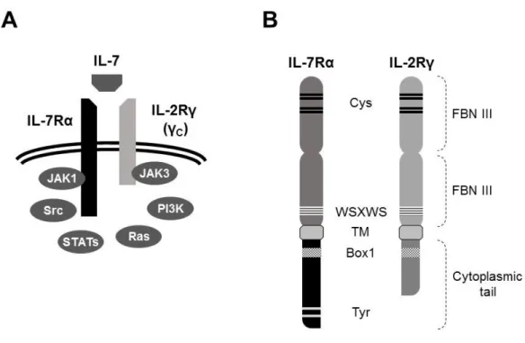
The IL-7R is a heterodimer consisting of the IL-2Rγ/γC, shared between the γC family of cytokines, and the IL-7Rα (CD127), shared between IL-7 and thymic stromal lymphopoietin (TSLP) [145, 193]. The heterodimerization of γC and IL-7Rα upon IL-7 binding gives the specificity to IL-7 [194, 195] and is required for receptor activation (Figure 3A) [196].
The γC subunit
As mentioned above the IL-2Rγ (γC/CD132) subunit is required for signaling of IL-2, IL-4, IL-7, IL-9, IL-15 and IL-21 [188]. The γC gene (IL2RG) is located in the X chromosome (Xq13.1) [197, 198], and inactivating mutations often result in X-linked severe combined immunodeficiency (X-SCID). In humans the X-SCID results in a T- B+ NK -phenotype, though B-cells are non-functional [194, 198]. In mice, the phenotype is more severe resulting in T- B- NK- phenotype [199, 200]. It is important to mention that Jak3-deficient mice have a phenotype that closely resembles that of γC loss [201-203].
The γC receptor belongs to the type I cytokine receptor family and the mature human protein has 374 aminoacid residues (Figure 3B). The extracellular domain possesses two tandem fibronectin type III domains that include two pairs of the conserved cysteine residues, characteristic of the family. A tryptophan-serine-X-tryptophan-serine (WSXWS) motif exists close to the transmembrane domain [204, 205]. The γC, as all type I cytokine receptors, does not possess endogenous tyrosine kinase activity, instead it relies on Jak family tyrosine kinases for signal transduction. The intracellular portion possesses, proximal to the transmembrane domain, a Box motif required for Jak3 binding and activation. The short cytoplasmic tail is apparently not directly involved in transducing downstream signaling [204, 206-208].
The IL-7Rα subunit
The IL-7Rα gene (IL7R) is located on the chromosome 5p13.2 and is composed of 8 exons. Exon 6 codes for the integral transmembrane domain. The canonical transcript is 4619 nucleotides long. Alternative splicing generates a soluble isoform lacking exon 6 and introducing a premature stop codon [209, 210]. Inactivating mutations on the IL7R gene result in a type of SCID. In humans, the SCID results from a T- B+ NK+ phenotype [26, 211, 212]. Mouse deficient in Il7r have impaired T- and B-cell development [24]. Importantly,
the presence of B-cell in human individuals and not in mice advocates for important differences in lymphopoiesis in both species. Notably, the phenotype of murine IL-7Rα deficiency is more severe than IL-7 deficiency. This has been attributed to be a consequence of TSLP signaling [213].
The IL-7Rα subunit is a type I cytokine receptor (Figure 3B). As such, it shares many structural similarities to the γC subunit described above. The mature form is 439 amino acid residues long. In the extracellular portion, it has two fibronectin type III domains, two paired cysteine residues and a WSXWS motif. The cytoplasmic tail contains a Box1 motif required for Jak1 binding and activity [205]. Additionally, the cytoplasmic tail contains at least 2 tyrosine residues (Y401, Y449) that have been shown to play a role in the activation of downstream signaling pathways [214-217].
As already mentioned, the IL-7Rα chain is also shared with the thymic stromal lymphopoietin/ cytokine receptor-like factor 2 (TSLPR/CRLF2) receptor for TSLP signaling [218, 219].
Interleukin-7
The IL-7 gene (IL7) is encoded in chromosome 8q12-13 [220] and requires glycosylation to be fully active [221]. Human and murine IL-7 share 65% aminoacid identity [222]. Human IL-7 can stimulate murine cells [223] and, conversely, murine IL-7 can stimulate human cells [224], although possibly with less potency in vivo [225]. IL-7 is produced by the stromal cells of the BM and thymus and by lymphatic endothelial cells [226-230]. IL-7 is a soluble factor, nevertheless it has been observed that it can bind to extracellular matrix (ECM)-associated glycosaminoglycan, heparan sulfate, and to fibronectin [231-233]. Similar to its receptor, IL-7 is essential for normal B- and T-cell development in mice [25].