R E S E A R C H
Open Access
Potentiation of a novel palladium (II) complex
lethality with bee venom on the human T-cell
acute lymphoblastic leukemia cell line (MOLT-4)
Zahra Safaeinejad, Mohammad Nabiuni
*and Zahra Nazari
Abstract
Background:Although honeybee venom (BV) has been reported to induce apoptosis in different types of cancerous cells, its synergistic effects with customary anti-cancer drugs remain largely unknown. In the present study, we evaluated the cytotoxic effect of BV alone (as a natural product) and the synergistic cytological effects of this component in combination with [Pd (bpy) (Pi-Pydtc)]NO3–a novel palladium complex on human T-cell lymphoblastic leukemia cells. To investigate the cytotoxic effect of the BV alone and in combination with palladium complex on MOLT-4 cells MTT assay was performed. In order to determine the apoptotic effects of BV separately and in combination with Pd (II) complex on these cells and its ability to induce apoptosis, morphological examination, flowcytometric analysis and caspase-3 colorimetric assay were done.
Results:We found that BV induced morphological changes, namely nuclear shrinkage, and inhibited MOLT-4 cell proliferation; both effects were dose- and time-dependent. Flow cytometry by Annexin-V antibody demonstrated that BV induced apoptosis in MOLT-4 cells. Furthermore, BV induced apoptosis independently of caspase-3 in these cells. In addition, we proved a clear synergistic effect of BV on [Pd (bpy) (Pi-Pydtc)]NO3. The apoptotic pathway activated by BV in combination with Pd complex was caspase-3-dependent.
Conclusions:These observations provide an explanation for the anti-proliferative properties of BV, and suggest that this agent may be useful for treating lymphoblastic leukemia alone or in combination with chemotherapy drugs pending further investigations on animal models as preclinical tests.
Keywords:Apoptosis, Bee venoms, Cytotoxicity, MOLT-4 cell line, Pd (II) complex
Background
Bee venom is a natural substance that contains only 0.1μg of dry venom [1]. The dry venom has a very com-plex mixture of such active peptides as melittin, apamin and adolapine, enzymes including hyaluronidase and phospholipase A2, biologically active amines such as his-tamine and epinephrine as well as non-peptide compo-nents with numerous medicinal properties [2]. Melittin, a hemolytic and strong cardiotoxic peptide, is the major active ingredient of BV. This main constituent of bee venom has been reported to induce apoptosis, and to produce anti-tumor effects [3,4]. Melittin, which makes up 50-60% of the dry venom, is a low-molecular-weight
protein (2846.46 Da), which is composed of 26 amino acids. It is found as a tetramer in the poison sac of the bee, but when influencing a cell, it acts as a monomer [5].
BV has been used as a traditional medicine to treat various diseases such as arthritis, rheumatism, back pain and skin diseases [2]. Besides, recent studies have reported that BV causes growth arrest and exerts cyto-toxic effects on various types of cancerous cells [6-11]. The cytotoxic effects mediated through the activation of PLA2 by melittin have been suggested to be the critical mechanism for the anti-cancer activity of BV [12].
It is well documented that induction of apoptosis is the most effective strategy by which anti-cancer agents target cancer cells [13]. Chemotherapy agents can induce apoptosis signaling through two major pathways. One is the mitochondrial (intrinsic) pathway and the other one is the death receptor (extrinsic) pathway. Cascading
* Correspondence:[email protected]
Department of Cell and Molecular Biology, Faculty of Biological Sciences, Kharazmi University, Tehran, Iran
intrinsic pathway activation of certain molecules finally provokes activation of downstream caspase-3, which is one of the key agents of apoptosis. Activated caspase-3 cleaves a wide array of substrates, such as poly(ADP-ri-bose) polymerase (PARP), a DNA repair enzyme, and in-evitably leads to cell death [14,15].
Cisplatin (cis-diammine dichloroplatinum II) is one of the most remarkable drugs which is used separately or in combination with other chemotherapy agents to treat different types of tumors [16,17]. Despite the success of cisplatin and platinum-based drugs, they have presented serious clinical side effects [18,19]. Therefore, much effort has been focused on identifying novel anti-tumor agents and examining new approaches to increase their damage to tumor cells at a lower concentration than conventional chemotherapy drugs [20].
The significant similarities between the coordination chemistry of palladium (II) and platinum (II) compounds have generated lines of research on Pd (II) complexes as anti-tumor components [21]. Recently we stated at the FAOBMB conference that [Pd (bpy) (Pi-Pydtc)]NO3, as a
novel palladium complex designed and synthesized by our research group, exerts clear anti-tumor effects on human lymphoblastic leukemia MOLT-4 cells [22].
In the present study, we first examined the cytotoxic effect of BV on the MOLT-4 cancerous cell line, then the synergistic effects of BV and the novel Pd (II), [Pd (bpy)(Pi-Pydtc)]NO3, on these cells. This investiga-tion employed the following techniques: MTT assay, morphological analysis, flow-cytometry assay and the caspase3 activity assay.
Methods
Bee venom collection and novel Pd (II) complex preparation
Venom from the Iranian honey bee (Apis mellifera) was prepared by placing bees on a 6-mm wire grid, which was electrically pulsed. The bees then produced venom that dropped onto a glass slide, from which it was col-lected and freeze-dried according to the method of Lariviere and Melzack [23], whereas the novel complex of the Pd (II) was designed and synthesized by our re-search group [24].
Cell culture
The human T-cell acute lymphoblastic leukemia MOLT-4 cells were purchased from the Pasteur Institute (Tehran, Iran). Cells were maintained in RPMI-1640 medium (Gibco, UK) and supplemented with 10% fetal bovine serum (Gibco, UK), penicillin at 100 units/mL, and streptomycin at 100μg/mL, in a humidified incuba-tor filled with 5% CO2 at 37°C. The medium was
re-placed every 48 hours.
MTT cytotoxicity assay
In order to determine the cytotoxic effects of BV separ-ately and in combination with Pd (II) complex on the MOLT-4 cells, cell viability was tested by MTT (3[4, 5-dimethylthiozol-2-yl]-2,5-diphenyl tetrazolium brom-ide) assay. The cells were first seeded into 24-well cul-ture plates (Nunc, Denmark) at a density of 1.0 × 10⁵ cells/mL and subsequently incubated in a humidified 5% CO2 environment for one hour. The cells were then
treated with BV at 1, 3, 6 and 8 μg/mL for 24 and 48 hours, the concentrations chosen as a result of pre-cipitation of the BV in the medium. Non-treated cells were used as controls.
MTT (100 μL of 5 mg/mL) (Sigma, USA) was added to each well and incubated at 37°C for four hours. The dark blue crystals were dissolved by adding 1000 μL of 0.04 M HCl/isopropanol. After an overnight incubation in darkness, optical density (O.D.) was read at a wave-length of 570 nm using a spectrophotometer. The O.D. values of the experimental groups were divided by those of the untreated control group, and the results were presented as the percentage of cell viability.
By calculating the minimum BV dosage that killed MOLT-4 cells, we exposed cells to the lowest lethal dos-ages of BV and Pd (II) complex simultaneously [1μg/mL BV/0.85 μM Pd (II) complex, 3 μg/mL BV/0.85 μM Pd (II) complex and 6.3 μg/mL BV/0.85 μM Pd (II) com-plex] for 24 hours. Cell survival was determined as de-scribed above.
Morphological analysis
To monitor the effect of BV alone and in combination with Pd (II) complex on MOLT-4 cells, the cells were treated with BV and BV/Pd (II) complex, then morpho-logically analyzed under an inverted microscope to see whether these components were able to induce conden-sation of their nuclei.
Apoptosis analysis by flow cytometry
FACSCalibur and the software Cell Quest (Becton Dickinson, USA).
Caspase activity assay
Caspase activity was determined by colorimetric assay using a caspase-3 activation kit (Abcam, UK) according to the manufacturer’s protocol. Briefly, cells were first treated with different concentrations of BV (3.15, 6.3 and 12.6 μg/mL) and BV/Pd (II) complex [0.5 μg/mL BV/0.425μM Pd (II) complex and 1μg/mL BV/0.85μM Pd (II) complex], and then lysed in lysis buffer. The supernatant was collected and incubated with the supplied reaction buffer, containing dithiothreitol and substrates, at 37°C for two hours. The reaction was mea-sured by changes in the absorbance at 405 nm using a microplate reader. The level of caspase enzymatic activ-ity in the cell lysate was proportional to the optical absorbance, which was read with an ELISA reader (Biotech, USA).
Statistical analyses
Statistical differences were determined by one-way ana-lysis of variance (ANOVA), with the results expressed as mean ± standard error of the mean (S.E.M.) for three in-dependent experiments (n = 3). Differences were consid-ered significant for p > 0.01.
Results
Cell viability assay
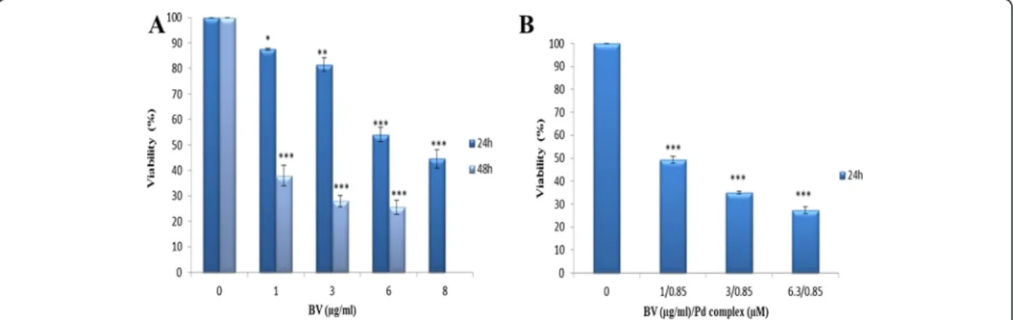
In order to determine the optimal dose and time of cyto-toxic effect of BV alone and in combination with this novel Pd (II) complex on MOLT-4 cells, an MTT assay was performed. The cells were treated with BV at vari-ous concentrations for 24 and 48 hours and with BV/Pd (II) complex for 24 hours. The respective viabilities of cells treated with BV at concentrations of 1, 3, 6 and 8 μg/mL for 24 hours were 87.5 ± 0.500, 81.5 ± 2.500,
54 ± 2.828 and 44.5 ± 3.5 in relation to the control value. The viabilities of cells treated with BV at the concentra-tions of 1, 3 and 6μg/mL for 48 hours were 38 ± 4, 28 ± 2.309 and 25.6 ± 2.728 relative to the control value, re-spectively (Figure 1A).
The viabilities of cells treated with BV/Pd (II) complex
at concentration of 1 μg/mL BV/0.85 μM Pd (II)
complex, 3 μg/mL BV/0.85 μM Pd (II) complex and 6.3 μg/mL BV/0.85 μM Pd (II) complex for 24 hours were 49.33 ± 1.435, 35 ± 0.5774, 27.33 ± 1.453 in relation to the control value, in that order (Figure 1B).
These results reveal that the cytotoxic effect of BV alone and in combination with Pd (II) complex on MOLT-4 cells is dose- and time dependent (Figure 1A and B). Based on these data, the respective 50% cyto-toxic concentrations (Cc50) of the BV after 24 and
48 hours of incubation were 6.3 and 0.6 μg/mL. The Cc50 value of BV in combination with Pd (II) complex
was 1μg/mL BV/0.85μM Pd (II) complex after 24 hours of incubation. The optimal dose and treatment time of BV alone and in combination with Pd (II) complex to be used in subsequent experiments were set according to Cc50values of these components at 24 hours.
Cellular morphological changes with BV and BV/Pd (II) complex
To examine the effects of BV and BV/Pd(II) complex on MOLT-4 cell morphology, cells were treated with BV and BV/Pd (II) complex and examined by phase-contrast microscopy. As shown in Figure 2, cells treated with BV (Figure 2B) or with BV/Pd complex (Figure 2C) displayed greater nuclear condensation than the control group (Figure 2A). This morpho-logical characteristic suggests that BV alone or in combination with Pd (II) complex induces apoptotic cell death in MOLT-4 cells.
Flow cytometry
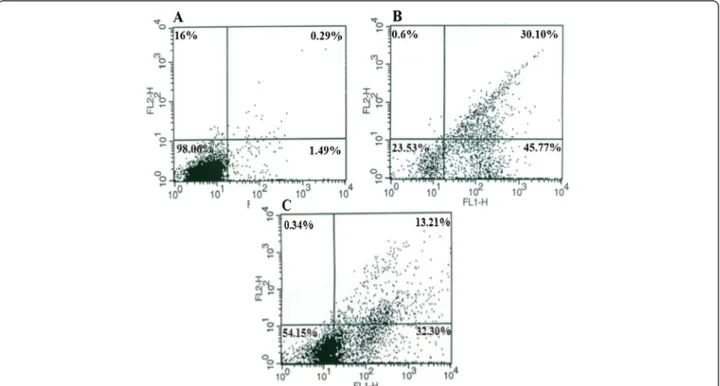
To prove that BV and BV/Pd(II) complex induce apoptosis in MOLT-4 cells, a flow cytometric ana-lysis with Annexin-V was performed (Figure 3). The results confirmed that the cells exposed to BV
alone or in combination with Pd (II) complex for 24 hours enter the early stage of apoptosis. Apop-tosis was induced in 32.30% of the cells exposed
simultaneously to the Cc50 value of these two
components.
Figure 2Effect of BV and BV/Pd (II) complex on the morphology of MOLT-4 cells.Photomicrographs from inverted microscope. Condensed nuclei obviously indicate apoptosis.(A)Controls,(B)cell treated with BV,(C)cells treated with BV/Pd (II) complex.
Figure 3Characterization of BV and BV/Pd (II) complex-induced apoptosis in MOLT-4 cells by flow cytometry.Cells were cultured(A)
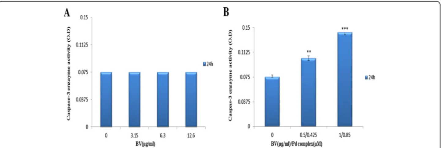
Caspase-3 enzyme activity
Caspase-3 enzyme activity was measured by a colorimet-ric assay. The enzyme activity assay revealed that caspase 3 was not affected by BV. The optical density of the samples exposed simultaneously to BV and Pd (II) complex increased from 0.075 to 0.1033 and from 0.075 to 0.14266 at 1/2 Cc50[0.5μg/mL BV/0.425 μM Pd (II)
complex] and Cc50 [1 μg/mL BV/0.85 μM Pd(II)
com-plex], respectively (Figure 4).
Discussion and conclusions
Although it has been previously reported that bee venom can inhibit human cancer cell growth through in-duction of apoptosis in many cancer cell lines such as prostate cancer, breast cancer and melanoma, there is no finding of the induction of apoptosis in human T cell acute lymphoblastic leukemia cells by BV [6-8]. Based on our knowledge, the present study is the first report about examination of the synergistic effect of BV with a palladium metal-based component.
Analysis of cytotoxicity by MTT assay proved that BV is both time- and dose-dependent in its cytotoxic effects, given that the Cc50 values of this component were 6.3
and 0.6 μg/mL after 24 and 48 hours, respectively. Due to the inconsistency in the MTT assay data–as a result of precipitation of the BV at high concentrations after 24 and 48 hours, and at low concentrations after 48 hours–only the concentrations less than 10 μg/mL were applied in these experiments. At these concentrations the findings were acceptable, except at 8μg/mL after 48 hours, which again resulted in precipitation of the BV.
The lethal dosage of BV in MOLT-4 cells is about 6.3μg/mL, which is lower than that for lung cancer cells reported by Jang et al. [9] (10 μg/mL), but exceeds that of leukemia U937 cells (about 2 μg/mL after 48 hours),
as well as human melanoma A2058 cells (2 μg/mL after one hour) [8,10]. However, BV required different dura-tions to induce cell death in these distinct types of cancerous cells. Such differences may be due to the bio-logical and genetic variations between the investigated cell types. Morphological analysis and the results of flow cytometry indicated that the type of cell death induced by BV is apoptosis.
The present data have also revealed that expression of caspase-3 protein in MOLT-4 cells exposed to BV is down-regulated. Ip et al. [11], when examining the effect of honey bee venom on human cervical epidermoid car-cinoma Ca Ski cells, observed that bee venom induced cell cycle arrest and apoptosis in these cells in caspase-dependent and caspase-incaspase-dependent pathways [11]. Tu et al. [8] also indicated that bee venom induces calcium-dependent but caspase-incalcium-dependent apoptotic cell death in human melanoma A2058 cells.
On the contrary, BV induced apoptosis in human leukemia U937 cells through down-regulation of the ERK and Akt signaling pathway, with Bcl-2 and caspase-3 as the key regulators [10]. A large amount of evidence indicates that apoptosis-induced factors (AIF) and endo-nuclease G (EndoG) act as major apoptosis agents in the caspase-independent cell death pathway [25-28].
In addition to the abovementioned effects, we proved that lethal effects of Pd complex were potentiated by adding a non-lethal dose of the bee venom. On the other hand, BV exerts a strong synergistic effect on the Pd (II) complex. Our preliminary data, which were presented at the FAOBMB Conference, indicated that 1.7 μM [Pd (bpy)(Pi-Pydtc)]NO3 produces a cytotoxic effect on the
MOLT-4 cells. It was also demonstrated that the lethal dose of this newly synthesized palladium complex can induce apoptosis in these cells [22].
Figure 4Results of caspase-3 enzyme activity assay.Cells treated(A)with BV and(B)with BV/Pd (II) complex. The optical density was measured at 405 nm. The OD values were not altered by increasing BV, while the OD values rose following a dose increase from 1/2Cc50BV/Pd (II) complex [0.5μg/mL BV/0.425μM Pd (II) complex] to Cc50BV/Pd (II) complex [1μg/mL BV/0.85μM Pd (II) complex] compared to the control.
In the present study, we demonstrated that when BV and palladium (II) complex were consumed simultan-eously, the combination of 1 μg/mL BV with 0.85 μM Pd (II) complex induces MOLT-4-cell apoptosis in a caspase-3-dependent manner. Orsolic [29], while investi-gating cytotoxic effects of bee venom applied alone or in combination with the DNA-damaging drug bleomycin on HeLa and V79 cells, found that bleomycin caused a dose-dependent decrease in cell survival. When used with a non-lethal dose of the BV, its lethal effect was po-tentiated. The author inferred that BV, by preventing re-pair of damaged DNA, increases bleomycin lethality and inhibited recovery from bleomycin-induced damage [29]. Because DNA is the main target of palladium metal-based complexes, we may conclude that BV is able to potentiate the lethality effect of [Pd (bpy)(Pi-Pydtc)]NO3
in this manner. In summary, the results of the present study suggest that the BV induces apoptosis in human lymphoblastic leukemia cells and, if further studies on animal models confirm these results, that bee venom may be used with customary chemotherapy agents to improve their cytotoxic effects.
Ethics committee approval
The present study was approved by the Ethics Committee of the Faculty of Biological Sciences at Kharazmi University.
Competing interests
The authors declare that there are no competing interests.
Authors’contributions
All authors contributed equally to this work. All authors read and approved the final manuscript.
Acknowledgments
The authors are grateful to the head of Biology Department and Cell and Developmental Biology Research Laboratory as well as Animal House Unit of the Faculty of Biological Sciences, at Kharazmi University for the financial support and to Dr. H. Mansouri Tourshizi for providing the novel palladium complex to our research lab. We also thank Dr. Imani for collaborating in the BV collection.
Received: 20 March 2013 Accepted: 3 September 2013 Published: 3 October 2013
References
1. Dotimas EM, Hider RC:Honeybee venom.Bee World1987,68(2):51–70. 2. Son DJ, Lee JW, Lee YH, Song HS, Lee CK, Hong JT:Therapeutic
application of anti-arthritis, pain-releasing, and anti-cancer effects of bee venom and its constituent compounds.Pharmacol Ther2007, 115(2):246–270.
3. Vento R, D’Alessandro N, Giuliano M, Lauricella M, Carabillò M, Tesoriere G: Induction of apoptosis by arachidonic acid in human retinoblastoma Y79 cells: involvement of oxidative stress.Exp Eye Res2000, 70(4):503–517.
4. Winder D, Gunzburg WH, Erfle V, Salmons B:Expression of antimicrobial peptides has an antitumour effect in human cells.Biochem Biophys Res Commun1998,242(3):608–612.
5. Kaiser T, Brennecke SP, Moses EK:Methylenetetrahydrofolate reductase polymorphisms are not a risk factor for preeclampsia/eclampsia in Australian women.Gynecol Obstet Invest2000,50(2):100–102.
6. Park MH, Choi MS, Kwak DH, Oh KW, Yoon do Y, Han SB, Song HS, Song MJ, Hong JT:Anti-cancer effect of bee venom in prostate cancer cells
through activation of caspase pathway via inactivation of NF-kB.
Prostate2011,71(8):801–812.
7. Ip SW, Liao SS, Lin SY, Lin JP, Yang JS, Lin ML, Chen GW, Lu HF, Lin MW, Han SM, Chung JG:The role of mitochondria in bee venom-induced apoptosis in human breast cancer MCF7 cells.In Vivo2008, 22(2):237–245.
8. Tu WC, Wu CC, Hsieh HL, Chen CY, Hsu SL:Honeybee venom induces calcium-dependent but caspase-independent apoptotic cell death in human melanoma A2058 cells.Toxicon2008,52(2):318–329.
9. Jang MH, Shin MC, Lim S, Han SM, Park HJ, Shin I, Lee JS, Kim KA, Kim EH, Kim CJ:Bee venom induces apoptosis and inhibits expression of cyclooxygenase-2 mRNA in human lung cancer cell line NCI-H1299.
J Pharmacol Sci2003,91(2):95–104.
10. Moon DO, Park SY, Heo MS, Kim KC, Park C, Ko WS, Choi YH, Kim GY:Key regulators in bee venom-induced apoptosis are Bcl-2 and caspase-3 in human leukemic U937 cells through downregulation of ERK and Akt.
Int Immunopharmacol2006,6(12):1796–1807.
11. Ip SW, Wei HC, Lin JP, Kuo HM, Liu KC, Hsu SC, Yang JS, Chiu TH, Han SM, Chung JC, Mei-Dueyang:Bee venom induced cell cycle arrest and apoptosis in human cervical epidermoid carcinoma Ca Ski cells.
Anticancer Res2008,28(2A):833–842.
12. Orsolic N:Bee venom in cancer therapy.Cancer Metastasis Rev2012, 31(1–2):173–194.
13. Kaufmann SH, Earnshaw WC:Induction of apoptosis by cancer chemotherapy.Exp Cell Res2000,256(1):42–49.
14. Hengartner MO:The biochemistry of apoptosis.Nature2000, 407(6805):770–776.
15. Cohen GM:Caspases: the executioners of apoptosis.Biochem J1997, 326(Pt 1):1–16.
16. Sun RW, Ma DL, Wong EL, Che CM:Some uses of transition metal complexes as anti-cancer and anti-HIV agents.Dalton Trans2007, 21(43):4884–4892.
17. Cooley ME, Davis LE, DeStefano M, Abrahm J:Cisplatin: a clinical review. Part I - Current uses of cisplatin and administration guidelines.
Cancer Nurs1994,17(3):173–184.
18. Martínez A, Lorenzo J, Prieto MJ, Font-Bardia M, Solans X, Avilés FX, Moreno V:Influence of the position of substituents in the cytotoxic activity of trans platinum complexes with hydroxymethylpyridines.Bioorg Med Chem2007,15(2):969–979.
19. Ronconi L, Giovagnini L, Marzano C, Bettìo F, Graziani R, Pilloni G, Fregona D:Gold dithiocarbamate derivatives as potential antineoplastic agents: design, spectroscopic properties, andin vitroantitumor activity.Inorg Chem2005,44(6):1867–1881.
20. Akdi K, Vilaplana RA, Kamah S, Navarro JA, Salas JM, González-Vílchez F: Study of the biological effects and DNA damage exerted by a new dipalladium-Hmtpo complex on human cancer cells.J Inorg Biochem 2002,90(1–2):51–60.
21. Mansoori-Torshizi H, Islami-Moghaddam M, Saboury AA:A microcalorime-try and spectroscopy study on the interaction of BSA with 2, 2’ -bipyridine octylglycinato Palladium (II) Nitrate.Acta Biochim Biophys Sin (Shangai)2003,35(10):886–890.
22. Nabiuni M, Divsalar A, Mansouri-Torshizi H, Safayinejad Z:Cytotoxic effect of honey bee venom and novel palladium complex against human T- cell acute lymphoblastic cell line (MOLT4) [abstract].InProceedings of FAOBMB Conference.Singapore; 2011.
23. Lariviere WR, Melzack R:The bee venom test: a new tonic-pain test.
Pain1996,66(2–3):271–277.
24. Mansouri-Torshizi H, Eslami-Moghadam M, Divsalar A, Saboury AA: DNA-binding studies of some potential antitumor 2, 2’–bipyridine Pt (II)/Pd (II) complexes of piperidinedithiocarbamate. Their synthesis, spectroscopy and cytotoxicity.Acta Chim Slov2011,58:811–822.
25. Joza N, Susin SA, Daugas E, Stanford WL, Cho SK, Li CY, Sasaki T, Elia AJ, Cheng HY, Ravagnan L, Ferri KF, Zamzami N, Wakeham A, Hakem R, Yoshida H, Kong YY, Mak TW, Zuniga-Pflucker JC, Kroemer G, Penninger JM:Essential role of the mitochondrial apoptosis-inducing factor in programmed cell death.Nature2001, 410(6828):549–554.
26. Li LY, Luo X, Wang X:Endonuclease G is an apoptotic DNase when released from mitochondria.Nature2001,412(6842):95–99.
of mitochondrial apoptosis-inducing factor.J Biol Chem2001, 276(19):16391–16398.
28. Kroemer G, Martin SJ:Caspase-independent cell death.Nat Med2005, 11(7):725–730.
29. Orsolic N:Potentiation of Bleomycin lethality in HeLa and V79 cells by bee venom.Arh Hig Rada Toksikol2009,60(3):317–326.
doi:10.1186/1678-9199-19-25
Cite this article as:Safaeinejadet al.:Potentiation of a novel palladium (II) complex lethality with bee venom on the human T-cell acute lymphoblastic leukemia cell line (MOLT-4).Journal of Venomous Animals and Toxins including Tropical Diseases201319:25.
Submit your next manuscript to BioMed Central and take full advantage of:
• Convenient online submission
• Thorough peer review
• No space constraints or color figure charges
• Immediate publication on acceptance
• Inclusion in PubMed, CAS, Scopus and Google Scholar
• Research which is freely available for redistribution