Dextrocardia with situs solitus and inversion apex-basis axis in lesser anteater
(Tamandua tetradactyla)
–
case report
[Dextrocardia com situs solitus e inversão de eixo ápice-base em tamanduá-mirim (Tamandua tetradactyla) – relato de caso]
V.L.C. Pinheiro1, L.C. Pereira2, A.R. Lima1, E. Branco1*
1Universidade Federal Rural da Amazônia – UFRA Belém, PA 2Área de Mina Bauxita – Estrada Mineiração Paragominas, PA
ABSTRACT
Dextrocardia is a rare cardiac anomaly where the heart is situated on the right antimeres of the thorax. This study had the objective of describing a case of dextrocardia with situs solitus and apex-basis axis
inversion in a lesser anteater (Tamandua tetradactyla) between five evaluated animals, all from the area
of Mine Bauxite Paragominas Para. The arterial system was filled with contrasted latex and the animals were fixed with 10% formaldehyde and a posterior dissection was done. The heart of an animal was found in right antimere with inversion of the base-apex axis. The right atrium was more developed then the left and the pulmonary veins arrived directly in the left ventricle. The main vases of the base were identified with some topographic alterations resulting in: aorta dorsal to the cava caudal vein, pulmonary artery dorsal and cranial to aorta, pulmonary veins ventral to the pulmonary artery, cava caudal vein in ventral plain and cava cranial vein in dorsal plan in relation to the other vessels. Internally there were four cardiac chambers, with absence of septal communication.
Keywords: dextrocardia, apex-basis axis inversion, lesser anteater
RESUMO
Dextrocardia é uma anomalia cardíaca rara no qual o coração está situado no antímero direito do tórax. Este estudo objetivou descrever um caso de dextrocardia com situs solitus e inversão do eixo ápice-base em um tamanduá-mirim (Tamandua tetradactyla), entre cinco animais avaliados, sendo todos da área da mina de bauxita Paragominas Pará. O sistema arterial foi preenchido com látex contrastado e os animais foram fixados com formol a 10% e seguido de dissecção posterior. O coração de um dos animais foi encontrado no antímero direito com inversão do eixo ápice-base. O átrio direito era mais desenvolvido do que o esquerdo e as veias pulmonares chegaram direto no ventrículo esquerdo. Foram identificados os principais vasos da base com alterações topográficas, resultando em: aorta dorsal à veia cava caudal, artéria pulmonar dorsal e cranial da aorta, veias pulmonares ventrais a artéria pulmonar, veia cava caudal em plano ventral e veia cava cranial em plano dorsal em relação a outros vasos. Internamente foram localizadas quatro câmaras cardíacas, com ausência de comunicação septal.
Palavra-chave: dextrocardia, inversão de eixo ápice-base, tamanduá-mirim
INTRODUCTION
The lesser anteater (Tamandua tetradactila) is a
Xenartra from the Myrmecophagidae family. Geographically, this species is distributed to the east of the Andes, in Venezuela, until the north of
Recebido em 31 de julho de 2012
Aceito em 7 de maio de 2013
*Autor para correspondência (corresponding author)
nocturnal and the feeding basis for these animals is insects, mainly ants and termites (Reis et al.,
2006; Cubas, 2007).
The dextrocardia is a cardiac anomaly characterized by the displacement of the biggest axis (base-apex) of the heart to the right side of the thorax with a reversion of the apical inclination, this occurs due to an anomalous rotation of the primitive tube of the heart to the right, being a rare anomaly in human beings, and not described in animals. This infirmity can occur separately (situs solitus) or in association
with situs inversus (complete inversion of all
visceral organs) presenting itself as an inverted image (Da Silva et al., 1992) due to cardiac
malformations (Bernasconi et al., 2005, Yusuf et al., 2009, Evans et al., 2010) or situs ambiguos
(the liver and the stomach are located in the centre of abdominal cavity and the stomach develops behind the liver) (Faig-Leite, 2008), but according to Perloff (1978), these infirmity can only be called assitus invesus, however without
necessarily occurring associated with cardiac malformations (Aiello et al., 2008).
The incidence of dextrocardia associated with
situs inversus in the general population is
1:10,000, while associated to situs solitus it is
1:30,000 in born living and only 1:900,000 in the adult population (McCasckie et al., 1991; Bohun et al. 2007). A similar situation described in the
literature and involving animals was only described by Castro et al. (2007).
Regarding the apex-basis axis inversion in a 180° of rotation, there is no data in literature describing animals or humans, which reinforces the unpublished aspect of this study.
CASUISTRY
Five adult animals were studied, two males and three females, from the area of Mine Bauxite Paragominas-Para, under authorization from SEMA-PA Nº 455/2009 and 522/2009, donated to the Laboratory of Animal Morphologic Research (LaPMA), by the Federal Rural University of the Amazonia (UFRA), after death due to accidental running over. The arterial system was filled with latex Neoprene 650, stained with red pigment (Xadrez; Sherwin Willian, São Paulo, Brazil ) and added radiopac
contrast (Bariogel; Cristalia, São Paulo, Brazil), in the ratio of 1:1, through the thoracic aorta.
The animals were fixed with 10% intramuscular, subcutaneous and intracavity applications of formaldehyde, keeping them in the same solution for seven days. After that the animals had been radiographed and dissected, the aortic arch was removed after the sternum.
In the dextrocardic animal (situs solitus) with
base-apex axis inversion the heart was removed by incision of the vessels of the basis.
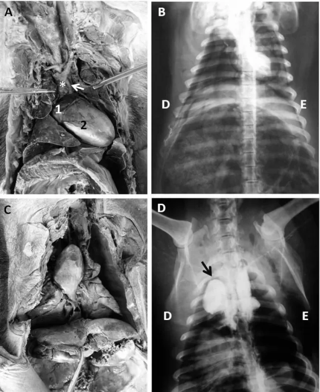
It was observed among between the animals studied (Figure 1 A and B) a female with dextrocardia with situs solitus, shown only an
inverted heart, while the and other organs had normal topography. Beyond the dextrocardia, the heart of this animal presented 180° rotation in relation to the bigger axis (apex-basis) (Figure 1 C and D).
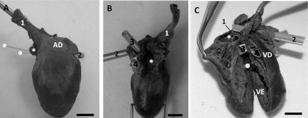
The four cardiac chambers were located, however, and the difference in size between the right and left atrium, in which the right was distended while the left was hypotrophic (Figure 2 A-C) called our attention.
The heart from the dextrocardic animal was located in the mediastinum between the 3rd and
4th intercostal spaces, whereas in other animals
the heart was located between the 3rd and 6th
intercostal spaces showing that the dextrocardic animal had a smaller heart (Figure 1 B and D).
Differing from the normal animals, the vessels on the base of the heart of the animal with dextrocardia situs solitus and apex-basis
Internally, the heart from the dextrocardic animal did not show any significant alteration, and it was possible to observe four chambers without septal communication. The wall of the left
ventricle (LV) was thicker than the right
ventricle (RV) similar to this organ’s normal
anatomy (Figure 1 C).
Figure 1. Cardiactopography of Tamandua tetradactyla. A and B – Animal with a pattern positioning of
the heart. Observed in A: cava cranial vein (*) and aorta (arrow), (1) right atrium, (2) right ventricle. C and D – Animal with dextrocardia with situs solitus and apex-basis invertion axis. Observed in C: cava
Figure 2. Heart of Tamandua tetradactyla with dextrocardia with situs solitus and apex-basis invertion
axis. A – Ventral view: (1) cava caudal vein cava caudal, (2) cava cranial vein, (RA) right atrium and (*) left atrium, (white pins) pulmonary veins. B – Dorsal view: (1) cava caudal vein, (2) cava cranial vein, (3) aorta, (4) pulmonary artery. C – Cardiac internal face: (1) cava caudal vein, (2) cava cranial vein, (3) aorta, (4) pulmonary artery, (white pin) pulmonary vein, (*) left atrium, (RV) right ventricule and (LV) left ventricule. Scale bar: (A) and (C) 0.5cm and (B) 1 cm.
DISCUSSION
Castro et al., (2007) described that the only
information found in the literature that approached dextrocardia in animals was about a brief report of Aristoteles (384-322 B.C), when described together with the transposition of other organs in two animals (situs inversus totalis).
Only in XV century did the dextrocardia reports become more frequent, however only in human beings, a fact that restricted animal investigations.
In humans, dextrocardia cases with situs inversus totalis, also seen with situs inversus (Perloff,
1978), in the heart and in other organs, are presented as a mirror image of the normal disposition. The cava veins, superior and inferior, are on the right atrium that is located to the left of the vertebral column, while the aorta is below on the right. The aortic arch places it on the right and the ascending aorta leaves the left ventricle in the caudo-cranial direction, from the right to the left (da Silva et al., 1992). Such
alterations had not been found in this animal because the heart presented an apex-based rotation axis. However, the pulmonary veins did not enter the left atrium, these entered in left ventricle, leading us to believe that during the bombardment, after the blood passes to the lungs for gaseous exchange, the arterial blood arrived
directly the left ventricle, not passing in the atrium and bicuspid valve.
Faig-Leite et al., (2008), described a case of a
human dextrocardic heart in a corpse of one year old, beyond the dextrocardia other anomalies were identified, like a great arterious duct. This condition was not found in the studied animal because there is no communication between the pulmonary artery and the aorta.
In general, authors classify these alterations as a direct consequence of dextrocardia associated to multiple cardiac malformations (Bernasconi et al., 2005; Yusuf et al., 2009; Evans et al., 2010).
This affirmation is refuted by Aiello et al.,
(2008), which affirmed that they must be treated only to determine the cardiac position in relation to the thorax, without the necessity of inferring on the occurrence of cardiac malformations, because they may or may not be associated. Our findings corroborate the named author, however, we did not find multiple malformations, only the shunting of pulmonary veins to the left ventricle as mentioned before.
Although there is literature that consecrates that in the dextrocardia there is a displacement of the main axis of the heart (basis-apex) to the right antimere (Bernasconi et al., 2005), there is no
rotation) of this axis where the cardiac apex is turned cranially and the basis caudally, such as described in this report.
CONCLUSION
The dextrocardia with situs solitus in itself is not
a common pathology in animals. The fact of the base-apex inverted axis makes this finding more uncommon.
REFERENCES
AIELLO, V.D.; CASTELLI, J.B.; BARRA, M.; MORHY, S.S. Anatomia cardíaca e análise segmentar sequencial nas Cardiopatias congênitas. In: CROTI, U.A.; MATTOS, S.S.; PINTO, JR. V.C.; AIELLO, V.D. (Eds).
Cardiologia e Cirurgia Cardiovascular Pediátrica. São Paulo: Roca, 2008. v.1, p.9-23.
BERNASCONI, A.; AZANCOT, A.; SIMPSON, J.M.; et al. Fetal dextrocardia: diagnosis and
outcome in two tertiary centres. Heart. v.91,
p.1590-1594, 2005.
BOHUN, C.M.; POTTS, J.E.; CASEY, B.M.; SANDOR, G.G.S. A population-based study of cardiac malformations and outcomes associated with dextrocardia. Am. J. Cardiol. v.100,
p.305-309, 2007.
CASTRO, C.S.; GARCIA, R.T.; CASTRO, V.M.; et al. Reconstrução mamária em paciente
com síndrome de Poland e situs inversus totalis:
relato de caso. Rev. Soc. Bras. Cir. Plast., v.22,
p.261-5, 2007.
CUBAS, Z.S.; SILVA, J.C.R.; CATÃO-DIAS, J.L. Xenarthra (Tamanduá, Tatu, Preguiça). Tratado de animais selvagens – medicina veterinária. São Paulo: Roca, 2006. p.402-406. DA SILVA, M.J.; ARIE, S.; GARCIA, D.P. et al. Dextrocardia in situs inversus totalis with
obstructive coronary disease. Its treatment by coronary angioplasty by the brachial approach.
Arq. Bras. Cardiol., v.59, p.303-307, 1992.
EVANS, W.N.; ACHERMAN, R.J.; COLLAZOS, J.C. et al. Dextrocardia: Practical Clinical Points
and Comments on Terminology. Pediatr. Cardiol., v.31, p.1-6, 2010.
FAIG-LEITE, F.S.; FAIG-LEITE, H. Relato de caso-Anatomia de um caso de Dextrocardia com Situs Solitus. Arq. Bras. Cardiol., v.9, p.e56-
e58, 2008.
MCCASCKIE, A.W.; THOMPSON, M.M.; UNDERWOOD, M.J.; PALLOT, D.J. A case of dextrocardia with normal situs. Acta Anat.,
v.142, p.288-292, 1991.
PERLOFF, J.K. The Clinical Recognition of Congenital Heart Disease, 3.ed. Philadelphia:
WB Saunders Co., 1978. p.19-42.
REIS, N.R.; PERACCHI, A.L.; PEDRO, W.A.; LIMA. I. Mamíferos do Brasil. SEMA/SETI/ UEL/UNIFIL/PPG Ciências Biológicas UEL/ EDIFURB/Schering-Plough, 2006. 439p. YUSUF, W.; DURAND, J.B.; LENIHAN, D.J.; SWAFFORD, J. Dextrocardia: An Incidental Finding. Texas Heart Instit. J., v.36, p.358-359,