419 1. Service of Pediatric Cardiovascular Surgery of São José do Rio
Preto – Hospital de Base –São José do Rio Preto Medical School, SP, Brazil.
Correspondence address: Ulisses Alexandre Croti
Hospital de Base – Faculdade de Medicina de São José do Rio Preto –
Ulisses Alexandre CROTI1, Domingo Marcolino BRAILE1, Marcelo Felipe KOZAK1, Lilian BEANI1
Rev Bras Cir Cardiovasc 2010; 25(3): 419-421 CLINICAL-SURGICAL CORRELATION
RBCCV 44205-1208
Ampliação da neopulmonar tardiamente à operação de Jatene
Enlargement of the neopulmonary after Jatene’s
operation
FAMERP – Avenida Brigadeiro Faria Lima, 5544 – São José do Rio Preto, SP, Brasil – CEP 15090-000
E-mail: [email protected]
Article received on August 9th, 2010 Article accepted on September 5th, 2010 CLINICAL DATA
7-year-old female child, 19 kg, height of 116 cm and asymptomatic.
Full-term baby with a diagnosis of transposition of the great arteries underwent Jatene’s operation with Lecompte maneuver, at the first week of life. In the immediate postoperative period, presented endocarditis due to Candida glabrata, which was treated medically.
In outpatient treatment it was noted an increase in gradient between the right ventricle and neopulmonary in routine echocardiographic examinations.
With 7 years of evolution, further surgery was indicated, although the patient did not use drugs.
The physical examination was completely normal except for a systolic ejection murmur at the left sternal border of +4 / 6+.
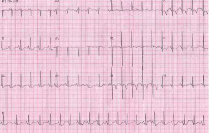
ELECTROCARDIOGRAM
Sinus rhythm, rate of 100 beats/min, SÂP + 60º, SÂQRS + 120°, QT 0,23, QTc 0,37. Atrial and right ventricular overload and change in anteroseptal repolarization (Figure 1).
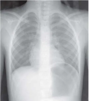
RADIOGRAPHY
Visceral situs solitus in levocardia. Diaphragmatic dome elevated to the left due to gastric bubble. Cardiothoracic ratio of 0.50. Heart size and pulmonary vasculature within normal limits (Figure 2).
420
CROTI, UA ET AL - Enlargement of the neopulmonary after Jatene’s operation
Rev Bras Cir Cardiovasc 2010; 25(3): 419-421
ECHOCARDIOGRAPHY
Situs solitus in levocardia, late postoperative arterial Jatene’s operation with stenosis of the pulmonary trunk (neopulmonary) located about 1 cm above the valve. The pulmonary valve annulus diameter was 17.2 mm, 13.8 mm pulmonary trunk, right pulmonary artery 6.3 mm and left 4.8 mm. On Doppler, the flow was turbulent and accelerated in the neopulmonary, compatible with maximum gradient of 116 mmHg and mean of 72.9 mmHg (Figures 3 and 4).
DIAGNOSIS
Reoperation was indicated by measurements made solely on the echocardiogram, which increased gradually, and the images of multi-detector computed tomography, which showed ring stenosis in the pulmonary trunk with discrete poststenotic dilatation. It is noteworthy that the child was totally asymptomatic, but there must always be concern to avoid the increase of the right ventricular mass, arrhythmias and sudden death [1,2].
OPERATION
The first arterial inversion procedure was performed at the beginning of our experience and occurred uneventfully in the operating room. Two points should be emphasized: the episode of endocarditis in the immediate postoperative period and the use of paches of bovine pericardium for reconstruction of the neopulmonary coronary sinus. Fig. 2 – Preoperative chest radiography with heart size within normal limits
Fig. 3 - Preoperative echocardiogram in subcostal view showing the right ventricular outflow tract, pulmonary valve annulus and the reduction in size of the pulmonary trunk
Fig. 4 - Continuous preoperative Doppler measuring the gradient at the local of supravalvular stenosis in the pulmonary trunk
421 After adequate heparinization, an arterial cannula was
inserted into the aorta and another (venous) into the right atrium. Cardiopulmonary bypass was started in normothermia without clamping the aorta and longitudinal
REFERENCES
1. Gontijo Filho B, Fantini FA, Lora HM, Martins C, Lopes RM, Hayden E, et al. Reconstrução da artéria pulmonar na operação de Jatene. Rev Bras Cir Cardiovasc. 2001:16(3):236-43.
2. Jatene MB, Jatene IB, Oliveira PM, Moysés RA, Souza LC, Fontes V, et al. Prevalência e abordagem cirúrgica da estenose supravalvar pulmonar pós-operação de Jatene para transposição das grandes artérias. Arq Bras Cardiol. 2008;91(1):17-24.
At reoperation, we proceeded to the dissection of the adhesions and individualization of the structures, identifying the location of the thrill properly and hence the stenosis of the neopulmonary supravalvular trunk (Figure 5/ Video 1).
Fig.6/Video 2 – Patch of bovine pericardium used in the pulmonary artery to enlarge the stenotic site
opening of the pulmonary trunk in the stenotic site. It was found a retraction ring in the site of the suture lines of the bovine pericardium patches with the autologous tissue of the native pulmonary artery. This ring was resected and a new bovine pericardium was previously implanted in the pulmonary trunk.
The CPB time was 18 minutes without myocardial ischemia.
In the immediate postoperative period, the child was uneventful and was discharged on the fourth day of hospital stay without use of medications and with echocardiogram demonstrating excellent enlargement of the neopulmonary trunk.
CROTI, UA ET AL - Enlargement of the neopulmonary after Jatene’s operation