Case Report
Key Words
dextrocardia; dissection; thoracic duct; heart septal defects.
We present a rare case of dextrocardia in the corpse of a female child, aged approximately one year old, presenting situs solitus. The cases of dextrocardia reported in the literature normally describe multiple associated cardiac malformations. In the present study, it is noteworthy the fact that a complete specular image of the heart and the vessels of the base of the heart was not found. There was no atrioventricular discordance or other intra and extracardiac malformations, which are commonly seen in cases of dextrocardia. A large ductus arteriosus was found, as well as the anomalous position of the right portion of the heart.
Anatomy of a Dextrocardia Case with
Situs Solitus
Fabíola Sawaguchi Faig-Leite1 e Horácio Faig-Leite2
Universidade Estadual Paulista Júlio de Mesquita Filho (UNESP) Campus de Botucatu1, Botucatu, SP; Universidade Estadual Paulista Júlio de Mesquita
Filho (UNESP) Campus de São José dos Campos2, São José dos Campos, SP - Brazil
Mailing Address: Fabíola Sawaguchi Faig Leite•
Rua Engenheiro João Fonseca dos Santos, 123 / 194, Vila Adyanna, 12.243-620, São José dos Campos, SP - Brazil.
E-mail: fasawaguchi@yahoo.com.br
Manuscript received January 11, 2008; revised manuscript received February 24, 2008; accepted March 10, 2008..;
Introduction
Dextrocardia is an embryologic malformation characterized by the displacement of the largest axis (base to apex) of the heart to the right side of the chest, with reversion of the apical inclination. This malformation is caused by an anomalous rotation of the primitive heart tube to the left, in which the bulboventricular loop bends to the left, in a specular image of the normal condition, which occurs around the 8th week
of embryonic life1,2.
The incidence of dextrocardia associated to situs inversus
in the general population is usually 1:10,000, whereas the one associated with situs solitus is 1:30,000 live births and only 1:900,000 in the adult population3,4. This high
incidence difference found in individuals with dextrocardia associated with situs solitus is due to the higher occurrence of associated cardiac and/or extracardiac diseases, such as tracheoesophageal fistula, pulmonary hypoplasia, imperforate anus, spina bifida and Kartagener syndrome3-6.
There is great discordance among authors regarding the classification of the several types of dextrocardia1,6-8. Among
the several studies, the one by Van Praagh et al6 is worth
mentioning, as it describes and classifies in details the different types of dextrocardia. Dextrocardia must be differentiated from the dextroversion, as the latter is not a congenital condition, in which the heart can change sides due to a disease
that pushes or pulls the organ into the right antimere, although the heart apex remains toward the left side2,5,7,9,10.
Case Report
Anatomical descriptionDextrocardia was identified in the corpse of a female child, probably one year of age, which belonged to the Laboratory of Anatomy of the Campus of São José dos Campos - UNESP.
The thorax of the anatomical piece was partially dissected, without the presence of the pericardium and the lungs. The sternum was in its normal position. The inspection of the heart and the vessels of the base of the heart did not disclose possible position alterations caused by the removal of the lungs. The entire heart and vessels of the base were intact and had not been dissected. The heart and its apex were completely turned to the right.
After the removal of the sternum and the dissection of the vessels of the base, it was observed that due to the rotation of the heart, its entire right portion was further displaced to the posterior part and this fact was so accentuated in the right auricle that, with the heart in the observed position, it was not possible to visualize it (Figure 1). This anomalous rotation of the heart made the pulmonary trunk emerge from the right ventricle abnormally, appearing much more superficial and extensive (Figure 1).
During the inspection of the pulmonary trunk, a large ductus arteriosus (Botallo’s ductus) was found (Figure 2). The heart apex was positioned along the heart axis and was turned to the right side, thus demonstrating the dextrocardia.
With the help of a stereoscopic magnifying glass, all afferent and efferent branches of the vessels of the base of the heart were dissected in order to look for anomalies. With the exception of the brachiocephalic trunk, which presented a more accentuated angulation than the normal one, no other variations regarding position or trajectory were observed (Figure 1).
The diaphragm muscle was removed and subsequently, the liver was dissected, which presented normal aspect and morphology. The abdominal cavity was open and the position of the viscera was normal, without any type of alteration, which characterizedsitus solitus.
After this phase, the heart was then carefully removed from its place and again inspected with a magnifying glass. The auricles and atriums were opened and analyzed and disclosed normal anatomical aspects. The atrioventricular concordance was anatomically normal. An incision was made in each ventricle with the objective of verifying its internal morphology and the emergence of its vessels. The two ventricles presented
Case Report
Faig-Leite et al Dextrocardia with Situs Solitus
Arq Bras Cardiol 2008;91(6):e64-e66
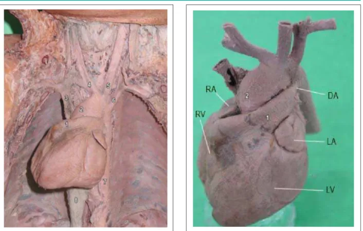
Figure 1 -Position of heart in the thorax: 1 - pulmonary trunk; 2 - ascending aorta; 3 - superior vena cava; 4 - brachiocephalic trunk; 5 - left common carotid artery; 6 - left subclavian artery; 7 - thoracic aorta; 8 - hepatic tissue covering the inferior vena cava.
Figure 2 - Isolated heart showing cardiac chambers: RA - right auricle; RV - right ventricle; LA - left auricle; LV - left ventricle; DA - ductus arteriosus; 1 - pulmonary trunk; 2 - ascending aorta.
normal internal morphology, atrioventricular valves and vessel emergence. The aorta and the pulmonary trunk presented normal semilunar valves.
Discussion
The literature presents a great diversity of types and classifications for dextrocardia cases. This diversity seems to be caused by the difficulty to establish criteria for a perfect classification6-8.
Through simple anatomical observation, it can be determined that there was no change in the heart position after the corpse underwent fixation.
This case is characterized as a dextrocardia case, as the heart apex was completely turned to the right antimere2-5,7,9. It
cannot be classified as a dextroversion case, as in the latter, the heart is located in the right hemithorax with the apex turned to the left antimere, different from what was observed in the present case5,9 (Figure 1).
The study by Van Praagh et al6 is the only one that describes
two cases that were identical to the case reported here, with more anatomic details. Both cases, as in the present one, constituted necropsies of children younger than one year with a type of dextrocardia classified by the authors as dextrocardia with normally related great vessels. In these cases, as well as in the present case, situs solitus was found, the great vessels were
normally related (with the bulboventricular loop to the right) and the absence of ventricular inversion was observed. The two cases described by these authors presented severe pulmonary alterations; as the lungs had been previously removed in the present case, it was not possible to study them.
Calcaterra et al7, studying several cases of dextrocardia,
described one case with the same anatomical variations found in the present case; the authors, however, did not search for extracardiac alterations in the necropsies, thus not allowing a more detailed comparison with the present case.
Normally, dextrocardia is more frequent when associated to situs inversus, with the presence or absence of associated cardiac and/or extracardiac malformations4,9. The case presented here is
more uncommon as it was associated to situs solitus and for the fact that it did not present these types of malformation, which allows us to state that this variant of heart position is not necessarily associated to anomalies in atrioventricular communications, in the relations between the heart and its afferent and efferent vessels or even in the heart function.
The diagnosis of dextrocardia can be attained during the physical examination, through an electrocardiogram or imaging examination4. Thus, the knowledge of this anomaly and its
variants is of crucial importance for physicians due to the risk of atypical angina presentations, its frequent association with other cardiac and/or extracardiac diseases and also, for constituting a malformation of which the abnormalities,
Case Report
Faig-Leite et al
Dextrocardia with Situs Solitus
Arq Bras Cardiol 2008;91(6):e64-e66
References
1. Macruz R, Mazzieri R, Mattar Jr J, Ebaid M. Más posições cardíacas. Arq Bras Cardiol. 1973; 26: 481-6.
2. Pego-Fernandes PM, Serro-Azul, JB, Matheus F, Maehara BS. Revascularização do miocárdio em paciente com situs inversus totalis. Arq Bras Cardiol. 2007; 88 (5): e103-e106.
3. McCasckie AW, Thompson MM, Underwood MJ, Pallot DJ. A case of dextrocardia with normal situs. Acta Anat. 1991; 142: 288-92.
4. Bohun CM, Potts JE, Casey BM, Sandor GGS. A population-based study of cardiac malformations and outcomes associated with dextrocardia. Am J Cardiol. 2007; 100: 305-9.
5. Leung AKC, Robson WLM. Dextrocardia with sinus solitus. Can Med Assoc J. 2006; 175 (3): 244.
6. Van Praagh R, Van Praagh S, Vlad P, Keith JD. Anatomic types of congenital dextrocardia; diagnostic and embryologic implications. Am J Cardiol. 1964; 13: 510-31.
7. Calcaterra G, Anderson RH, Lau KC, Shinebourne EA. Dextrocardia-value of segmental analysis in its categorization. Br Heart J. 1979; 42: 497-507.
8. Stanger P, Rudolph AM, Edwards JE. Cardiac malpositions; an overview based on study of sixty-five necropsy specimens. Circulation. 1977; 56 (2): 159-72.
9. Garg N, Agarwal BL, Modi N, Radhakrishnan S, Sinha N. Dextrocardia: an analysis of cardiac structures in 125 patients. Int J Cardol. 2003; 88: 143-55.
10. Lucchese FA, Becker AE, Macartney FJ, Meier MA, Jimenez MQ, Shinebourne EA, et al. Classificação das cardiopatias congênitas. Arq Bras Cardiol. 1980; 35 (5): 427-34.
despite their complexity, are currently possible of undergoing surgical correction8,9.
Potential Conflict of Interest
No potential conflict of interest relevant to this article was reported.
Sources of Funding
There were no external funding sources for this study.
Study Association
This study is not associated with any graduation program.