2013/2014
Pedro Manuel Correia Rodrigues
Minimally Invasive Repair of Pectus Excavatum:
A 13-year Experience At a Tertiary Surgical Center
Mestrado Integrado em Medicina
Área: Cirurgia Pediátrica
Trabalho efetuado sob a Orientação de:
Doutor Tiago Alexandre Henriques-Coelho
Trabalho organizado de acordo com as normas da revista:
Journal of Pediatric Surgery
Pedro Manuel Correia Rodrigues
Minimally Invasive Repair of Pectus Excavatum:
A 13-year Experience At a Tertiary Surgical Center
Ao meu irmão Francisco,
MINIMALLY INVASIVE REPAIR OF PECTUS EXCAVATUM:
A 13-YEAR EXPERIENCE AT A TERTIARY SURGICAL CENTER
Pedro Correia-Rodrigues 1, Ruben Lamas-Pinheiro 1, Tiago Henriques-Coelho 1,2,*
1
Pediatric Surgery Department, Centro Hospitalar São João, Porto, Portugal
2
Pediatrics Department, Faculty of Medicine, Porto, Portugal
* CORRESPONDING AUTHOR:
Tiago Henriques-Coelho
Pediatric Surgery Department, Centro Hospitalar São João
Alameda Professor Hernâni Monteiro, 4200-319 Porto, Portugal
Tel.: 00351225512100
2
A
BSTRACTBackground/Purpose: Pre-surgical automatic and personalized bar bending for pectus excavatum (PE)
correction allows a correct size and shape of the bar using 3D computerized tomography (CT) scan. This
study retrospectively reviews the experience at a tertiary pediatric center for surgical correction of PE
and analyses the impact of the pre-bended bar in Nuss procedure (NP).
Methods: Patients who underwent a NP from January 2000 to December 2013 were included. Data
regarding demographics, previous PE correction, anesthesia, surgery and complications were obtained
from clinical files. Statistical analysis was performed between patients who received pre-surgical
automatic bended (AB group) or classic manual bended (MB group) bars. Data are presented as median
(range).
Results: A total of 139 (78% male) patients were operated. Median age at the time of surgery was 14.7
years (range, 7-30 years). Ten patients (7%) had been previously submitted to Ravitch procedure. Since
2007, the automatic pre-bended bar was used in 96 patients (69%). MB and AB groups were identical for
gender, age and symmetry of the defect, but patients in MB group had a higher median Haller index. A
thoracic epidural catheter was placed in almost every patient (98%). In AB group, surgery lasted less
time, the hospital length of stay was shorter and complication rate was lower. There was no mortality.
Complications included pneumothorax, skin erosion, bar displacement, wound infection and bar
infection. The bar was removed after a longer period in the AB group.
Conclusion: The actual surgical technique using pre-bended bars is safe and quick, with a low
complication rate.
3
1.
I
NTRODUCTIONPectus Excavatum (PE) is the most common congenital chest wall deformity and several procedures
have been described to manage this deformity. Donald Nuss introduced the minimally invasive repair of
PE (MIRPE) technique in 1998 [1]. Since then, there has been a worldwide significant increase in the
number of patients with PE treated by the Nuss procedure (NP). It is currently a first-line approach for
PE in many centers, regarding being a much less radical operation with better cosmetic results than
previous techniques such as Ravitch procedure [2]. Other innovative approaches are under evaluation,
such as vacuum treatment [3], custom-made silicone implants [4], the pectoscope [5], pectus
tunneloscopy [6] and the magnetic mini-mover [7].
Original Nuss procedure describes an intraoperative manual bar bending assisted by a template that
reproduces the patients’ thorax morphology. This laborious protocol is time-consuming and often
results in imperfections that could adversely compromise the correction success [8, 9]. In order to
overcome these disadvantages, a new system that allows pre-surgical automatic and personalized
modeling and bending of the bar prosthesis was described to predict the correct size and shape for each
patient, based on 3D computerized tomography (CT) scan images [10]. Our group already described the
advantages of personalized prosthesis modeling and bending. In the present study, we review the
experience in a tertiary center for surgical correction of PE comparing the pre-bended bar group with
4
2.
M
ATERIAL ANDM
ETHODS2.1. Study Design
This retrospective observational study enrolled 139 patients submitted to PE surgical correction by the
Nuss procedure from January 2000 to December 2013, at Pediatric Surgery Department of Centro
Hospitalar São João in Porto, Portugal. The Health Ethics Committee of Centro Hospitalar São João
approved the study. Data was obtained from paper and digital clinical files of the selected patients.
Criteria for surgical correction was based not only on objective parameters as the Haller index or
physiologic compromise, but also on the psychological effects and body-image distortion associated
with PE deformity. The preoperative protocol included electrocardiogram, echocardiogram and
computerized tomography (CT) scan. Follow-up period after the procedure ranged from a minimum of 2
months and a maximum of 45 months.
2.2. Evaluated Parameters
Patients submitted to Nuss procedure were divided in two groups: those that underwent MIRPE
procedure with an intra-operative manual bended (MB) metal bar (historic controls) and those with
pre-surgical automatic bended (AB) metal bar personalized in accordance to their 3D CT scan. Demographic
data, deformity characterization, surgical data and complications were collected. Regarding
demographic data, gender, age at surgery and history of previous surgery were acquired. Morphologic
characterization of the deformity by CT scans included Haller index (HI) and symmetry (Type 1 –
symmetric; Type 2 - asymmetric). Surgical data included: type of bar bending (manual or pre-bended),
number of bars placed, duration of surgery, duration of anesthesia, type of postoperative analgesia,
5 occurring intra-operatively or during the initial hospital stay), late complications (during follow-up),
average time with the bar and mean age at bar removal.
2.3. Automatic Bar Bending
The selection of the prosthesis size and shape was based on 3D reconstruction of the thoracic grade
from 2D DICOM (Digital Imaging and Communications in Medicine) slices of preoperative chest CT data
scan. A sequence of automatic image processing techniques simulated the most appropriate surgical
prosthesis to the patient. After this simulation, the system bends the bar with a precision of
micrometers using an electromechanical apparatus with real-time monitoring and control [10].
2.4. Nuss Procedure
The Nuss procedure was performed under general endotracheal anesthesia. A thoracic epidural block
for intra-operative and post-operative pain control was used. A Foley catheter was placed. All patients
received a pre-operative course of prophylactic antibiotic regimen. Patient was supine positioned with
both arms abducted at the shoulders. Bilateral 1.5-2.5 cm transverse thoracic incisions between the
anterior and midaxillary lines were used. A 5mm 30 degree thoracoscope was inserted one or two
intercostal spaces below the right thoracic incision and the chest was insufflated with CO2 until 6 mmHg.
Under endoscopic guidance, a subcutaneous-substernal-subcutaneous tunnel was raised anteriorly from
both incisions to the top of the pectus ridge. In MB group the pre-operative chest measurement was
reconfirmed and a bar was selected for bending into the desired chest-wall curvature (this step was
absent in AB group). The sterilized convex metal bar was inserted and advanced across the mediastinum
in the retro-sternal space under thoracoscopic vision. The bar was then flipped and positioned in the
6 tube was placed using the incision for 5mm trocar. After surgery a chest radiography was performed to
confirm adequate lung expansion and to reveal the final positioning of the bar.
The prosthesis was removed after two to three years. This procedure was performed under general
anesthesia using the previous incisions as an outpatient procedure.
2.5. Pain Control Protocol
An epidural catheter was placed in the region of T5-7 to assure analgesia in the dermatomes affected by
the surgery. Infusion comprised a local anesthetic (bupivacaine or levobupivacaine) and an opioid
(morphine). During hospital stay, the epidural catheter was left in place for pain management for 3-6
days. In cases of epidural failure, intravenous patient controlled analgesia (PCA) and oral analgesia was
used.
2.6. Statistical Analysis
Data were analyzed with IBM SPSS Statistics ® 21.0. To characterize variables, descriptive statistics was
used and normality of data was tested by Shapiro-Wilk test and Normal Q-Q Plots. Parametric and
nonparametric comparisons were performed as appropriate. Variables median age at surgery, median
Haller index, surgery duration, anesthesia duration, post-operative lengths of stay and median period
with the bar for both groups were analyzed using Wilcoxon-Mann-Whitney U test. Chi-square and
Fisher’s Exact test were used for categorical variables such as gender, epidural catheter, need for PICU
7
3.
R
ESULTS3.1. Demographic Data and Deformity Characterization
Data are summarized in Table 1. A total of 139 patients underwent PE surgical correction by Nuss
procedure. From these, 43 patients received a manual bended bar (MB group) and 96 patients received
an automatically pre-bended bar (AB group) after 2007. Eleven patients (8%) had scoliosis. One patient
was diagnosed with Poland syndrome and two with Marfan syndrome. Regarding previous surgery, one
patient was submitted to a neonatal thoracotomy for a diaphragmatic hernia and ten patients (7%) had
been previously submitted to Ravitch procedure. None of the patients were previously submitted to a
Nuss procedure. There was a male preponderance (78%) and patients’ age ranged from 7 to 30 years
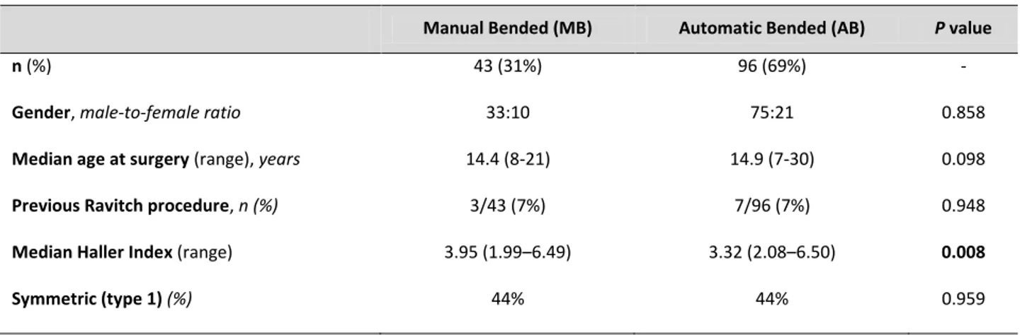
(median age 14.7 years). Both groups were identical for gender, age and symmetry of the defect, but
MB patients had a higher Haller index.
3.2. Surgical Data
Data are summarized in Table 2. Only one bar was used. Thoracoscopy was always used. There was no
need for blood transfusion. Surgery duration in the group of patients with automatic bended bar was
significantly lower than in the MB group in average 48 minutes (120 vs. 72, p<0.001). Figure 1 represents
surgery duration along the years evaluated. A statistically significant decrease in the intra-operative time
was found. Length of stay was significantly reduced around 2 days (7 vs. 5, p<0.001) with the
introduction of the automatic pre-bended bars after 2007. A thoracic epidural catheter was used in all
8
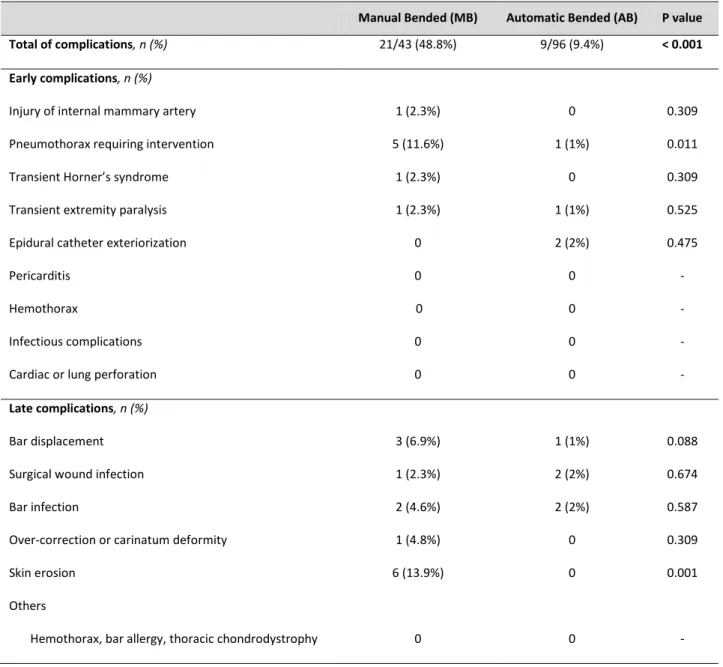
3.3. Complications
Table 3 summarizes early and late complications. There was no mortality. In general, there were less
complications in the AB group (9.4% vs. 48.8%, p<0.001). Regarding early complications, patients in the
MB group had a higher rate of pneumothorax (11.6% vs. 1%, p<0.05) requiring intervention
(percutaneous drainage, chest tube insertion or endotracheal intubation). Incidental findings on
postoperative control x-ray with spontaneous resolution and no need for intervention, such as residual
pneumothorax, pleural effusion or subcutaneous emphysema, were not considered. In MB group, there
was one episode of internal mammary artery injury, without repercussions. Epidural catheter-related
complications occurred in both groups: in MB patients, there was a case of transient Horner’s syndrome that reverted after removal of the catheter and there were two cases of catheter exteriorization in the
AB group. Besides transient extremity paralysis was reported in both groups, patients recovered
completely. Regarding late complications, there were bar displacements requiring intervention (MB – 3
vs. AB – 1, p=0.088), surgical wound infections (MB – 1 vs. AB – 2, p=0.674), bar infection (MB – 2 vs. AB – 2, p=0.587) or over-correction (MB – 1 vs. AB – 0, p=0.309). There was a significantly higher rate of skin
erosion in the MB group (MB – 6 vs. AB – 0, p<0.001).
3.4. Bar Removal
From a total of 139 patients, 91 (65.5%) are already without bar. The bar was removed later in the AB
group [median period with the bar, 28 months (range, 2-45 months) vs. 32 months (range, 18-45
months), p<0.001]. There was no statistical difference in median age at removal [16.9 (range 10.3-24.2)
9
4.
D
ISCUSSIONPatients with PE may occasionally experience symptoms, such as complaining of decreased tolerance
performing extenuating exercises. Other physical disabilities include recurrent upper respiratory tract
infections such as pneumonia, poor feeding, retarded growth, poor posture and even scoliosis.
Nevertheless, in most cases, the reasons for seeking medical care are related to psychosocial features
surrounding body image. Loss of self-esteem and social activities avoidance are common in this group of
patients. Moreover, there is evidence that surgical repair of PE considerably improves body image and
cardiorespiratory limitations on physical activity, improving patients’ quality of life [11]. Some groups presently use HI values greater than 3.20 to 3.25 as the main criterion for surgical correction [12]. Our
group adopted a patient-based approach, considering aesthetic parameters and assessing all physical
and psychological effects of the deformity, such as body image distortion.
Nuss procedure was mainly intended for pre-pubertal child. Besides optimal age for repair is still
unclear, it is currently recommended for patients between 8 and 12 years [1, 13]. A balance between
the younger patients’ chest softness and malleability and the older patients’ maturation to understand
and cooperate is desirable. Our average ages were higher – 14.4 and 14.9 years for MB and AB groups respectively. We believe this is an advantage: at first, rib cage flexibility is still preserved; furthermore,
this prevents recurrence of PE due to early correction. Even though, only further studies can validate the
impact of this hypothesis.
Several diagnostic tests can be performed before patients undergo MIRPE procedure. CT scan is
routinely requested in our department. High-resolution assessment of the deformity allows
automatically personalization of the bar bending process. It also reports cardiac or lung compression
and displacement showing the significant internal morbidity of what is often described as a purely
10 is still unknown [14]. Chest fast MRI was recently suggested for pre-operative workup and Piccolo and
colleagues [15] obtained accurate measurements of the chest avoiding CT scan radiation exposure.
Our center has been through important changes in the MIRPE procedure since 2007. A new concept of
morphology-based, patient approach was developed by creating the i3DExcavatum system [10].
Meticulous assessment of individual chest wall morphology and calculation of bar size and shape for
automatic bending is based on 3D reconstruction of costal grade; on the other hand, classic manual
bending is based on external shape of the thorax. Even for asymmetric patients, this resulted in the
appropriate size and better shape of the bar [10]. Smooth and precise bending of the bar and enhanced
fitting with uniform strength distribution applied by the prosthesis over the ribs are main advantages.
When compared with the Nuss traditional method, this system consistently showed better results [10].
In our experience, we found a statistically significant reduction in surgery time after the introduction of
automatic bending in 2007. This is one of the major advantages of this new approach. Per year analysis
revealed a consistent tendency of surgery time reduction along the years and from 2000 to 2013, the
procedure duration was reduced to half of time. However, we cannot exclude the learning curve in the
first years of implementation of the NP in our center. It is important to notice that complication rates
were lower with a faster procedure, revealing the safety and effectiveness of this modelling system.
Differences between soft tissue thicknesses of left and right thoracic wall sides exist in every patient,
irrespectively to their PE symmetry [16]. Furthermore, this tissue varies with age, sex and body mass
index. Accordingly, a particular advantage might exist in two groups: asymmetric or female patients.
Park and colleagues [17] began to adopt asymmetric bar shaping techniques and a symmetric repair was
achieved successfully. Even though, they recognized how challenging and laborious is to manually bend
the bar for different thoracic configurations. Software such as i3DExcavatum can precisely assist bar
modeling in order to achieve uniform strength exerted by the prosthesis on the ribs in this group of
11 MIRPE procedure. Manual bending of the bar can account additional errors due to breast tissue. In our
series, around 56% of the patients exhibited one form of asymmetric chest wall morphology based on
their chest CT. Regardless chest wall morphology, symmetrically shaped bars are applied in the classic
Nuss technique.
Bar displacement leads to imperfect correction or complete failure of the procedure. Nuss technique
suggests simple fixation of the bar to adjacent subcutaneous tissue. In 2002, Croitoru and Nuss
proposed a lateral stabilizer as a solution for this serious problem and reported a reduction from 15% to
5% in bar migration rate [18]. Pre-bended bar used by our group have a unilateral stabilizer incorporated
in the bar. Instead of using lateral stabilizers, a multiple point pericostal fixation technique was
presented by Park and colleagues [17] as a better approach, with lower rates of displacement – around 0.5% - and less difficulty on insertion and removal. However, this is a time-consuming step.
Vergunta et al [19], along with Dr. Nuss and Dr. Croitoru groups, support the use of 2 bars in severe or
older patients and already routinely use them in most patients. Pressure distribution over a wider area
and increased mechanical stability is provided using two bars placed above and below the midpoint of
the deformity, with one stabilizing plate for each, on opposite sides of the chest. On the other hand,
they may cause over correction in some patients. Every patient enrolled in the present study only
received one bar. We admit that patients with Grand Canyon type of PE can benefit from two bars.
Even being a minimally invasive procedure, the Nuss technique is associated with important
post-operative pain and in some patients this was described as a crucial limiting factor. In our institution, all
patients that underwent the MIRPE procedure received thoracic epidural catheter anesthesia during
hospital stay. With this approach, postoperative pain was successfully managed with a very high success
rate. Epidural analgesia kept the patients comfortable and stable and reduces bar displacement. It is
12 stably fixed and comfortably located and that there is no major trauma to the chest wall after the
procedure [17].
Cardiac or lung injury is one of the major complications of MIRPE procedure. It can occur during
retrosternal tunnel dissection, Nuss bar introduction or bar removal because anterior mediastinal space
is very narrow. In our center, we use thoracoscopy (VATS) to aid dissection and no cases of cardiac injury
were described, except for one patient with internal mammary artery injury. Darlong [6] recently
presented an innovative technique for real-time endoscopic vision of the retrosternal tunnel blind spot,
named pectus tunneloscopy. This comprises the use of a hollow transparent tube for bar conduit along
with thoracoscope insertion. As no additional cost, time or skin incision is needed, this can be a safe
route to avoid rare but fatal cardiopulmonary injuries. Other approaches comprise the use of a more
dorsally placed laparoscopic dissector instead of the Nuss introducer [20] or even a modified bilateral
13
5.
C
ONCLUSIONDuring a 13-year period, MIRPE procedure was safely performed in pediatric patients, even after
previous Ravitch surgery and with associated musculoskeletal disorders. Our department was very
successful with thoracic epidural catheter anesthesia, crucial for a better post-operative pain control.
Nuss procedure using automatic pre-bended bars improved the outcomes, significantly reducing the
time-consuming step of manual bending during surgery. After some years of learning curve, the actual
14
R
EFERENCES1. Nuss D, Kelly RE, Croitoru DP, et al: A 10-year review of a minimally invasive technique for the
correction of pectus excavatum. J Pediatr Surg 1998;33(4):545-52.
2. Nasr A, Fecteau A, Wales PW: Comparison of the Nuss and the Ravitch procedure for pectus
excavatum repair: a meta-analysis. J Pediatr Surg 2010;45(5):880-6.
3. Schier F: Vacuum treatment of pectus excavatum. Eur J Cardiothorac Surg 2006;30(4):687-8.
4. Snel BJ, Spronk CA, Werker PM, et al: Pectus excavatum reconstruction with silicone implants:
long-term results and a review of the english-language literature. Ann Plast Surg
2009;62(2):205-9.
5. Park HJ: Minimally Invasive Surgery for Pectus Excavatum: Park Technique. Journal of Clinical
and Analytical Medicine, 2011;2(3):84-90.
6. Darlong LM: Pectus tunneloscopy: making Nuss procedure for pectus excavatum safe. Interact
Cardiovasc Thorac Surg 2013;17(2):233-6.
7. Harrison MR, Gonzales KD, Bratton BJ, et al: Magnetic mini-mover procedure for pectus
excavatum III: safety and efficacy in a Food and Drug Administration-sponsored clinical trial. J
Pediatr Surg 2012;47(1):154-9.
8. Park HJ, Lee SY, Lee CS, et al: The Nuss procedure for pectus excavatum: evolution of techniques
and early results on 322 patients. Ann Thorac Surg 2004;77(1):289-95.
9. Lai JY, Wang CJ, Chang PY: The measurement and designation of the pectus bar by computed
15 10. Vilaça JL, Rodrigues PL, Soares TR, et al: Automatic Prebent Customized Prosthesis for Pectus
Excavatum Minimally Invasive Surgery Correction. Surg Innov (in press).
11. Kelly RE Jr, Cash TF, Shamberger RC, et al: Surgical repair of pectus excavatum markedly
improves body image and perceived ability for physical activity: multicenter study. Pediatrics
2008;122(6):1218-22.
12. Glinkowski W, Sitnik R, Witkowski M, et al: Method of pectus excavatum measurement based on
structured light technique. J Biomed Opt 2009;14(4):044041.
13. Fonkalsrud EW: Current management of pectus excavatum. World J Surg 2003;27(5):502-8.
14. Paterson A, Frush DP, Donnelly LF: Helical CT of the body: are settings adjusted for pediatric
patients? AJR Am J Roentgenol 2001;176(2):297-301.
15. Lo Piccolo R, Bongini U, Basile M, et al: Chest fast MRI: an imaging alternative on pre-operative
evaluation of Pectus Excavatum. J Pediatr Surg 2012;47(3):485-9.
16. Rodrigues PL, Direito-Santos B, Moreira AH, et al: Variations of the soft tissue thicknesses
external to the ribs in Pectus Excavatum patients. J Pediatr Surg 2013;48(9):1878-86.
17. Park HJ, Jeong JY, Jo WM, et al: Minimally invasive repair of pectus excavatum: a novel
morphology-tailored, patient-specific approach. J Thorac Cardiovasc Surg 2010;139(2):379-86.
18. Croitoru DP, Kelly RE Jr, Goretsky MJ, et al: Experience and modification update for the
minimally invasive Nuss technique for pectus excavatum repair in 303 patients. J Pediatr Surg
2002;37(3):437-45.
19. Vegunta RK, Pacheco PE, Wallace LJ, et al: Complications associated with the Nuss procedure:
16 20. Noguchi M, Kondoh S, Fujita K: A simple and safe technique for manipulation of retrosternal
dissection in the nuss procedure. Eplasty 2014;14:e8.
21. Cheng YL, Lee SC, Huang TW, et al: Efficacy and safety of modified bilateral
thoracoscopy-assisted Nuss procedure in adult patients with pectus excavatum. Eur J Cardiothorac Surg
17
L
EGENDSFigure 1. Intra-operative time progression of Nuss procedure along the years.
There was a statistically significant decrease in intra-operative time (vertical axis) during the years
evaluated (horizontal axis). The vertical line in the year 2007 indicates the introduction of the
Table 1. Demographic data and deformity characterization.
Manual Bended (MB) Automatic Bended (AB) P value
n (%) 43 (31%) 96 (69%) -
Gender, male-to-female ratio 33:10 75:21 0.858
Median age at surgery (range), years 14.4 (8-21) 14.9 (7-30) 0.098
Previous Ravitch procedure, n (%) 3/43 (7%) 7/96 (7%) 0.948
Median Haller Index (range) 3.95 (1.99–6.49) 3.32 (2.08–6.50) 0.008
Table 2. Surgical data.
Manual Bended (MB) Automatic Bended (AB) P value
Surgery duration, median (range), minutes 120 (60-195) 72 (45-136) < 0.001
Anesthesia duration, median (range), minutes 155 (70-120) 133 (75-215) < 0.001
Epidural catheter, n (%) 42 (98%) 94 (98%) 0.928
Need for PICU, n (%) 28 (65%) 1 (1%) < 0.001
Table 3. Early and late complications.
Manual Bended (MB) Automatic Bended (AB) P value
Total of complications, n (%) 21/43 (48.8%) 9/96 (9.4%) < 0.001
Early complications, n (%)
Injury of internal mammary artery 1 (2.3%) 0 0.309
Pneumothorax requiring intervention 5 (11.6%) 1 (1%) 0.011
Transient Horner’s syndrome 1 (2.3%) 0 0.309
Transient extremity paralysis 1 (2.3%) 1 (1%) 0.525
Epidural catheter exteriorization 0 2 (2%) 0.475
Pericarditis 0 0 -
Hemothorax 0 0 -
Infectious complications 0 0 -
Cardiac or lung perforation 0 0 -
Late complications, n (%)
Bar displacement 3 (6.9%) 1 (1%) 0.088
Surgical wound infection 1 (2.3%) 2 (2%) 0.674
Bar infection 2 (4.6%) 2 (2%) 0.587
Over-correction or carinatum deformity 1 (4.8%) 0 0.309
Skin erosion 6 (13.9%) 0 0.001
Others
A
GRADECIMENTOSO espaço limitado desta secção e o seu teor meramente textual não me permite expressar todos os
agradecimentos àqueles que, ao longo do meu Mestrado Integrado em Medicina, contribuiram para que
cumprisse os meus objectivos e concluísse com sucesso esta etapa da minha formação académica.
Deixo, assim, apenas algumas palavras a algumas figuras que destaco pela sua importante contribuição
para o presente trabalho.
Ao Professor Doutor Tiago Alexandre Henriques-Coelho, expresso o meu profundo agradecimento pelas
oportunidades e por todo o apoio e orientação prestada não apenas no decorrer do desenvolvimento
deste trabalho, mas também ao longo de todo o meu percurso académico. Agradeço a sua exigência e o
estímulo da vontade constante de aprender, de melhorar e de inovar.
Ao Dr. Ruben Lamas-Pinheiro, pela sua simpatia e disponibilidade e pela sua contribuição para o
presente trabalho.
A todo o Serviço de Cirurgia Pediátrica, por me ter proporcionado as condições necessárias para a
elaboração da minha dissertação e por permitir a minha integração numa equipa de elevada qualidade.
À minha família, em especial aos meus pais e ao meu irmão, pelo carinho, força e confiança transmitidos
para a realização deste projecto e ao longo de toda a minha formação.
Guide for Authors
Submitting the manuscript
In regard to preparation of clinical research papers, the Journal has a list of recommended guidelines that should
be addressed. This form can be obtained from the Journal website www.jpedsurg.org. Authors are requested to fill
out the checklist on the form and submit it along with your manuscript.
All manuscripts (including figures) must be submitted to the Journal of Pediatric Surgery through our Web site
(http://ees.elsevier.com/jpedsurg/). Submission items should include separate files for a cover letter, title page,
abstract, manuscript text, references, legends for table/figure, tables, and figures. Revised manuscripts should also
be accompanied by a unique file (separate from the cover letter) with responses to reviewers' comments. The
preferred order of files for electronic submission is as follows: cover letter, response to reviews (revised
manuscripts only), title page, manuscript file(s), table(s), figure(s). Files should be labelled with appropriate and
descriptive file names (e.g., SmithText.doc, Fig1.eps, Table3.doc). Upload text, tables and graphics as separate files.
Do not import figures or tables into the text document; submit them as separate files. Complete instructions for
electronic artwork submission can be found via the journal home page. All manuscripts must be submitted
double-spaced in English. Please visit http://ees.elsevier.com/jpedsurg/ to submit your manuscript electronically. The
website guides authors stepwise through the creation and uploading of the various files. Note that original source
files, not PDF files, are required. Once the submission files are uploaded the system automatically generates an
electronic (PDF) proof, which is then used for reviewing. All correspondence, including the Editor's decision and
request for revisions, will be by e-mail. All pediatric surgical image manuscripts, operative technique manuscripts,
Letters to the Editor, and Replies to Letters to the Editor should be submitted to Dr. Grosfeld online.
Correspondence concerning abstracts, book reviews, notices, reports of meetings, and other announcements
should be addressed to Dr. Grosfeld by mail or e-mailed to [email protected].
Original articles are preferred and are accepted for publication on the condition that they are contributed solely to
Reports are no longer accepted by the Journal of Pediatric Surgery. Authors have the option of submitting to the
new open access, online-only companion journal, Journal of Pediatric Surgery Case Reports
(http://www.jpscasereports.com). Dr. Grosfeld welcomes submissions to the Pediatric Surgical Images Section
that display the classic radiologic or pathologic childhood surgical disease or showcase new developments in
imaging relevant to pediatric surgery. Plain radiography, Ultrasound, CT (including 3-D/helical), MR (including MRA,
MRCP) and scintigraphy images may be used. Text and image reproductions are to be up no more than 2-3 journal
pages (approximately 5-7 typewritten pages). Content should focus on the radiographic diagnosis and include 2 to
3 images along with a supporting clinical photograph when appropriate. References should be limited to a
maximum of 10-12. A brief unstructured abstract and up to 6 keywords should accompany the submitted work.
Authors submitting a manuscript do so on the understanding that if it is accepted for publication, copyright in the
article, including the right to reproduce the article in all forms and media, shall be assigned exclusively to the
publisher.
The Journal of Pediatric Surgery subscribes in general to the "Uniform Requirements for Manuscripts Submitted to
Biomedical Journals"(N Engl J Med 336:309-315, 1997).
Preparing the manuscript
Manuscripts must be submitted electronically, preferably in Microsoft Word. References and figure legends must
appear at the end of the manuscript. Please refrain from using endnotes as references or automatic list numbering
because these features are lost in conversion: simply type the reference number in parentheses in the text and
type the reference list. Formatting, such as Greek letters, italics, super and subscripts may be used: the coding
scheme for such elements must be consistent throughout. Authors are responsible for applying for permission for
both print and electronic rights for all borrowed materials and are responsible for paying any fees related to the
applications of these permissions.
Please be sure to include an accurate mailing address (including US zipcode, or postal code for other countries)
telephone and fax numbers, and an email address for editorial communications and for reprint requests. All
A brief structured abstract of the paper with the headings Background/Purpose, Methods, Results, and Conclusions
should precede the body of the paper, to run no more than 200 words, and to replace any summary section at the
end of the article. Following the abstract should appear several words for the purposes of indexing to be titled:
KEY WORDS. The body of the paper should lead off with a minimum of 2 to 5 sentences, setting the general train of
thought, before any headings.
Measurements should be in the metric system.
Illustrations and tables
Figures and tables should be cited in order in the text; their position should be marked in the margin of the
manuscript. Arabic numbering should be used for both figures and tables. Legends for illustrations should be
typewritten, double-spaced, on a separate sheet, and included at the end of the manuscript. A legend must
accompany each illustration.
Electronic Submission of Illustrations: Figures may be submitted in electronic format. Images should be provided
in EPS or TIF format. Graphics software such as Photoshop and Illustrator, not presentation software such as
PowerPoint, CorelDraw, or Harvard Graphics, should be used to create the art. Color images must be CMYK, at
least 600 DPI, with a digital color proof, not a color laser print or color photocopy (this proof will be used at press
for color reproduction).Gray scale images should be at least 1200 DPI and accompanied by a proof. Combinations
of gray scale and line art should be at least1200 DPI and accompanied by a proof. Line art (black and white or
color) should be at least 1200 DPI and accompanied by a proof. Please include hardware and software information,
in addition to the file names.
Each table should be provided in a separate file and appropriately numbered. Legends should appear with the
same sheets as the tables. The contributor must bear all costs connected with printing color illustrations.
References
References should be compiled at the end of the article according to the order of citation in the text, not
information must be accurate. Abbreviations for titles of medical periodicals should conform to those used in the
latest edition of Index Medicus. Give inclusive page numbers.
Examples of references
Journal article, one author:
1. Valayer J: Conventional treatment of biliary atresia: Long-term results. J Pediatr Surg 1996;31:1546-1551.
Journal article, two or three authors:
2. Atwell JD, Spargo PM: The provision of safe surgery for children. Arch Dis Child 1992;67:345-349.
Journal article, more than three authors:
3. Seo T, Ito T, Ishiguro Y, et al: New neonatal extracorporeal membrane oxygenation circuit with a self-regulating
pump. Surgery 1994;115:463-472.
Journal article, in press:
4. Coran AG: The hyperalimentation of infants. Biol Neonate (in press)
Complete book:
5. Rowe MI, O'Neill JA, Grosfeld JL, et al: Essentials of Pediatric Surgery. St Louis, MO, Mosby Year-Book, 1995
Chapter of book:
6. Skandalakis JE, Gray SW, Ricketts R: The esophagus, in Skandalakis JE, Gray SW (eds): Embryology for Surgeons.
Baltimore, MD, Williams & Wilkins, 1994, pp 65-112
Paper presented at a meeting:
7. Bealer JF, Vanderwall K, Adzick NS, et al: A new treatment option for patients with congenital diaphragmatic
hernia. Presented at the 14th annual meeting of the International Fetal Medicine and Surgery Society, Newport, RI,
May 3-6, 1996
Proofreading
Contributors are provided with galley proofs and are asked to proofread them for typesetting errors. Important
changes in data are allowed, but authors will be charged for excessive alterations in proof. Galley proofs should be
Reprints
Reprints of articles will be furnished to contributors when ordered in advance of publication. An order form,
showing cost of reprints, is sent with proofs. Individuals wishing to obtain reprints of an article that appeared in
the Journal of Pediatric Surgery can do so by contacting the author at the address given in the journal.
Open Access
This journal offers authors two choices to publish their research;
1. Open Access
* Articles are freely available to both subscribers and the wider public with permitted reuse
* An Open Access publication fee is payable by authors or their research funder
2. Subscription
* Articles are made available to subscribers as well as developing countries and patient groups through our access
programs (http://www.elsevier.com/access)
* No Open Access publication fee
All articles published Open Access will be immediately and permanently free for everyone to read and download.
Permitted reuse is defined by your choice of one of the following Creative Commons user licenses:
Creative Commons Attribution-Non Commercial-Share Alike (CC BY-NC-SA): for non-commercial purposes, lets
others distribute and copy the article, to create extracts, abstracts and other revised versions, adaptations or
derivative works of or from an article (such as a translation), to include in a collective work (such as an anthology),
to text and data mine the article, as long as they credit the author(s), do not represent the author as endorsing
their adaptation of the article, do not modify the article in such a way as to damage the author's honor or
reputation, and license their new adaptations or creations under identical terms (CC BY NC SA).
Creative Commons Attribution-Non Commercial-No Derivs (CC-BY-NC-ND): for non-commercial purposes, lets
others distribute and copy the article, and to include in a collective work (such as an anthology), as long as they
credit the author(s) and provided they do not alter or modify the article.
Creative Commons Attribution (CC-BY): available only for authors funded by organizations with which we have
Elsevier has established agreements with funding bodies. This ensures authors can comply with funding body Open
Access requirements, including specific user licenses, such as CC-BY. Some authors may also be reimbursed for
associated publication fees. http://www.elsevier.com/fundingbodies.
To provide Open Access, this journal has a publication fee which needs to be met by the authors or their research
funders for each article published Open Access. Your publication choice will have no effect on the peer review
process or acceptance of submitted articles.
The Open Access publication fee for this journal is $USD 2,500, excluding taxes.
Learn more about Elsevier's pricing policy http://www.elsevier.com/openaccesspricing.
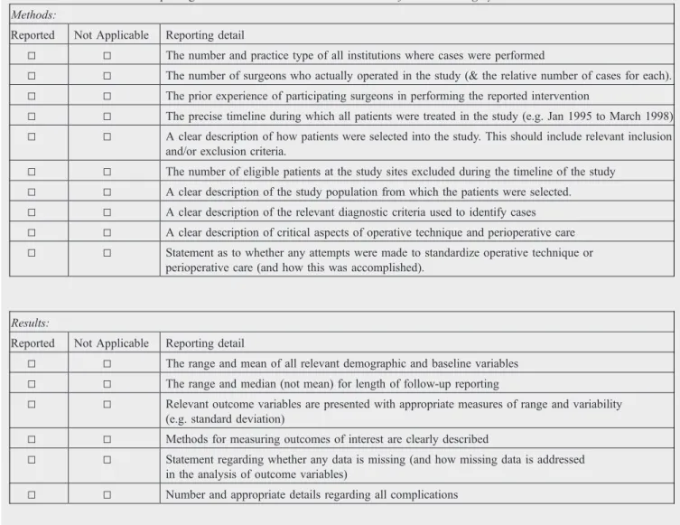
Table 1 Guidelines for the reporting of clinical research data in theJournal of Pediatric Surgery
Methods:
Reported Not Applicable Reporting detail
5 5 The number and practice type of all institutions where cases were performed
5 5 The number of surgeons who actually operated in the study (& the relative number of cases for each).
5 5 The prior experience of participating surgeons in performing the reported intervention
5 5 The precise timeline during which all patients were treated in the study (e.g. Jan 1995 to March 1998)
5 5 A clear description of how patients were selected into the study. This should include relevant inclusion and/or exclusion criteria.
5 5 The number of eligible patients at the study sites excluded during the timeline of the study
5 5 A clear description of the study population from which the patients were selected.
5 5 A clear description of the relevant diagnostic criteria used to identify cases
5 5 A clear description of critical aspects of operative technique and perioperative care
5 5 Statement as to whether any attempts were made to standardize operative technique or perioperative care (and how this was accomplished).
Results:
Reported Not Applicable Reporting detail
5 5 The range and mean of all relevant demographic and baseline variables
5 5 The range and median (not mean) for length of follow-up reporting
5 5 Relevant outcome variables are presented with appropriate measures of range and variability (e.g. standard deviation)
5 5 Methods for measuring outcomes of interest are clearly described
5 5 Statement regarding whether any data is missing (and how missing data is addressed in the analysis of outcome variables)
5 5 Number and appropriate details regarding all complications
Additional details for studies reporting more than one treatment group (e.g. controls):
Reported Not Applicable Reporting detail
5 5 Mean and range for all relevant demographic and baseline variables for all treatment groups.
5 5 The range and median (not mean) for length of follow-up reporting for each treatment group.
5 5 A precise timeline during which all patients were treated for each group
5 5 Outcome variables being compared between groups are presented with appropriate measures of variability (e.g. standard deviation)
5 5 Measures of type II error (P-values) for comparison statistics are presented with actual values ifP= .01 or larger
(e.g.P= NS andPb.05 are not acceptable)
5 5 A description of how patients were selected into each treatment group
5 5 A statement is made as to whether the same surgeons operated on patients from different treatment groups
Manuscripts concerning clinical research should follow a uniform set of reporting guidelines. The guidelines, listed above, were developed from sound clinical research principles and are designed to improve the reporting accuracy of clinical data pertaining to surgical conditions.