The unique regulation of iron-sulfur cluster biogenesis
in a Gram-positive bacterium
Joana A. Santosa,b, Noelia Alonso-Garcíaa, Sandra Macedo-Ribeiroa,1, and Pedro José Barbosa Pereiraa,1
aInstituto de Biologia Molecular e Celular (IBMC), Universidade do Porto, 4150-180 Porto, Portugal; andbInstituto de Ciências Biomédicas de Abel Salazar
(ICBAS), Universidade do Porto, 4050-313 Porto, Portugal
Edited by Gregory A. Petsko, Weill Cornell Medical College, New York, NY, and approved April 28, 2014 (received for review December 6, 2013)
Iron-sulfur clusters function as cofactors of a wide range of proteins, with diverse molecular roles in both prokaryotic and eukaryotic cells. Dedicated machineries assemble the clusters and deliver them to the final acceptor molecules in a tightly regulated process. In the prototypical Gram-negative bacterium Escherichia coli, the two existing iron-sulfur cluster assembly systems, iron-sulfur cluster (ISC) and sulfur assimilation (SUF) pathways, are closely intercon-nected. The ISC pathway regulator, IscR, is a transcription factor of the helix-turn-helix type that can coordinate a [2Fe-2S] cluster. Redox conditions and iron or sulfur availability modulate the liga-tion status of the labile IscR cluster, which in turn determines a switch in DNA sequence specificity of the regulator: cluster-con-taining IscR can bind to a family of gene promoters (type-1) whereas the clusterless form recognizes only a second group of sequences (type-2). However, iron-sulfur cluster biogenesis in Gram-positive bacteria is not so well characterized, and most organisms of this group display only one of the iron-sulfur cluster assembly systems. A notable exception is the unique Gram-posi-tive dissimilatory metal reducing bacteriumThermincola potens, where genes from both systems could be identified, albeit with a diverging organization from that of Gram-negative bacteria. We demonstrated that one of these genes encodes a functional IscR homolog and is likely involved in the regulation of iron-sulfur cluster biogenesis inT. potens. Structural and biochemical character-ization ofT. potensand E. coliIscR revealed a strikingly similar architecture and unveiled an unforeseen conservation of the unique mechanism of sequence discrimination characteristic of this distinc-tive group of transcription regulators.
Rrf2-like regulator
|
transcription regulation|
helix-turn-helix motif|
DNA recognition
|
specificity modulationI
ron-sulfur (Fe/S) proteins play crucial roles for the functioning of both prokaryotic and eukaryotic cells, being required for biological functions ranging from electron transport to redox and nonredox catalysis, and from DNA synthesis and repair to sens-ing in regulatory processes (1). The main role of the Fe/S cluster assembly machineries is to mobilize iron and sulfur atoms from their storage sources, assemble the two components into an Fe/S cluster, and then transfer the newly formed cluster to the final protein acceptors (2). InEscherichia coli, there are two of these Fe/S cluster‘‘factories,’’the ISC (iron-sulfur cluster) and SUF (sulfur assimilation) systems, whose corresponding genes are organized in two operons, iscSUA-hscBA-fdx and sufABCDSE, respectively (2, 3). Deletion mutants of the ISC system display a variety of growth defects due to loss of Fe/S cluster-containing enzyme activity and disruption of sulfur metabolism whereas failure of both the ISC and SUF systems leads to synthetic lethality (4, 5).InE. coli, the ISC machinery is considered the housekeeping system responsible for the maturation of a large variety of Fe/S proteins whereas the SUF system is triggered under stress con-ditions, such as oxidative stress or iron starvation (6). ISC pathway regulator (IscR) is a [2Fe-2S] cluster-containing tran-scription factor with a single predicted helix-turn-helix motif, first identified for its role in regulating expression of the ISC
biogenesis pathway (7) and subsequently found to control the expression of more than 40 genes inE. coli(7, 8). According to the currently accepted model for Fe/S cluster biogenesis, under conditions unfavorable for Fe/S cluster formation, the labile IscR cluster is lost, and IscR-mediated repression of the isc (iron-sulfur cluster) operon is alleviated. At the same time, apo-IscR activates the suf (sulfur assimilation) operon to further com-pensate for damage or loss of Fe/S clusters (9, 10). Once the demand for Fe/S biogenesis is met, higher levels of cluster-con-taining holo-IscR exist, causing an increased repression of the ISC pathway. Moreover, under iron limitation, the ISC and SUF machineries are unable to maintain the levels of holo-IscR, and therefore this feedback mechanism allows IscR to sense Fe/S demand and enables E. colito respond appropriately to stress conditions (11).
There are two classes of IscR binding sites in theE. coli ge-nome: a type-1 site deduced fromiscR,yadR, andyhgIpromoter regions and a type-2 site compiled from the IscR sites upstream of the hyaA, ydiU, andsufA promoters (8). Interestingly, IscR binds type-1 promoters solely in its holo-form whereas binding to type-2 promoters was shown to be independent of the presence of the Fe/S cluster (12). InE. coli, IscR mutation E43A enabled specific recognition of type-1 promoters by apo-IscR, likely mimicking the interaction mode of the cluster-bound form of the protein (13).
Significance
Iron-sulfur clusters are ubiquitous cofactors of proteins in-tervening in disparate biological processes. Iron-sulfur cluster biosynthesis pathways are tightly regulated in Gram-negative bacteria. One of the participating transcription factors, iron-sulfur cluster pathway (ISC) regulator (IscR), can itself bind an iron-sulfur cluster. Depending on its ligation status, IscR recognizes and binds to distinct promoters, therefore modu-lating cluster biosynthesis. This unique protein at the crossroad between the ISC and sulfur assimilation (SUF) iron-sulfur clus-ter biosynthetic pathways was thought to be restricted to Gram-negative bacteria. We demonstrated the existence of a func-tional IscR in the unique Gram-positive bacteriumThermincola potens. Structural and functional analysis of T. potens and
Escherichia coliIscR unveiled a conserved mechanism of pro-moter discrimination, along with subtle structural differences that explain their distinct DNA sequence recognition specificity.
Author contributions: J.A.S., S.M.-R., and P.J.B.P. designed research; J.A.S., N.A.-G., and P.J.B.P. performed research; J.A.S., N.A.-G., S.M.-R., and P.J.B.P. analyzed data; and J.A.S., S.M.-R., and P.J.B.P. wrote the paper.
The authors declare no conflict of interest. This article is a PNAS Direct Submission.
Data deposition: The atomic coordinates and structure factors have been deposited in the Protein Data Bank,www.pdb.org(PDB ID codes4CHUand4CIC).
1To whom correspondence may be addressed. E-mail: [email protected] or sribeiro@
ibmc.up.pt.
This article contains supporting information online atwww.pnas.org/lookup/suppl/doi:10. 1073/pnas.1322728111/-/DCSupplemental.
BIO
CHEMISTRY
PNAS
Fe/S cluster biosynthetic machineries. Most Gram-positive bac-teria carry only asufoperon, containing genes coding for SufU and the SufBCD complex (14, 15), but nosufEorsufA-related genes, even if in some casessufAcan be found elsewhere in the genome (14, 16).
Thermincola potens(strain JR) is an anaerobic, thermophilic, Gram-positive dissimilatory metal-reducing bacterium (DMRB), isolated from a thermophilic microbial fuel cell (17). It is of the first Gram-positive DMRB for which there is a complete genome sequence, which revealed an unusual abundance of multiheme c-type cytochromes (17, 18). Using homology searches, we iden-tified a series of genes with sequence similarity to both E. coli SUF and ISC machineries in theT. potensgenome, including a gene locus coding for a putative IscR protein. Taken together, our results both identify and characterize a unique Fe/S bio-genesis regulator in Gram-positive bacteria. Through structural and biochemical analysis of bothT. potensandE. coliapo-IscR proteins and their E43A mutants, we were able to unveil subtle structural features important for DNA recognition and binding specificity.
Results
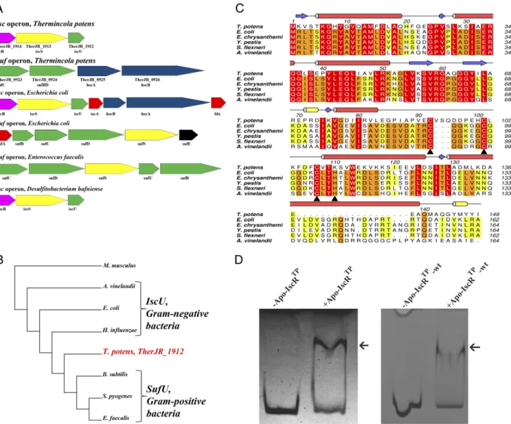
Unique Fe/S Cluster Biogenesis inT. potens.In Gram-positive bac-teria, there is conservation of the sufoperon, often present as sufCDSUB, which is the only machinery for Fe/S cluster bio-synthesis in the majority of these organisms (14, 19). Surpris-ingly, homology searches on the Gram-positive DMRBT. potens JR genome (17) allowed identifying two gene loci with sequence similarity to E. coli SUF and ISC machineries (Fig. 1A). In T. potens, there are ORFs coding for homologs of the tran-scription factor IscR (TherJR_1914, 37% identical to theE. coli protein) (7), the cysteine desulfurase IscS (TherJR_1913) (4, 20), and the scaffold IscU (TherJR_1912) (21) from the ISC pathway. An additionalsuf-like operon inT. potenscomprises homologs of sufC (TherJR_0923), sufB, and sufD (TherJR_0924) from the E. coliSUF pathway, and the chaperones hcsA (TherJR_0925) and hcsB (TherJR_0926) from theE. coli iscoperon (22–24).
Compared with other Gram-positive bacteria, namely from the Firmicutes phylum, some unique features of the T. potens suf operon become evident. InT. potens, the sufoperon does not code for cysteine desulfurase (SufS) homologs although there are elsewhere in the T. potens genome two additional genes coding for putative cysteine desulfurases (TherJR_0460 and TherJR_3003) homologous to CsdA/SufS, which can function as complementary sulfur sources for Fe/S cluster biogenesis, possibly through the recruitment of the SUF machinery (25). TheT. potens sufoperon is also devoid of homologs of SufU, recently reported to be a zinc-dependent sulfurtransferase in Bacillus subtilis(26), but encodes a sufBD protein, which to-gether with sufC was shown to act as scaffold in Gram-nega-tive bacteria (27, 28). Furthermore, thesufoperon inT. potens includes the hscAandhscBgenes coding for the chaperones responsible for transferring preformed clusters from the scaf-fold IscU to final acceptors and that, in E. coli, are cotran-scribed with the isc and not with the sufoperon (4). Addi-tionally, genes coding for A-type carriers are absent from the T. potensgenome.
IscU is a highly conserved protein that functions as scaffold for cluster assembly and subsequent transfer. Preserved features include the cluster ligands (three cysteines and one histidine), an aspartate residue that plays a critical role in cluster transfer to apo-proteins and the LPPVK motif recognized by the chaperone
insertion signature specific of SufU-type proteins. Accordingly, phylogenetic analysis of IscU and SufU protein sequences places the protein encoded by the TherJR_1912 gene between the Gram-negative IscU-type and the SufU-like proteins from Gram-positive bacteria (Fig. 1B).
As previously reported for Clostridium perfringens (30), searches for Fe/S cluster biogenesis operons in Gram-positive bacteria with completely sequenced genomes, namely the DMRBs Desulfitobacterium hafnienseandDesulfotomaculum reducens, revealed that they possess a single ISC gene locus (iscRSU) but no SUF apparatus. In contrast, in other Gram-positive bacteria (e.g.,E. faecalis), only the SUF pathway can be found. Therefore, contrary to other Gram-positive bacteria described so far,T. potens not only has two gene loci coding for the two Fe/S cluster bio-synthesis machineries present inE. coli(ISC and SUF), but these systems display a unique organization.
TheT. potensTherJR_1914 Gene Codes for IscR.IscR is a [2Fe-2S]
cluster-containing transcriptional regulator encoded by the first gene of theiscRSUA-hscBA-fdxoperon that regulates both ISC and SUF systems inE. coli and other Gram-negative bacteria (10, 11). Apart from the sequence-unrelated SufR found in cya-nobacteria (31, 32), no IscR homolog was described in Gram-positive bacteria, with the possible exception of some species of theClostridium genus for which functional data are still lacking (30). InT. potens, the TherJR_1914 gene encodes a protein of the Rrf2 family of transcriptional regulators, sharing only 37% identity withE. coliIscR but with full conservation of the cysteine residues known to coordinate the [2Fe-2S] cluster (Cys92, 98, 104, E. coli numbering) (Fig. 1C) (10).
Clusterless (apo) IscR fromE. coliwas shown to activatesuf operon expression during stress conditions, such as iron starva-tion (6). The as-purified apo form of the protein encoded by T. potensgene TherJR_1914 (apo-IscRTp-wt) was found to bind to the upstream region of the putativesufoperon, between genes TherJR_0922 and TherJR_0923 (Fig. 1D). A similar behavior was observed for a triple mutant (C92/101/107S) of IscRTp (apo-IscRTp) (Fig. 1D) where all putative cluster-binding cysteine residues (Fig. 1C) were replaced by serine. Given the structural similarity between cysteine and serine and the requirement for homogeneous sample for downstream functional and structur-al assays, this variant was used in structur-all experiments where the apo form of IscRTp was required. The ability of apo-IscRTp-wt and apo-IscRTpto bind the promoter region of thesufoperon suggests that IscRTpcan function as an Fe/S cluster regulator in this organism, with the apo form involved in the regulation of the suf operon expression, as observed for E. coli. Although such regulators have been found and characterized in a number of Gram-negative bacteria (7, 8, 33–35), the characterization of orthologous proteins from Gram-positive species has not yet been reported. Therefore,T. potenshas a unique organization and regulation of Fe/S cluster assembly genes, among Gram-positive bacteria.
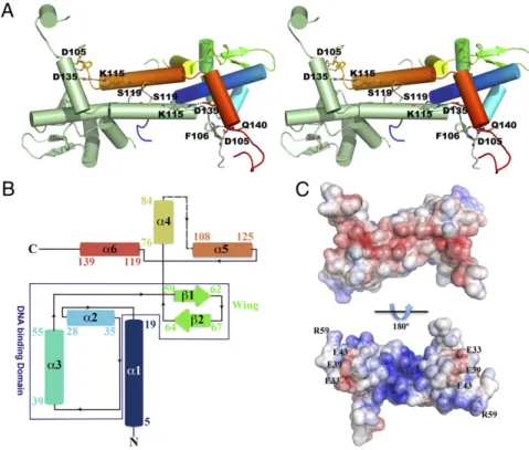
are predominantlyα-helical and, whereas the N-terminal region is formed by three consecutiveα-helices preceding the charac-teristic wing β-hairpin, the C-terminal domain is exclusively α-helical (Fig. 2B). Within each monomer, the N-terminalα-helix (α1) interfaces with the wing-helix subdomain (α2-α3-β1-β2) and with the N-terminal end of the dimerization helix (α5) from the adjacent monomer. The dimerization helix, which comprises two of the putative iron-sulfur cluster binding res-idues (101 and 107), is further stabilized by close contacts with helixα6 from the adjacent monomer. Residues 86–100, encom-passing part of the putative iron-sulfur cluster-binding segment (Fig. 2B), are disordered in both monomers and could not be modeled (Fig. 2A andB). The structures of the monomers are
nearly identical, superposing with an rmsd of 0.17 Å for 133 aligned Cαatoms.
Apo-IscRTpdimer formation involves mostly interactions be-tween residues from helix α5 of each monomer, but residues from helices α1 and α4 further stabilize the homodimer. The extensive dimerization interface between the two helices α5 is hydrophobic, except for a single hydrogen bond between the side chains of neighboring Ser119 residues. Further intermonomer polar interactions are established between the side chain of Gln140 at the C terminus of helix α6 and Asp105 OD1 and Phe106 O (Fig. 2A).
In agreement with its DNA-binding function (Fig. 1D), the electrostatic surface potential of apo-IscRTpis highly polarized
Fig. 1. Identification of an Fe/S cluster biosynthesis regulator inT. potens. (A)T. potenspossesses a unique organization of genes involved in Fe/S cluster biosynthesis, with bothiscandsufoperons. Colors denote gene function conservation between Gram-negative (E. coli), Gram-positive (E. faecalis), and DMRB Gram-positive bacteria (T. potensandD. hafniense). (B) The amino acid sequence ofT. potensscaffold protein reveals features characteristic of the IscU-proteins from Gram-negative bacteria. Neighbor-joining phylogenetic analysis of conserved protein sequences of putative IscU-type or SufU-type IscU-proteins in both Gram-positive (T. potens,Streptococcus pyogenes,E. faecalis,B. subtilis)and Gram-negative bacteria (E. coli,Azotobacter vinelandii,Haemophilus influenza), usingMus musculusas outgroup. The sequences were aligned with three distinct alignment algorithms as implemented in ADOPS (57). The resulting cladogram places theT. potensscaffold protein between IscU proteins from Gram-negative bacteria and SufU proteins from other Gram-positive bacteria. (C) TheT. potensTherJR_1914 gene codes for a protein that is highly homologous to IscR from Gram-negative bacteria. Strictly conserved amino acids are highlighted in red, and increasing residue conservation is represented by a color gradient from green to red. Alignment prepared with ClustalW (58) and colored with Aline (59). (D)T. potensapo-IscR recognizes the upstreamsufoperon region between genes TherJR_0922 and TherJR_0923. Incubation of either apo-IscR Cys-to-Ser mutant (apo-IscRTp) or its as-purified wild-type version (apo-IscRTp-wt) with the putativesufpromoter region (sufsequence) (Table
S1) resulted in a mobility-shift (arrow).
BIO
CHEMISTRY
PNAS
(Fig. 2C), being predominantly negative at the solvent-exposed face of helicesα5 andα6, and positively charged at the opposite, putative DNA-binding side. This asymmetric surface-charge distribution promotes initial DNA positioning by nonspecific electrostatic contacts, before fine-tuning through the estab-lishment of base- and shape-specific interactions. Negatively charged residues (Glu33, Glu39, and Glu43) protrude from this positively charged surface (Fig. 2C), resemblingE. coliIscR (13). Among these residues, Glu39 is unique in T. potens IscR (replaced by a leucine residue in closely related molecules) (Fig. 1C).
T. potens IscR belongs to the Rrf2-like family of transcrip-tional regulators and displays highest structural similarity with the global cysteine regulator CymR from B. subtilis (PDB ID code 2Y75) (36) andS. aureus(PDB ID code 3T8T) (37) as well as with the recently determined structure ofE. coliIscR (PDB ID code 4HF0) (13), superposing with these models with an rmsd of 1.5–2.3 Å. The conserved DNA-binding helix-turn-helix motif can also be superposed to the corresponding domain of more distantly related proteins, albeit with somewhat higher rmsd values (Table 1). The DNA-binding winged-helix motif is structurally similar in the selected structures (Fig. 3 Aand B), and most structural differences occur in the dimerization helixα5
and in the length and orientation of helixα4, which precedes the iron-sulfur cluster-binding region.
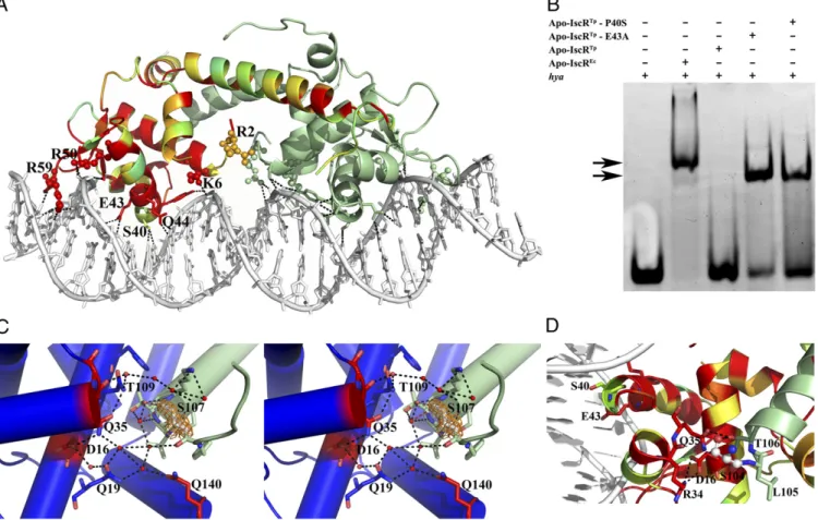
Molecular Details of IscR-DNA Interaction.To better grasp the fine molecular details of specific promoter sequence recognition by IscR, we determined the structure of apo-IscR fromE. coli (C92/98/104S triple mutant with the putative cluster-binding cysteine residues mutated to serine; apo-IscREc) in complex with theE. coli hya (hydrogenase-1) promoter sequence (8, 12, 31). The asymmetric unit contains the apo-IscREc biological dimer bound to a 26-bp double-stranded oligonucleotide with a single nucleotide overhang at the 5′end of each strand (Fig. 4A and Table S1). This structure is very similar to the recently reported model ofE. coliapo-IscR (C92/98/104A triple mutant) in com-plex with DNA (PDB ID code 4HF1) (13). Overall, the protein main chains of the two models superpose with an rmsd of 0.5 Å for 124 aligned Cα atoms. DNA binding induces a small con-certed movement of the wing-helix motif within each apo-IscREc monomer and of the dimerization helix α5 of the adjacent monomer (Fig. S2AandB) (13). In the complex, helixα3 of the winged-helix motif from each apo-IscREcmonomer is presented to the major groove of the corresponding DNA half-site whereas theβ-hairpin inserts into the minor groove (Fig. 4A).
sented as dashed lines. (B) Topology diagram of the apo-IscRTp monomer. Secondary structure element
colors match those ofA. (C) Solid-surface represen-tation of the apo-IscRTpdimer, with mapped
elec-trostatic surface potential contoured from+5 (blue) to−5 (red) kbTe−1[kb, Boltzmann’s constant; T, tem-perature (K); e, charge of an electron].
Table 1. Structural similarity betweenT. potensIscR and other winged-helix transcription regulators
Protein PDB ID code rmsd, Å
No. of aligned Cαatoms
Amino acid
sequence identity, %* Z-score
B. subtilisCymR 2y75 1.5 119 55 17.2
E. coliIscR (unliganded) 4hf0 2.0 116 41 16.8
E. coliIscR (DNA complex) 4hf1 2.3 120 40 16.3
S. aureusCymR 3t8t 2.3 119 46 15.9
Putative transcriptional regulator fromL. innocua
3lwf 3.8 124 48 14.9
B. cereusprotein BC1842 1ylf 2.7 118 20 13.9
S. aureus,Staphylococcus aureus;L. innocua,Listeria innocua;B. cereus,Bacillus cereus.
As observed for apo-IscRTp, the apo-IscREcbiological dimer is highly polarized with a clustering of basic residues on the DNA-interacting surface (Fig. S2C). Although the positive electrostatic surface might be required to orient the protein toward pro-ductive binding with the DNA duplex, in the apo-IscREc-DNA complex very few protein-DNA interactions involve basic side chains. A notable exception is Arg59 that protrudes from the β-hairpin wing and wedges into the symmetrically related AT-rich regions of the narrow groove (Fig. 4A). Accordingly, the E. coliapo-IscR R59A mutant is unable to bind to either type-1
or type-2 promoter sequences, demonstrating the relevance of this residue for DNA recognition (13). Further interactions with the minor grove are established by residues at the tip of the wing (Gly60-Pro61) that slot between deoxyribose moieties and by Pro27 that packs against phosphodiester linkages.
Apo-IscREc residues contributing to the partial negative charge within the DNA-binding surface (Glu33, Asp30, and Glu43) (Fig. S2C) are structurally equivalent to the acidic resi-dues identified on the equivalent side of apo-IscRTp. However, only Glu43 contacts directly the bound oligonucleotide. Together Fig. 3. IscR fromT. potensis structurally similar to other winged-helix transcriptional regulators. Su-perposition of apo-IscRTpmonomer (green) with (A)
freeE. coliapo-IscR C92/98/104A (blue; PDB ID code 4HF0) and (B) freeB. subtilisCymR (magenta; PDB ID code 2Y75) highlighting the overall structural conservation.
Fig. 4. Binding of IscR to type-2 promoter sequences. (A) Bidentate binding of apo-IscREc(C92/98/104S) to thehyapromoter DNA sequence. In one of the
monomers, residues are colored according to conservation, where red corresponds to positions strictly conserved betweenE. coliandT. potensIscR. Residues at the DNA-interacting interface are highlighted as sticks and basic residues as spheres. Hydrogen bonds between apo-IscREcand DNA are represented as
dotted lines. (B) Electrophoretic mobility-shift assay analysis of apo-IscR binding to theE. coli hyapromoter. Arrows denote observed band-shifts. (C) Ste-reoscopic view of the intricate network of hydrogen bonds in apo-IscRTp(one monomer of the functional dimer is colored green and the other one blue)
centered on the cluster-binding residue 107 (light gray). The 2Fo−Fcelectron density map around residue 107 is represented as an orange mesh. Water
molecules and strictly conserved residues in closely related IscR molecules are colored red. (D) In apo-IscREc(C92/98/104S), serine 104 (ball and stick) participates
in a network of polar interactions with neighboring residues (sticks), cross-linking helicesα1,α2, andα5. The corresponding cysteine residue in the wild-type protein could be part of a sensing mechanism for the presence of the Fe/S cluster.
BIO
CHEMISTRY
PNAS
Subtle Structural Differences Modulate DNA Sequence Recognition Specificity. Overall, there is a striking conservation of the DNA-binding interface (Figs. 1C and 4A). Apo-IscRTp differs from the E. colihomolog only at four positions within the in-teraction surface, which could result in altered DNA binding affinity and specificity: Ser27 (Pro27 in apo-IscREc), Pro40 (Ser40 in apo-IscREc), and Ala61-Gln62 (Pro61-Gly62 in apo-IscREc). The replacement of Pro27 by a serine is likely to in-crease the flexibility of the linker between the first twoα-helices although a large change in DNA affinity is not predictable. In contrast, the substitution of Pro61-Gly62 by an Ala-Gln di-peptide can impact the conformation of the wingβ-hairpin and interfere with the tight packing of this structural element within the minor groove. In particular, the residue at position 40 is likely to play a key role in sequence-specific recognition of DNA. In the apo-IscREc-DNA complex, the protein packs very tightly within the major groove, leaving limited space for bulkier resi-dues (Fig. 4A). Although a proline could be accommodated at the N terminus of helix α3 without helical disruption, the resulting steric hindrance might prevent the placement and base readout of the conserved Glu43-Gln44 and/or contacts of the residues interacting with the phosphate backbone (Tyr9, Ser38, Tyr41). Substitution of the purines interacting with Ser40 (G20′ and A19) prevents binding ofE. coli apo-IscR to thehya pro-moter sequence, highlighting the importance of this residue for base-specific recognition (12). Further, mutation of Ser40 to alanine inE. coliIscR decreases binding to thehyapromoter by 90% compared with the wild-type protein, a decrease that is sequence-dependent and more pronounced for type-2 sites (13). The influence of Pro40 in apo-IscRTpinteraction with DNA is evidenced by its inability to bind the E. coli hyapromoter se-quence (Fig. 4Band Table S2). Replacement of Pro40 in apo-IscRTpby the structurally equivalent amino acid inE. coliIscR (apo-IscRTpP40S) is sufficient to allow binding to the heterol-ogous promoter (Fig. 4BandTable S2). The presence of a serine residue at position 40 is likely to alleviate the tight packing of IscR within the major groove of DNA, reducing steric hindrance and allowing binding. Accordingly, the IscRTp E43A mutant, where the shorter alanine side chain can provide room for po-sitional adjustments of this region, also recognized thehya se-quence (Fig. 4B) with an affinity comparable with that of the E. coliprotein, as assessed by microscale thermophoresis (Table S2). Taken together, these results suggest that substitution of Ser40 by a proline in apo-IscRTp prevents base recognition through steric hindrance, an impairment lifted by introducing less bulky residues at either position 40 or 43.
Position of the Cluster-Binding Residues. In contrast to previous studies withE. coliIscR, where all putative cluster-binding cys-teine residues were mutated to alanine to obtain homogeneous clusterless protein (10, 12, 13), in T. potens IscR, the corre-sponding residues were mutated to serine, which is a closer structural match. In allE. coliandT. potensIscR structures, the region involved in iron-sulfur cluster association is partially dis-ordered, but the serine residues replacing Cys107 in apo-IscRTp and Cys104 in apo-IscREc are clearly visible in the electron density maps (Fig. 4CandD). In contrast to what is observed for the Cys-to-Ala mutant structure ofE. colifree apo-IscR where the two visible cluster ligands (Ala104 and His107) are on the outer face of the longer dimerization helix α5 (13), in apo-IscRTp, the equivalent Ser107 is part of the coil region preceding
bond network interfacing the two monomers of the functional dimer and involving the side chains of Gln19, Asp16, and Gln35 from the adjacent monomer (the last two residues strictly con-served across IscR molecules) (Fig. 1C). Of particular relevance is the involvement of Asp16 side chain in a salt bridge with Arg34 within the DNA-binding helix-turn-helix motif. The cluster-binding segment is further stabilized by a polar contact with Gln140 of the adjacent monomer, which also connects the cor-responding helicesα1 andα6. Altogether, this polar interaction network tightly connects the cluster-binding segment at the N-terminal portion of helix α5 from one monomer with the N-terminal helix α1, helix α6, and helix α2 from the adjacent monomer. Particularly, this network suggests an interconnection between structural changes in the cluster-binding segment and functional effects at the DNA-binding interface.
The geometry of the hydrogen bonds established by the mu-tated Ser107 in apo-IscRTp, and the rotational freedom of Thr109, evidenced by the two discrete conformations of its side-chain in the current crystal structure, are compatible with the existence of similar hydrogen bonds involving Cys107 in the cluster-free wild-type IscR. Indeed, the cysteine side chain thiol group is a moderately good hydrogen bond donor, sometimes crucial for protein activity and function (38–41).
In the crystal structure of the apo-IscREc-DNA complex, Ser104 (structurally equivalent to Ser107 in apo-IscRTp) is also well defined in the electron density maps. In one of the mono-mers, Ser104 is part of helixα5, as previously reported for the triple Cys-to-Ala mutant structure (13). However, in the other monomer of the apo-IscREcdimer, this residue hydrogen bonds to the conserved Thr106 (Thr109 inT. potens IscR), which in turn engages in a network of direct polar contacts cross-linking the dimerization helixα5 to the C terminus of the adjacent helix α2 (Arg34, Gln35) and to helixα1 (Asp16) (Fig. 4D). This hy-drogen bond network involves direct interactions between the amino acid side chains, in contrast to what is observed in apo-IscRTp, where solvent molecules mediate some of the contacts. In the Cys-to-Ala triple mutant ofE. coliIscR, this arrangement of polar contacts is preserved, with the expected exception of residue 104, which is there an alanine (13).
The predicted function of residue 107/104 (inT. potens and E. coli, respectively) in iron-sulfur cluster binding, as well as its location between the dimerization helix of one monomer and the first helix of the helix-turn-helix DNA-binding motif of the neighboring subunit, suggest a possible role as a central nano-switch, whereby cluster binding-induced movement could trigger a global motion involving both the dimer interface and the DNA-binding region from the opposite monomer. The resulting struc-tural changes could explain the observed alteration in DNA binding specificity upon cluster association (13).
48% identity to theE. coli iscpromoter sequence and containing a−10 element and a consensus−35 hexamer of the Eσ70-binding site with a 20-bp spacer region (13).
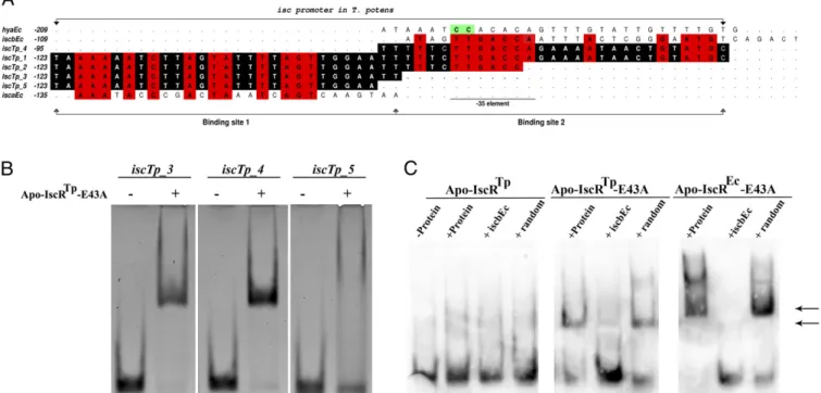
Similar to what is observed forE. coliapo-IscR andisc, apo-IscRTp does not bind iscTp_1 (Fig. S3A). Using an enzymatic system under oxygen-depleted atmosphere (42), an Fe/S cluster could be reversibly reconstituted in wild-type apo-IscRTp, yield-ing the holo form of the protein (reconstituted IscRTp-wt; R-IscRTp-wt), as judged by the appearance of absorption maxima at 320 and 420 nm (Fig. S3B). A dose-dependent structural change ofiscTp_1DNA could be identified by circular dichroism spec-troscopy, upon R-IscRTp-wt binding (Fig. S3C). These results demonstrate that, as expected for a bona fide IscR, the enzy-matically reconstituted Fe/S cluster-bound form of IscRTp-wt binds to theT. potens iscpromoter region (Fig. S3C). As seen for E. coliIscR (13), the single point mutant apo-IscRTpE43A binds specifically to the iscTp_1 sequence, seemingly forming two distinct complexes—with either one or two IscR dimers binding to the target sequence—as suggested by the two observed DNA band shifts (Fig. S3A). This finding is further supported by the observation of a single complex with the 3′-trimmed iscTp_1 sequence, termediscTp_2(Fig. 5A,Table S1, andFig. S3A). In E. coli, DNase footprinting led to the identification of two IscR binding sites within theiscpromoter region,iscraandiscrb(8). Two highly homologous regions could be identified in the T. potens iscpromoter,iscTp_3andiscTp_4(Fig. 5AandTable S1), to which apo-IscRTpE43A displays specific binding (Fig. 5Band Table S2). Further, removal of the two 3′-end nucleotides of iscTp_3, yielding the shorter iscTp_5 (Fig. 5A and Table S1), effectively prevents binding of apo-IscRTpE43A (Fig. 5Band Table S2), in good agreement with the observed bidentate
binding of IscR to the minor groove of AT-rich segments at the termini of its recognition sequence (Fig. 4A).
In line with the structural similarity ofE. coliand T. potens IscR proteins and the considerable conservation ofiscpromoter sequences, there is cross-recognition between the transcriptional regulator ofT. potensand theE. colipromoter. Although apo-IscRTp does not bind E. coli iscrb (iscbEc, Table S1), this se-quence is specifically recognized by the E43A mutant (Fig. 5C andTable S2), as observed forE. coliapo-IscR (13). Therefore, the unique mechanism of promoter-sequence discrimination by IscR seems to be conserved between these organisms.
Discussion
We performed a detailed analysis of the product of gene TherJR_1914 fromT. potens, undoubtedly establishing its func-tional relationship with the Fe/S cluster-binding transcription regulator IscR, known to control Fe/S cluster biogenesis in sev-eral Gram-negative bacteria. The identification of an IscR ho-molog inT. potenswas unprecedented: most other Gram-positive bacteria studied so far do not code for any IscR-like proteins or have an iscoperon, and the rare cases where anisc operon is present (e.g., the DMRBD. hafnienseor the bacteriumC. per-fringens) (30) lack the SUF machinery.
The combination of biochemical and structural studies, on T. potensIscR and its homolog from E. coli, revealed also an unforeseen conservation of the unique mode of IscR promoter sequence recognition and discrimination. Despite extensive conservation of the DNA-binding surface, apo-IscRTp was un-able to recognize the heterologous hya promoter fromE. coli. Residue at position 40 played a pivotal role in this process be-cause relief of steric hindrance (P40S mutant) was sufficient to Fig. 5. Modulation of apo-IscRTpspecificity by a single point mutation. (
A) IscR binding sites in type-1 [T. potens isc(iscTp) andE. coli isca(iscaEc) andiscb
(iscbEc)] and type-2 [E. coli hya(hyaEc)] promoters. Numbers refer to the most upstream base of each IscR site relative to the corresponding start codon. Conserved bases between theiscpromoters are highlighted in red whereas bases conserved between the fiveT. potens iscpromoter sequences are shaded black. The highly conserved CC motif in type-2 promoters is colored green (12). (B) There are two independent binding sites for apo-IscRTpE43A in the T. potens iscpromoter. Purified apo-IscRTpE43A (7.5μM) was incubated withiscTp_3,iscTp_4andiscTp_5sequences, analyzed by nondenaturing PAGE, and
visualized by ethidium bromide staining. An arrow denotes the DNA band-shift upon complex formation. (C) Cross-recognition of theE. coli iscpromoter by
T. potensIscR. Purified proteins (apo-IscRTp, apo-IscRTpE43A, or apo-IscREcE43A) were incubated with DIG-labelediscrbpromoter sequence (iscbEc) (Table S1)
and analyzed by nondenaturing PAGE. An arrow denotes bands indicative of DNA–IscR complex formation. Where indicated, coldiscrbor a similarly sized random sequence (random) was added in 100-fold molar excess as competitor.
BIO
CHEMISTRY
PNAS
is recognized by holo-IscR. In the absence of the Fe/S cluster, the strictly conserved E43 residue is pivotal for discriminating be-tween type-1 and type-2 promoters by establishing specific in-teractions with an invariant CC dinucleotide in type-2 sequences. In fact, mutation of this residue to an uncharged alanine seems to mimic the cluster-induced specificity switch of IscR, allowing the clusterless regulator to recognize and to bind to type-1 promoter sequences (13). In apo-IscRTp, the E43A mutation promotes binding to two sequences upstream of the T. potens iscRSUoperon. These regions are highly homologous to theE. coli iscsequences recognized by both holo- and mutant apo-IscR E43A. Given that one of these sequences (iscTp_4) contains a−35-element sequence, we propose thatT. potensholo-IscR may act as a repres-sor of Fe/S biogenesis by hindering RNA polymerase binding. As a whole, our results suggest a conserved regulation mechanism by IscR, where Fe/S cluster binding to this transcription regu-lator enables recognition of type-1 promoters, a process that is mimicked by the E43A mutation.
By using structurally relevant mutants, where serine replaces all putative cluster-coordinating cysteine residues, a network of polar interactions could be identified, connecting the cluster-binding region of one subunit to the DNA-contacting interface of its neighbor in the functional IscR dimer. Any perturbation resulting from cluster ligation could therefore be allosterically transmitted to the nucleic acid-binding region that comprises the conserved E43. Regulation through Fe/S cluster ligation allows T. potensIscR to act as a sensor and to be a central player of an auto-regulatory loop responsible for keeping the appropriate levels of cellular Fe/S cluster formation and delivery.
Methods
Protein Expression and Purification.A syntheticiscrgene, encoding the same amino acid sequence as TherJR_1914 from theT. potensgenome, except for a Gly-Leu insertion immediately downstream from the N-terminal methio-nine, was ordered from Eurofins MWG Operon. TheE. coli iscrgene (b2531) fragment spanning nucleotides+4 to+489 of the IscR ORF was amplified from anE. coliK12 colony using specific primers. Both ORFs were cloned into the NdeI and XhoI sites of the expression vector pET30a (IscRTp-wt) or into
the Acc65I and NcoI sites of the expression vector pETZ2_1a (43). The latter constructs were used to obtain the triple mutants (C92/101/107S for
T. potensor C92/98/104S forE. coli) corresponding to the clusterless forms of the proteins (apo-IscRTpand apo-IscREc) by site-directed mutagenesis.
The N-terminal His6-tagged apo-IscRTpwas overexpressed inE. coliBL21
(DE3) cells and theE. coliprotein inE. coliBL21 Star (DE3) (Life Technolo-gies). Briefly, cells were grown in LB medium at 37 °C until OD600=0.7. At this point, the temperature was decreased to either 25 °C (apo-IscREc) or 30 °C
(apo-IscRTp), and the expression was induced with the addition of 0.5 mM
isopropylβ-D-1-thiogalactopyranoside (IPTG). Cells were harvested by
centri-fugation after 4 h and lysed by incubation (60 min on ice with shaking) with 25 μg/mL chicken egg white lysozyme (Sigma). Clarified protein extracts in 20 mM sodium phosphate (pH 7.5), 0.5 M NaCl, 10 mM imidazole, 5% (vol/vol) glyc-erol, 150 mM arginine, and 2.5 mMβ-mercaptoethanol (buffer A) were loaded onto a HisTrap HP column (GE Healthcare) preequilibrated in the same buffer, and bound proteins were eluted with buffer A containing 125 mM imidazole. The IscR-containing fractions were pooled, and the His6and the solubility tags
were removed by incubation with tobacco etch virus (TEV) protease at 4 °C concomitantly to an overnight dialysis against 20 mM sodium phosphate (pH 7.5), 0.2 M NaCl, 10 mM imidazole, 5% (vol/vol) glycerol, 150 mM arginine, and 2.5 mMβ-mercaptoethanol. Pure recombinant IscR was separated from the expression tag and noncleaved material by a second immobilized-metal af-finity chromatography (IMAC) step, in the same conditions as described above. The buffer was further exchanged for 10 mM Hepes (pH 7.5), 800 mM KCl, and 5% (vol/vol) glycerol using a HiPrep 26/10 (GE Healthcare) desalting column. The protein was either used immediately or flash-frozen in liquid nitrogen and
26/10 column (GE Healthcare). TheE. colicysteine desulfurase IscS used in reconstitution assays was expressed and purified as described previously (42).
Fe/S Cluster Reconstitution.Reconstitution of the Fe/S cluster of apo-IscRTp-wt
was performed under oxygen-depleted atmosphere (<3 ppm O2) in an an-aerobic chamber (Belle Technology). All buffers used were sparged with nitrogen gas for 20 min and kept in the anaerobic chamber for at least 12 h before use. Small volumes of aerobically purified proteins (apo-IscRTp-wt and E. coliIscS), as well as sodium dithionite, cysteine, and iron (II) sources were equilibrated in the same low-oxygen conditions for 1 h.
An apo-IscRTp-wt solution (25μM) was mixed with ammonium iron (II)
sulfate (Sigma) andL-cysteine (Sigma) in 20-fold and eightfold molar excess,
respectively. The reaction [in 50 mM Tris (pH 8), 150 mM NaCl, 5 mM DTT] was started after 10 min by addition of 2μME. coliIscS, and Fe/S cluster formation was followed by monitoring absorbance at 420 nm. Upon re-action completion, R-IscRTp-wt was purified by IMAC (His-Buster Nickel spin
columns; Amocol). Absorption spectra (250–750 nm) were recorded imme-diately after purification and upon sample reduction with 2 mM sodium dithionite. Control reactions were performed omittingL-cysteine
(non-reconstituted IscRTp-wt; NR-IscRTp-wt).
Electrophoretic Mobility-Shift Assay. Complementary oligonucleotides (Sigma) containing the sequence of theE. coli hyaor of theT. potens isc
promoter (Table S1) were annealed into double-stranded DNA by heating a 50-μM solution to 95 °C for 5 min in a water bath, followed by slowly (overnight) cooling to room temperature. For electrophoretic mobility-shift assay (EMSA) analysis using the complete sequence of the T. potens suf
promoter region (Table S1), the sequence upstream of the sufC gene (TherJR_0923) was amplified by PCR using a synthetic template (Eurofins). DNA solutions (1μM) were incubated with 7.5–10μM purified protein at room temperature for 20 min in binding buffer [40 mM Tris·HCl (pH 8.0), 100–150 mM KCl, 5% (vol/vol) glycerol, and 1 mM DTT], and the resulting complexes were resolved on 8% (wt/vol) nondenaturing polyacrylamide gels using 1×TAE (40 mM Tris·HCl, 20 mM acetic acid, and 1 mM EDTA) as run-ning buffer. DNA was detected by either ethidium bromide stairun-ning or chemiluminescent detection. For chemiluminescent detection, annealed DNA probes were end-labeled with digoxigenin using recombinant ter-minal transferase (Roche). The labeled probes (1.5μmol) were mixed with 11.25μmol purified IscR in 15μL of binding buffer [40 mM Tris·HCl (pH 8.0), 10% (vol/vol) glycerol, 1 mM DTT, and 50 mM KCl]. For competition reactions, labeled probe was added after incubating the protein for 10 min with 100-fold molar excess competitor DNA. The samples were separated in 8% (wt/vol) nondenaturing polyacrylamide gels and elec-troblotted onto positively charged nylon membrane (GE Healthcare), and the digoxigenin-labeled probes were detected with anti-digox-igenin-AP antibody and the chemiluminescent substrate disodium 2-chloro-5-(4-methoxyspiro [1,2-dioxetane-3,2′-(5′
-chloro)tricyclo[3.3.1.1(3,7)]decan]-4-yl)-1-phenyl phosphate (CDP-Star; Roche).
Circular Dichroism Measurements.Binding of wild-type IscRTpto DNA was
monitored by circular dichroism (CD) spectroscopy (44). CD spectra were recorded at 20 °C in 1-nm steps on a temperature-controlled Jasco J-815 spectropolarimeter, continuously purged with nitrogen gas. For each sam-ple, the smoothed average of four spectra was considered. Briefly, either anaerobically reconstituted (R-IscRTp-wt) or nonreconstituted (NR-IscRTp-wt)
and purified apo-IscRTp-wt was added incrementally to a sealed 10-mm-path
quartz cuvette containing theiscTp_1sequence (Table S1) in binding buffer, and CD spectra (260–320 nm) were recorded.
Microscale Thermophoresis Assays.Interactions between IscR variants (apo-IscRTp, apo-IscRTpE43A, apo-IscRTpP40S, and apo-IscREcE43A) and the
dif-ferent DNA sequences (iscTp_3,iscTp_4,iscTp_5,hyaEc, andiscbEc) (Fig. 5A
andTable S1) were assessed using microscale thermophoresis (45) with a
20]. Ligand dilutions were prepared in assay buffer without Tween 20 and mixed with each protein sample at a volume ratio of 1:1. Measurements with apo-IscRTp, apo-IscRTpP40S, and apo-IscRTpE43A were performed in
standard capillaries whereas hydrophilic capillaries were used for measure-ments with apo-IscREcE43A. For each interaction, data from at least two
independent runs were averaged, and the average curve was fitted with NTAnalysis software (NanoTemper Technologies).
Crystallization of apo-IscRTpand apo-IscREc:hyaComplex.Initial crystallization
conditions for apo-IscRTp were screened at 20 °C using the sitting-drop
method with commercial sparse-matrix crystallization screens. Drops con-sisting of equal volumes (1μL) of protein (at 20 mg/mL) and precipitant solution were equilibrated against a 300-μL reservoir. Crystals were obtained after 2 d using 0.1 M Bis-Tris (pH 6.5) and 3 M NaCl as precipitant. Before data collection, crystals were cryoprotected by immersing them briefly in a 1:1 mixture of precipitant solution and 40% (vol/vol) of 2 mg/mL NDSB-201 (3-(1-pyridino)-1-propane sulfonate) solution in ethylene glycol and flash-cooled in liquid nitrogen (46). Selenomethionyl apo-IscRTpcrystallized in
the same conditions and was cryoprotected following the procedure described above.
A 3.8-fold molar excess of apo-IscREcwas mixed with double-stranded
oligonucleotide (prepared as described in Electrophoretic Mobility-Shift Assay) comprising region−30 to−55 of theE. coli hyapromoter sequence with a single-base 5′overhang (hya_26_OH) (Table S1) and incubated at room temperature for 30 min. The complex was either used immediately or flash frozen in liquid nitrogen and stored at−80 °C. Initial crystallization conditions were established at the High Throughput Crystallization Labo-ratory of the European Molecular Biology LaboLabo-ratory, using the sitting-drop method. Crystals were obtained at 20 °C, from 0.2-μL drops composed of identical volumes of complex solution [350μM protein and 92μM oligonu-cleotide in 40 mM Tris·HCl (pH 8.0), 150 mM KCl, 10% (vol/vol) glycerol,
1 mM DTT] and of precipitant [0.1 M citric acid (pH 4.0 or 6.0), 1 M lithium chloride, 20% (wt/vol) PEG 6000]. Better and larger crystals could be obtained from the condition at pH 4.0 using the hanging-drop vapor diffusion method. The optimized crystals were cryoprotected in the same conditions as the apo-IscRTpcrystals.
Data Collection and Processing.X-ray diffraction data were collected from cooled (100 K) single crystals at synchrotron beam lines ID29 (apo-IscRTpand
Se-Met apo-IscRTp) (47) and ID23-EH2 (apo-IscREc:hya complex) (48) of the
European Synchrotron Radiation Facility. The apo-IscRTpdata were recorded
on a Pilatus 6M detector (Dectris) using a wavelength of 0.9763 Å (native dataset) or 0.9792 Å (Se-Met dataset). For the native data, 1,200 images were collected in 0.1° oscillation steps with 0.1-s exposure per frame whereas, for the Se-Met data, 3,600 images were recorded in 0.1° oscillation steps with 0.037-s exposure per frame. The apo-IscREc:hya complex data
were recorded on a MX-225 detector (Marresearch) using a wavelength of 0.8726 Å. One hundred images were collected in 0.95° oscillation steps with 5.43-s exposure per frame. Diffraction data were integrated with XDS (49), scaled with XSCALE (50), and reduced with utilities from the CCP4 program suite (51). Data collection statistics are summarized in Table 2.
Structure Solution and Refinement.The structure of apo-IscRTpwas solved by
single-wavelength anomalous diffraction using the anomalous signal of se-lenium-substituted crystals with the SHELXC/SHELXD/SHELXE pipeline (52) and the HKL2MAP GUI (53). The resulting electron density maps were readily interpretable. The structure of the apo-IscREc:hya complex was solved by
molecular replacement with PHASER (54) using a truncated version of the refined apo-IscRTpstructure as search model. For both structures, alternating
cycles of model building with COOT (55) and of refinement with PHENIX (56) were performed until model completion. For the apo-IscRTpstructure, the
final model comprises residues Gly-3 to Gly85 and Ser101 to Ile149 for subunit
Table 2. Statistics of data collection, processing, and refinement
Dataset T. potensIscR* (native) T. potensIscR* (Se-Met) E. coliIscR-DNA complex*
Crystallographic analysis
Wavelength, Å 0.9763 0.9792 0.8726
Space group P41 P41 P212121
Unit cell dimensions, Å a=b=53.6; c=118.4 a=b=53.4; c=118.7 a=49.0; b=75.8; c=173.4
Resolution range, Å 53.6–1.60 (1.69–1.60) 48.7–2.47 (2.61–2.47) 46.0–2.49 (2.62–2.49)
Reflections (measured/unique) 196,159/43,750 (28,394/6,325) 111,297/11,861 (13,614/1,647) 87,182/23,387 (12,463/3,244)
Completeness, % 99.7 (98.7) 99.1 (94.1) 99.3 (96.3)
Multiplicity 4.5 (4.5) 9.4 (8.3) 3.7 (3.8)
Rmerge† 0.046 (1.299) 0.190 (1.573) 0.103 (0.911)
Rpim‡ 0.024 (0.688) 0.064 (0.555) 0.061 (0.531)
〈I/σ(I)〉 14.3 (1.6) 7.3 (1.4) 8.6 (1.5)
Monomers per asymmetric unit 2 2 2
Mathews coefficient, Å3·Da−1 2.53 2.52 3.13
Solvent content, % 51.4 51.2 60.7
Structure refinement
Resolution range, Å 48.8–1.60 — 46.0–2.49
Rfactor§/Free Rfactor¶ 0.202/0.220 — 0.207/0.251
Unique reflections (work/test set) 41,642/1,973 — 22,057/1,193
Water molecules 156 — 15
Total no. of atoms 2,363 — 2,999
No. of macromolecule atoms 2,205 — 2,984
rmsd bond lengths, Å 0.011 — 0.008
rmsd bond angles, ° 1.09 — 1.38
Average overall B factor, Å2 38.1
— 77.7
Ramachandran favored, % 97.5 — 96.0
Ramachandran outliers, % 0.0 — 0.4
PDB entry 4cic — 4chu
*Values in parentheses correspond to the outermost resolution shell. Each dataset was recorded from a single crystal. †R
merge=PhklPijIi(hkl)–〈I(hkl)〉j/PhklPiIi(hkl), whereIi(hkl) is the observed intensity and〈I(hkl)〉is the average intensity of multiple observations of
symmetry-related reflections. ‡R
pim=Phkl[1/(N–1)]1/2PijIi(hkl)–〈I(hkl)〉j/PhklPiIi(hkl), whereIi(hkl)is the observed intensity and〈I(hkl)〉is the average intensity of multiple observations of symmetry-related reflections.
§R
factor=PjjFoj−jFcjj/PjFoj, wherejFojandjFcjare observed and calculated structure factor amplitudes, respectively. ¶
Free Rfactoris the cross-validationRfactorcomputed for a randomly chosen subset of 5% of the total number of reflections, which were not used during
refinement.
BIO
CHEMISTRY
PNAS
Synchrotron Radiation Facility (ESRF) for provision of synchrotron radiation facilities and thank the ESRF staff for help with data collection. Microscale
funding from the European Community’s Seventh Framework Programme (FP7/2007-2013) under BioStruct-X (Grant Agreement 283570).
1. Beinert H, Holm RH, Münck E (1997) Iron-sulfur clusters: Nature’s modular,
multi-purpose structures.Science277(5326):653–659.
2. Ayala-Castro C, Saini A, Outten FW (2008) Fe-S cluster assembly pathways in bacteria. Microbiol Mol Biol Rev72(1):110–125.
3. Fontecave M, Choudens SO, Py B, Barras F (2005) Mechanisms of iron-sulfur cluster assembly: The SUF machinery.J Biol Inorg Chem10(7):713–721.
4. Takahashi Y, Nakamura M (1999) Functional assignment of the ORF2-iscS-iscU-iscA-hscB-hscA-fdx-ORF3 gene cluster involved in the assembly of Fe-S clusters inEscherichia coli.J Biochem126(5):917–926.
5. Takahashi Y, Tokumoto U (2002) A third bacterial system for the assembly of iron-sulfur clusters with homologs in archaea and plastids.J Biol Chem277(32): 28380–28383.
6. Outten FW, Djaman O, Storz G (2004) A suf operon requirement for Fe-S cluster as-sembly during iron starvation inEscherichia coli.Mol Microbiol52(3):861–872. 7. Schwartz CJ, et al. (2001) IscR, an Fe-S cluster-containing transcription factor, represses
expression ofEscherichia coligenes encoding Fe-S cluster assembly proteins.Proc Natl Acad Sci USA98(26):14895–14900.
8. Giel JL, Rodionov D, Liu M, Blattner FR, Kiley PJ (2006) IscR-dependent gene expres-sion links iron-sulphur cluster assembly to the control of O2-regulated genes in Escherichia coli.Mol Microbiol60(4):1058–1075.
9. Lee KC, Yeo WS, Roe JH (2008) Oxidant-responsive induction of the suf operon, en-coding a Fe-S assembly system, through Fur and IscR inEscherichia coli.J Bacteriol 190(24):8244–8247.
10. Yeo WS, Lee JH, Lee KC, Roe JH (2006) IscR acts as an activator in response to oxidative stress for the suf operon encoding Fe-S assembly proteins.Mol Microbiol61(1):206–218. 11. Giel JL, et al. (2013) Regulation of iron-sulphur cluster homeostasis through tran-scriptional control of the Isc pathway by [2Fe-2S]-IscR inEscherichia coli.Mol Micro-biol87(3):478–492.
12. Nesbit AD, Giel JL, Rose JC, Kiley PJ (2009) Sequence-specific binding to a subset of IscR-regulated promoters does not require IscR Fe-S cluster ligation.J Mol Biol387(1): 28–41.
13. Rajagopalan S, et al. (2013) Studies of IscR reveal a unique mechanism for metal-dependent regulation of DNA binding specificity.Nat Struct Mol Biol20(6):740–747. 14. Riboldi GP, Verli H, Frazzon J (2009) Structural studies of theEnterococcus faecalis
SufU [Fe-S] cluster protein.BMC Biochem10:3.
15. Riboldi GP, de Oliveira JS, Frazzon J (2011)Enterococcus faecalisSufU scaffold protein enhances SufS desulfurase activity by acquiring sulfur from its cysteine-153.Biochim Biophys Acta1814(12):1910–1918.
16. Albrecht AG, et al. (2010) SufU is an essential iron-sulfur cluster scaffold protein in Bacillus subtilis.J Bacteriol192(6):1643–1651.
17. Byrne-Bailey KG, et al. (2010) Complete genome sequence of the electricity-producing
“Thermincola potens”strain JR.J Bacteriol192(15):4078–4079.
18. Carlson HK, et al. (2012) Surface multiheme c-type cytochromes fromThermincola potensand implications for respiratory metal reduction by Gram-positive bacteria. Proc Natl Acad Sci USA109(5):1702–1707.
19. Riboldi GP, de Mattos EP, Frazzon J (2013) Biogenesis of [Fe-S] cluster inFirmicutes: An unexploited field of investigation.Antonie van Leeuwenhoek104(3):283–300. 20. Zheng L, Cash VL, Flint DH, Dean DR (1998) Assembly of iron-sulfur clusters.
Identi-fication of an iscSUA-hscBA-fdx gene cluster fromAzotobacter vinelandii.J Biol Chem 273(21):13264–13272.
21. Yuvaniyama P, Agar JN, Cash VL, Johnson MK, Dean DR (2000) NifS-directed assembly of a transient [2Fe-2S] cluster within the NifU protein.Proc Natl Acad Sci USA97(2):599–604. 22. Rangachari K, et al. (2002) SufC hydrolyzes ATP and interacts with SufB from
Ther-motoga maritima.FEBS Lett514(2-3):225–228.
23. Saini A, Mapolelo DT, Chahal HK, Johnson MK, Outten FW (2010) SufD and SufC ATPase activity are required for iron acquisition during in vivo Fe-S cluster formation on SufB.Biochemistry49(43):9402–9412.
24. Silberg JJ, Tapley TL, Hoff KG, Vickery LE (2004) Regulation of the HscA ATPase re-action cycle by the co-chaperone HscB and the iron-sulfur cluster assembly protein IscU.J Biol Chem279(52):53924–53931.
25. Trotter V, et al. (2009) The CsdA cysteine desulphurase promotes Fe/S biogenesis by recruiting Suf components and participates to a new sulphur transfer pathway by recruiting CsdL (ex-YgdL), a ubiquitin-modifying-like protein.Mol Microbiol74(6): 1527–1542.
26. Selbach BP, et al. (2014) Fe-S cluster biogenesis in Gram-positive bacteria: SufU is a zinc-dependent sulfur transfer protein.Biochemistry53(1):152–160.
27. Chahal HK, Dai Y, Saini A, Ayala-Castro C, Outten FW (2009) The SufBCD Fe-S scaffold complex interacts with SufA for Fe-S cluster transfer. Biochemistry48(44): 10644–10653.
28. Tian T, He H, Liu XQ (2014) The SufBCD protein complex is the scaffold for iron-sulfur cluster assembly inThermus thermophilesHB8.Biochem Biophys Res Commun443(2): 376–381.
29. Hoff KG, Ta DT, Tapley TL, Silberg JJ, Vickery LE (2002) Hsc66 substrate specificity is directed toward a discrete region of the iron-sulfur cluster template protein IscU. J Biol Chem277(30):27353–27359.
30. André G, et al. (2010) Global regulation of gene expression in response to cysteine availability inClostridium perfringens.BMC Microbiol10:234.
31. Shen G, et al. (2007) SufR coordinates two [4Fe-4S]2+, 1+clusters and functions as
a transcriptional repressor of the sufBCDS operon and an autoregulator of sufR in cyanobacteria.J Biol Chem282(44):31909–31919.
32. Wang T, et al. (2004) The sufR gene (sll0088 inSynechocystis sp.strain PCC 6803) functions as a repressor of the sufBCDS operon in iron-sulfur cluster biogenesis in cyanobacteria.J Bacteriol186(4):956–967.
33. Choi YS, et al. (2007) Identification ofPseudomonas aeruginosagenes crucial for hydrogen peroxide resistance.J Microbiol Biotechnol17(8):1344–1352.
34. Runyen-Janecky L, et al. (2008) Role and regulation of iron-sulfur cluster biosynthesis genes inShigella flexnerivirulence.Infect Immun76(3):1083–1092.
35. Zeng J, Zhang K, Liu J, Qiu G (2008) Expression, purification, and characterization of iron-sulfur cluster assembly regulator IscR fromAcidithiobacillus ferrooxidans. J Microbiol Biotechnol18(10):1672–1677.
36. Shepard W, et al. (2011) Insights into the Rrf2 repressor family—the structure of CymR, the global cysteine regulator ofBacillus subtilis.FEBS J278(15):2689–2701. 37. Ji Q, et al. (2012)Staphylococcus aureusCymR is a new thiol-based oxidation-sensing
regulator of stress resistance and oxidative response.J Biol Chem 287(25): 21102–21109.
38. Gregoret LM, Rader SD, Fletterick RJ, Cohen FE (1991) Hydrogen bonds involving sulfur atoms in proteins.Proteins9(2):99–107.
39. Zhou P, Tian F, Lv F, Shang Z (2009) Geometric characteristics of hydrogen bonds involving sulfur atoms in proteins.Proteins76(1):151–163.
40. Fuentes-Prior P, Salvesen GS (2004) The protein structures that shape caspase activity, specificity, activation and inhibition.Biochem J384(Pt 2):201–232.
41. Turk V, et al. (2012) Cysteine cathepsins: From structure, function and regulation to new frontiers.Biochim Biophys Acta1824(1):68–88.
42. Prischi F, et al. (2010) Of the vulnerability of orphan complex proteins: The case study of theE. coliIscU and IscS proteins.Protein Expr Purif73(2):161–166.
43. Olichon A, Surrey T (2007) Selection of genetically encoded fluorescent single domain antibodies engineered for efficient expression inEscherichia coli.J Biol Chem282(50): 36314–36320.
44. Carpenter ML, Kneale GG (1994) Circular dichroism for the analysis of protein-DNA interactions.Methods Mol Biol30:339–345.
45. Jerabek-Willemsen M, Wienken CJ, Braun D, Baaske P, Duhr S (2011) Molecular interaction studies using microscale thermophoresis.Assay Drug Dev Technol9(4): 342–353.
46. Pereira PJ, et al. (2008) Mycobacterium tuberculosis glucosyl-3-phosphoglycerate synthase: Structure of a key enzyme in methylglucose lipopolysaccharide biosynthesis. PLoS ONE3(11):e3748.
47. de Sanctis D, et al. (2012) ID29: A high-intensity highly automated ESRF beamline for macromolecular crystallography experiments exploiting anomalous scattering. J Synchrotron Radiat19(Pt 3):455–461.
48. Flot D, et al. (2010) The ID23-2 structural biology microfocus beamline at the ESRF. J Synchrotron Radiat17(1):107–118.
49. Kabsch W (2010) XDS.Acta Crystallogr D Biol Crystallogr66(Pt 2):125–132. 50. Kabsch W (2010) Integration, scaling, space-group assignment and post-refinement.
Acta Crystallogr D Biol Crystallogr66(Pt 2):133–144.
51. Collaborative Computational Project, Number 4 (1994) The CCP4 suite: Programs for protein crystallography.Acta Crystallogr D Biol Crystallogr50(Pt 5):760–763. 52. Sheldrick GM (2010) Experimental phasing with SHELXC/D/E: Combining chain tracing
with density modification.Acta Crystallogr D Biol Crystallogr66(Pt 4):479–485. 53. Pape T, Schneider TR (2004) HKL2MAP: A graphical user interface for macromolecular
phasing with SHELX programs.J Appl Cryst37:843–844.
54. McCoy AJ, et al. (2007) Phaser crystallographic software.J Appl Cryst40(Pt 4):658–674. 55. Emsley P, Cowtan K (2004) Coot: Model-building tools for molecular graphics.Acta
Crystallogr D Biol Crystallogr60(Pt 12 Pt 1):2126–2132.
56. Adams PD, et al. (2010) PHENIX: A comprehensive Python-based system for macro-molecular structure solution.Acta Crystallogr D Biol Crystallogr66(Pt 2):213–221. 57. Reboiro-Jato D, et al. (2012) ADOPS—Automatic Detection Of Positively Selected
Sites.J Integr Bioinform9(3):200.
58. Thompson JD, Gibson TJ, Higgins DG (2002) Multiple sequence alignment using ClustalW and ClustalX.Curr Protoc BioinformaticsChapter 2: Unit 2 3.
59. Bond CS, Schüttelkopf AW (2009) ALINE: A WYSIWYG protein-sequence alignment editor for publication-quality alignments.Acta Crystallogr D Biol Crystallogr65(Pt 5): 510–512.