Universidade de Lisboa
Faculdade de Motricidade Humana
MANUAL LYMPHATIC DRAINAGE IN
CHRONIC VENOUS DISEASE
Tese elaborada com vista à obtenção do Grau de Doutor em Motricidade Humana na especialidade de Reabilitação
Orientador: Professor Doutor Paulo Alexandre Silva Armada da Silva
Júri:
Presidente
Reitor da Universidade de Lisboa
Vogais:
Doutor Carlos Fernando Pereira Alves
Professor Associado com Agregação Convidado e Jubilado Faculdade de Ciências Médicas da Universidade Nova de Lisboa Doutora Maria Margarida Marques Rebelo Espanha
Professora Associada
Faculdade de Motricidade Humana da Universidade de Lisboa Doutor Paulo Alexandre Silva Armada da Silva
Professor Auxiliar
Faculdade de Motricidade Humana da Universidade Nova de Lisboa Doutor Carlos Manuel da Costa Almeida
Professor Auxiliar Convidado
Faculdade de Medicina da Universidade de Coimbra Doutor Eduardo Manuel Brazete Carvalho Cruz Professor Coordenador
Escola Superior de Saúde do Instituto Politécnico de Setúbal
Rute Sofia dos Santos Crisóstomo
This work was partially supported by PhD grant by the Portuguese Fundação para a Ciência e Tecnologia, number SFRH/BD/62673/2009.
NOTE:
This thesis is based on the following studies:
Crisóstomo, R. S., Candeias, M. S., & Armada-da-Silva, P. A. (2014). The use of ultrasound in the evaluation of the efficacy of calf muscle pump function in primary chronic venous disease. Phlebology, 29(4), 247-256. doi: 10.1177/0268355512471757 (Annex 1).
Crisóstomo RS, Candeias MS, Ribeiro AM, Martins CD, Armada-da-Silva PA. (2013). Manual lymphatic drainage in chronic venous disease: A duplex ultrasound study. Phlebology. doi: http://dx.doi.org/10.1177/026835551350278 (Annex 2).
Crisóstomo RS, Candeias MS, Armada-da-Silva PA. Venous flow during manual lymphatic drainage applied to different regions of the lower extremity. Physiotherapy, (submitted).
DEDICATORY
This work is dedicated to my family: Bárbara, António, Pedro, Liliana, Paulo, Ema, Joaquina and Joaquim.
ACKNOWLEDGMENTS
I would like to thank Physiotherapist Diana Arraia and Cardiopneumologist Miguel Candeias (also a co-author in three of the studies in this thesis), for helping in data collection, for staying with me from the moment when this research was only a dream, and for helping me to make it true.
Also, I would like to give a very special thanks to participants in Lisbon and in Castelo Branco that kindly volunteered to participate in the studies making up this thesis.
I would like to thank Aida Paulino, M.D. in General Surgery, and Nurses Maria Conceição Branco and Manuel Machado for referring patients to this study from Unidade Local de Saúde de Castelo Branco.
I would like to thank Professor Isabel Salavessa for reviewing the English of some of the studies and Mrs. Edite Santos for helping me with the formatting of this thesis. Also thanks to those from Escola Superior de Saúde Dr. Lopes Dias, Instituto Politécnico de Castelo Branco (where three studies were carried out), who always pleased me with nice and supportive words, and helped me to make this work come true.
I would like to thank Faculdade de Motricidade Humana from Universidade de Lisboa and Laboratório de Biomecânica e Morfologia Funcional, for all suport.
I would like to thank my students and ex-students and colleagues, who also shared with me the enthusiasm for studying manual lymphatic drainage and chronic venous disease, and especially to Physiotherapist Margarida Ribeiro, a co-author in one study, and Physiotherapist Mylène Martins.
I want to thank Physiotherapist Daniela Costa e Catarina Martins, co-authors in some of the studies, for all their support as colleague and friends, and for treating all patients in the last study. Also thanks to Cardiopneumologist Tânia Fernandes (co-author in the last study), for helping in data collection.
To my best friends that stay, talk, laugh and cry or not with me in the right moments, and to those that for some moments in the last four years were important to me, but the life let me apart from them, thank you. João Costa, Veronika Kozlova, Stela Frazão and Pedro André a special thanks to you.
ABSTRACT
Title: Manual lymphatic drainage in chronic venous disease
Objective: To assess the efficacy of manual lymphatic drainage (MLD) in the treatment of patients with chronic venous disease (CVD).
Design: Three cross-sectional studies and one single-blind randomized controlled trial, were performed.
Methods: A total of 108 participants with CVD and 62 healthy participants were assessed in four studies. The first study assessed calf muscle pump function (CMPF) and architecture of gastrocnemius muscles by ultrasound in CVD and healthy participants; the second and third studies were performed with duplex ultrasound to assess venous hemodynamics during MLD; the fourth study, a randomized controlled study, assessed for efficacy of the MLD in CVD management.
Results: Ultrasound measures demonstrate changes in CMPF efficacy along a series of contractions as well as between CVD and healthy participants, although the method suffers from bias. MLD maneuvers increase superficial and deep venous flow, mostly when applied along the anatomical course of the major lower limb veins, but without differences between different MLD maneuvers. MLD decreases the symptoms and clinical severity (related to venous edema) of CVD, and improve dimension of pain of health-related quality of life in this condition, after four weeks of treatment and the effect is maintained after 4 weeks of follow up.
Conclusions: MLD applied with skin-stretching along the course of venous vessels increases venous return, and may be used as a conservative coadjutant option to treat patients with CVD.
RESUMO
Título: Drenagem linfática manual na doença venosa crónica
Objetivo: Avaliar a eficácia da drenagem linfática manual (DLM) no tratamento de utentes com doença venosa crónica (DVC).
Desenho do estudo: Foram realizados três estudos transversais e um estudo prospetivo, controlado e com ocultação simples.
Metodologia: Nos 4 estudos foram avaliados 108 participantes com DVC e 62 participantes saudáveis. No primeiro estudo foi avaliada a bomba muscular venosa da perna (BMVP) e a arquitetura dos músculos gémeos por ultrassonografia, em participantes com DVC e saudáveis. No segundo e terceiro estudos foram avaliadas por ultrassonografia vascular as variações hemodinâmicas venosas durante a DLM. No quarto estudo, o estudo prospetivo, avaliou-se a eficácia da DLM no tratamento de doentes com DVC.
Resultados: A avaliação por ultrassonografia identificou alterações na eficácia da BMVP durante uma série de 10 contrações nos participantes com DVC e nos participantes saudáveis, contudo, este método apresentou uma fiabilidade pobre. As manobras de DLM aumentaram o fluxo venoso, sobretudo quando aplicadas na localização anatómica das principais veias do membro inferior. A DLM aliviou sintomas e reduziu a dimensão da dor da qualidade de vida relacionada com a saúde e severidade clinica da DVC (sobretudo edema) no fim de 4 semanas de tratamento. Parte destes resultados mantiveram-se após 4 semanas de follow-up.
Conclusão: Tracionar a pele durante a DLM, ao longo do percurso das veias, aumenta o retorno venoso e revela-se como uma potencial estratégia conservadora e coadjuvante no tratamento de doentes com DVC.
INDEX
TABLE INDEX ... xvii
FIGURE INDEX ...xix
LIST OF ABBREVIATORS ...xxi
1 INTRODUCTION ... 1
2 REVIEW OF LITERATURE ... 5
2.1 Health and social impact of CVD ... 5
2.1.1 Epidemiology ... 5
2.1.2 Functional and HRQL implications of CVD ... 6
2.1.3 Socio-economic impact of CVD ... 7
2.2 Pathology of CVD ... 9
2.2.1 Etiology and anatomical location of CVD ... 10
2.2.2 Physiopathology hypothesis for CVD ... 12
2.2.3 Symptoms in CVD ... 16
2.2.4 Signs of CVD ... 20
2.3 CVD diagnosis ... 23
2.4 Calf muscle pump function ... 25
2.4.1 The three venous muscle pumps of the lower limb ... 26
2.4.2 Impairment of calf muscle pump and functional capacity ... 28
2.4.3 Air-plethysmography: the gold standard assessment of calf muscle pump ... 30
2.4.4 Ultrasound assessment of hemodynamic component of calf muscle pump ... 31
2.4.5 Ultrasound assessment of muscle component of calf muscle pump .. 32
2.5 Manual lymphatic drainage ... 35
2.5.1 Brief history of manual lymphatic drainage ... 35
2.5.2 Manual lymphatic drainage - Leduc method ... 37
2.5.3 Therapeutic efficacy of manual lymphatic drainage ... 38
2.5.4 Decongestive lymphatic therapy ... 40
2.5.5 Contraindications/Precautions ... 42
2.5.6 Manual lymphatic drainage in CVD ... 43
2.6 The role of conservative treatments of CVD ... 44
3.1 Objectives of the study ... 48
3.2 Hypothesis ... 49
4 INSTRUMENTS AND METHODS ... 51
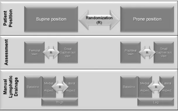
4.1 Studies outline ... 51
4.2 Ethics ... 52
4.3 Participants ... 54
4.4 Diagnose and Severity of disease ... 56
4.5 Duplex scanning: diagnose of CVD ... 57
4.6 CEAP classification ... 58
4.7 Severity of disease: Venous Clinical Severity Score ... 61
4.8 Health-related quality of life and symptoms quantification ... 63
4.9 Symptoms ... 64
4.10Vascular ultrasound assessments for venous hemodynamic ... 65
4.10.1 Popliteal vein assessment for calf muscle pump function ... 65
4.10.2 Venous hemodynamics during manual lymphatic drainage ... 68
4.11Calf muscles architecture ... 75
4.12Leg volume ... 76
4.13Ankle isokinetic dynamometer assessment ... 78
4.14Educational session... 79
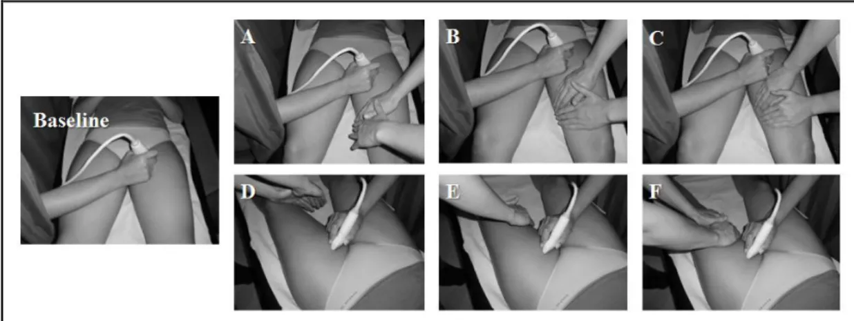
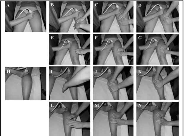
4.15Manual lymphatic drainage sequence and method ... 80
4.16Statistical analysis... 82
5 RESULTS ... 85
5.1 The use of ultrasound in the evaluation of the efficacy of calf muscle pump function in primary chronic venous disease... 92
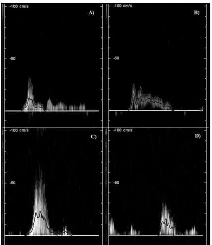
5.1.1 Venous dynamic blood flow ... 92
5.1.2 Gastrocnemius muscle architecture ... 94
5.1.3 Test-retest reliability of calf muscle pump and gastrocnemius architecture ultrasound measures ... 95
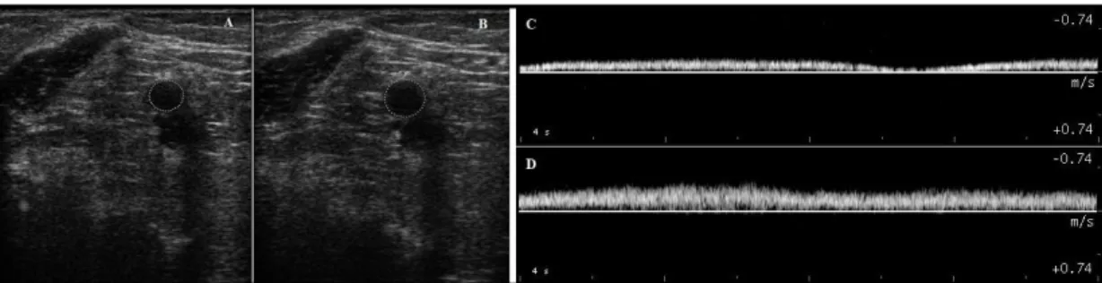
5.2 Venous flow at superficial and deep venous system during manual lymphatic drainage ... 96
5.2.1 Baseline measures of venous blood flow ... 96
5.2.2 Call-up and Reabsorption maneuver ... 101
5.2.3 Severity of CVD and MLD maneuvers ... 105
5.3.2 Manual lymphatic drainage at the leg ... 106
5.3.3 Clinical severity of chronic venous disease and response to MLD 109 5.3.4 Reliability ... 109
5.4 Efficacy of manual lymphatic drainage in chronic venous disease patients ... 110
5.4.1 Health-related quality of life ... 110
5.4.2 Severity of the disease, symptoms and leg volume ... 112
5.4.3 Ankle muscles strength ... 113
6 DISCUSSION ... 117
6.1 Ultrasound assessment of calf muscle pump function ... 117
6.2 Hemodynamic effects of manual lymphatic drainage ... 120
6.2.1 Call-up versus Reabsorption maneuver ... 121
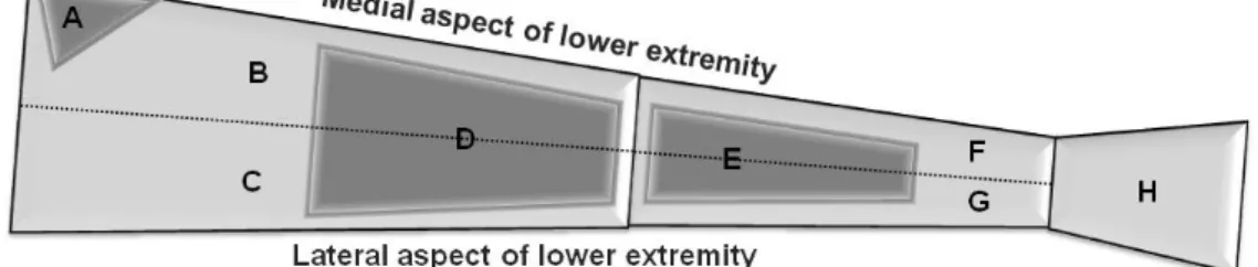
6.2.2 The effect of manual lymphatic drainage when applied to different regions of the lower limb ... 123
6.2.3 The effect of chronic venous disease severity on manual lymphatic drainage efficacy in venous return ... 125
6.3 Therapeutic efficacy of manual lymphatic drainage for treatment of patients with chronic venous disease ... 128
7 CONCLUSION ... 133
7.1 Clinical notes ... 134
7.2 Limitations ... 135
7.3 Future directions ... 136
8 REFERENCES ... 137
9 APPENDIXES ... 153
9.1 Appendix 1 -Informed Consent of Study I ... 155
9.2 Appendix 2 -Informed Consent of Study II ... 159
9.3 Appendix 3 -Informed Consent of Study III ... 163
9.4 Appendix 4 -Informed Consent of Study IV ... 167
9.5 Appendix 5 - Characterization Questionnaire ... 171
10 ANNEXES ... 173
10.1Annex 1 - Article: The use of ultrasound in the evaluation of the efficacy of calf muscle pump function in primary chronic venous disease ... 175
10.2Annex 2 - Article: Manual lymphatic drainage in chronic venous disease: A duplex ultrasound study ... 187
TABLE INDEX
Table 1 - Assessment of venous blood flow during calf muscle pump ... 34
Table 2 - Objectives and Procedures of the studies ... 53
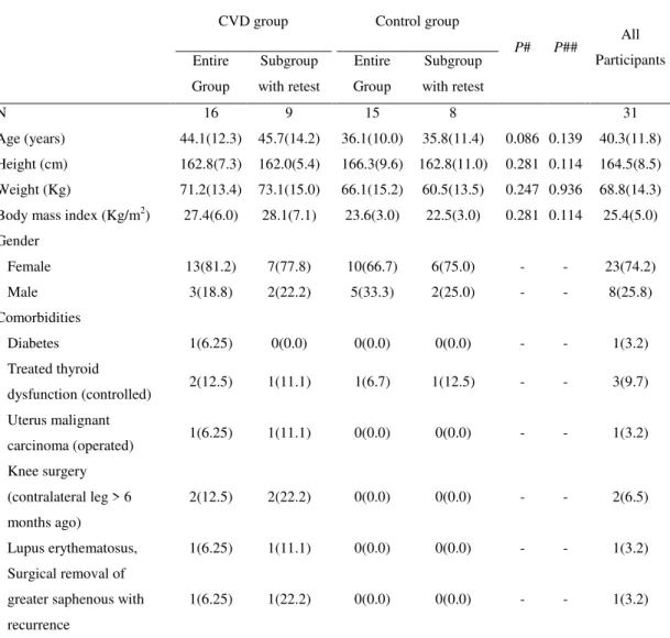
Table 3 - Demographic and clinical data of Study I ... 86
Table 4 - Clinical data of CVD group of Study I ... 87
Table 5 - Demographic and clinical data of Study II ... 88
Table 6 - Demographic and clinical data of Study III ... 89
Table 7 - Clinical data of CVD group in Study III ... 90
Table 8 - Demographic data in Study IV ... 90
Table 9 - Clinical data of Study IV ... 91
Table 10 - Popliteal vein hemodynamics in CVD and control groups ... 94
Table 11 - Differences between CVD and control group in gastrocnemius muscle architecture ... 97
Table 12 - Test-retest reliability of venous hemodynamics and muscle architecture measures ... 98
Table 13 - Venous blood flow in femoral vein ... 100
Table 14 - Venous blood flow in great saphenous vein ... 101
Table 15 - Venous blood flow during manual lymphatic drainage at the thigh ... 107
Table 16 - Venous blood flow during manual lymphatic drainage on the leg ... 108
Table 17 - Test-retest reliability for duplex ultrasound assessments ... 109
Table 18 - Severity of chronic venous disease, symptoms and leg volume ... 111
Table 19 - Calf muscle dynamometer isokinetic performance for ankle dorsiflexion ... 114
FIGURE INDEX
LIST OF ABBREVIATORS
CEAP - Clinical Etiological Anatomical Classification CIVIQ - Chronic Venous Insufficiency Questionnaire
CMPF - Calf Muscle Pump Function CVD - Chronic Venous Disease FV - Femoral Vein
GSV - Great Saphenous Vein
HRQL - Health-related quality of life
IC - Interval of Confidence
ICC - Intraclass Correlation Coefficient
MLD - Manual Lymphatic Drainage PV - Popliteal Vein
SSV - Small Saphenous Vein
1 INTRODUCTION
In Portugal the prevalence of chronic venous disease (CVD) in males is 17.8% and in females is 34.1%, i.e., around 2.5 million cases, with around 70.000
new cases every year (Capitão et al., 1995).
Chronic venous insufficiency represents the most severe cases of CVD, and is related to a physiopathology that associates venous hypertension, vein valve damage,
venous obstruction, calf muscle pump impairment, inflammations of tissues (skin, subcutaneous tissue and muscle) and veins, alterations of morphology and function
of veins, with abnormal venous reflux, venous edema, changes of the skin and subcutaneous tissues, with ulcer representing the more severe stage of this condition
(Eklof et al., 2004; Meissner et al., 2007b; Panny et al., 2009; Qiao et al., 2005; Rowland, 2001; Simka, 2007; Yamaki et al., 2010). It is estimated that 100.000 Portuguese citizens present an open (or active) venous ulcer (Oliveira et al., 2003),
and 280.000 an open or healed venous ulcer, with 10% of healed ulcer at risk for recurrence (Bradbury, 2010; Pina et al., 2005).
The first problem referred from patients are the symptoms that, together with varicose veins, is present form mild to severe cases of CVD, strongly contributing to a the negative self-esteem that also characterizes this disease (Boisseau, 2007;
Bradbury et al., 1999; Campbell et al., 2007; Cesarone et al., 2010; Darvall et al., 2012; Eklof et al., 2009; Koupidis et al., 2008). The diminished health-related quality
important that the previous view of this condition as an aesthetic problem has been abandoned for years.
The socioeconomic burden of this condition is very high. The indirect costs are substantial and are associated with the symptoms, functional impairment,
emotional disturbances and negative impact in HRQL (Lamping et al., 2003). The direct costs of CVD treatment are almost entirely related to its high prevalence, morbidity, and chronicity (Labropoulos et al., 2009; Lamping et al., 2003). In
developed countries, around 1-3% of the heath costs are due to CVD (Eklof et al., 2004). However, when patients with less severe stages of the disease are diagnosed
and treated early, the physiopathology course of the disease can be prevented or even receded (Nullen, 2010).
Manual lymphatic drainage (MLD) is a low pressure form of skin-stretching
massage, described as a conservative treatment option for the treatment of patients with CVD (Steins & Jünger, 2000) as a coadjutant of other treatments, like stockings and surgery for example (Molski et al., 2013; Molski et al., 2009). It is assumed that
this technique may have an effect on superficial venous flow, when a special maneuver the so-called call-up maneuver from the Leduc method (Leduc & Leduc,
2000) is applied, although to a limited extent (Leduc et al., 2011).
This technique has been proposed as an option in the treatment of venous lymphedema associated to CVD (Mortimer, 2000; Raju et al., 2012). It is suggested
by the literature that this technique should be applied in the course of the great saphenous vein (GSV) to treat patients with CVD, but the capacity of MLD to
reduce edema or lymphedema, MLD associated with other treatments, the so-called lymphatic decongestive therapy, may have an important role in improving health and
the functional status in patients with edema associated to sport injury or related to breast cancer surgery, just to mention two common situations (Ebert et al., 2013;
Huang et al., 2013; Vairo et al., 2009). However, the real efficacy of MLD to treat patients with CVD remains unknown.
The importance of calf muscle pump function (CMPF) in CVD development
is well established (Araki et al., 1994; Kan & Delis, 2001; Moloney et al., 2007; O'Brien et al., 2012; Panny et al., 2009; Recek, 2013; Shiman et al., 2009; Simka,
2007; Yang et al., 1999). Whether MLD has any role in improving calf muscle pump function in CVD patients has not been explored before. Also, it seems very important that low cost and reliable methods for dynamic evaluation muscle pump function in
CVD are developed, taking into account its critical importance.
The major aim of the work reported in this thesis was to evaluate the efficacy of MLD in the treatment of patients with CVD. To this end, the efficacy of MLD
maneuvers (call-up and reabsorption) in increasing venous flow in both superficial and deep venous system of the lower limbs was evaluated. Also, the reproducibility
of ultrasound evaluation of the CMPF was assessed in order to ascertain its potential role in testing the efficacy of MLD.
For this purpose, after this brief introduction where an overall view of the
work is presented we proceed to the "Review of the Literature" section, where major concepts on which our investigation relied are explored, like the social and health
function, the principles of MLD and the evidence supporting the use of this therapy as a conservative treatment of CVD, and the role of the conservative treatments in
the intervention in this disease. In the section "Scope of the Problem and Hypothesis", the principles and objectives of this thesis are presented and our four
basic hypotheses are established. The fourth section, "Instruments and Methods", describes the design for each of the four studies comprising this thesis, as well as the detail related with the participants, instruments and methodologies, and statistical
analysis. The section "Results" presents the results and outcomes of the four studies in a descriptive manner and guided by the major objectives highlighted in previous
sections. In the "Discussion" section offers an interpretation of the results based on existing evidence and taking into account the hypotheses that were formulated and divided in three subsections: "Ultrasound assessments of calf muscle pump
function", "Hemodynamic effects of manual lymphatic drainage" and "Therapeutic efficacy of manual lymphatic drainage for treatment of patients with chronic venous disease". The "Conclusion" section presents the synthesis of the results of the thesis
2 REVIEW OF LITERATURE
2.1 Health and social impact of CVD
2.1.1 Epidemiology
Chronic venous insufficiency represents the most severe stages of CVD and is characterized by the presence of edema, alterations of the skin and venous ulcer,
either healed or active, and comprises the C3-6 of clinical etiological anatomical pathological (CEAP) classification (Eklof et al., 2009). Despite its frequency in the population, the prevalence of CVD is still underestimated. Epidemiological data
estimate that this condition affects 1-17% of men and 1-40% of the women (Robertson et al., 2008), accounting to around 2.5 million people in the USA
(Koupidis et al., 2008). The estimated prevalence of CVD varies according to its severity, being around 10%, 9%, 1.5% and 0.5% for CEAP clinical levels C3, C4, C5 andC6, respectively (Gloviczki et al., 2011). In Portugal, approximately 20.7-36.8%,
of men and 40.8-62.4% of women aged 24 years or over suffer from CVD (Capitão et al., 1995), and around 50% of the total number of skin ulcers is of venous origin
(Pina et al., 2005). In Europe, the prevalence of varicose veins (C2, from clinical CEAP classification) vary in the range 7-40% in men and 25-32% in women (Robertson et al., 2008). Variation in estimations of CVD prevalence are likely
2.1.2 Functional and HRQL implications of CVD
Patients with CVD display impaired functional capacity (de Moura et al., 2012; Koupidis et al., 2008) and diminished HRQL (Andreozzi et al., 2005; Beebe-Dimmer et al., 2005; Furtado et al., 2008; Vasquez & Munschauer, 2008). The
severity of CVD, HRQL scores, the clinical signs, and venous ultrasound findings are usually correlated (Passman et al., 2011).
The impact of CVD on HRQL is primarily seen in the physical items and in the emotional role, but in advance stages (presence of venous ulcers) the mental
dimension might also become involved (Andreozzi et al., 2005). In its most severe stages, the impact of CVD in HRQL is similar to that of other chronic diseases, such as diabetes, cancer, chronic pulmonary disease, or other like heart failure (Andreozzi
et al., 2005).
Patients with venous ulcer present severe pain, which is in relationship with
impaired tissue healing ability, diminished HRQL, and lowered self-esteem and social interactions (Koupidis et al., 2008). The psychological effects of CVD may not be strictly related to ulceration itself but rather with the symptoms (80.5-69.4%),
altered appearance and esthetical concerns (66.7%), lack of sleep (66.6%), functional impairment (58.3%), and disappointment with treatment outcomes (50%) (Koupidis
et al., 2008). Also, patients with uncomplicated varicose veins often have severe symptoms that adversely affect their HRQL, irrespectively of the severity of the disease, refuting the view that this disease is mostly an aesthetic problem (Darvall et
swelling, heaviness, and fatigue (Darvall et al., 2012; Eklof et al., 2009). A recent survey reveals that 14.9% of the general Greek population refer symptoms and/or
present signs related to CVD (Dimakakos et al., 2013).
Concerning the lower extremities, one-third of people with CVD report health
or cosmetic problems that turn going out of home and to participate in social activities a burden, and they avoid wearing clothes exposing their legs or going on vacations to very warm places (Dimakakos et al., 2013). According to self-reports,
functional status is diminished in these patients (de Moura et al., 2012; Passman et al., 2011) also because of some physical dysfunctions, like abnormalities in gait (van
Uden et al., 2005), impaired balance, peripheral neuropathy (Newland et al., 2009; Shiman et al., 2009), weak calf muscles (plantar and dorsiflexors muscles) (Panny et al., 2009; Qiao et al., 2005; van Uden et al., 2005), or diminished ankle range of
motion (Dix et al., 2003; Furtado et al., 2008; Panny et al., 2009). These dysfunctions are also associated with impaired muscle pump function in the lower extremity (Shiman et al., 2009), an important risk factor for venous ulceration (Kan et al.,
2001).
2.1.3 Socio-economic impact of CVD
Severe CVD has a significant economic impact, mostly due to raised
morbidity. During the last decade neither CVD-associated and inflation-adjusted mean hospital charge, nor length of hospital stay decreased, possibly as a result of poor advancements in prevention and treatment of this disease (Tsai et al., 2005). In
mechanical debridement and, in frequent cases, antibiotic therapy (Tsai et al., 2005). Western European countries spend 1.5-2% of their annual health budget in the
treatment of this disease (Sandor, 2004). The economic burden is not just associated with clinical visits and outpatient treatments, but also with travel time, loss of work
hours for patients and family, and physiological impairment related to analgesic and antibiotic use (Tsai et al., 2005). Limb amputation is a radical outcome of this disease: three in four amputations of venous origin are undertaken in ambulatory
regimen, and many of these cases are also related to comorbidities, for instance diabetes and arterial vascular disease (Tsai et al., 2005). Other important
complications, such as hemorrhage, thrombosis and pulmonary embolism, also compound the unhealthy profile of these patients (Dimakakos et al., 2013). Deep venous thrombosis may cause chronic conditions like post-thrombotic syndrome and
CVD, increasing the costs of the treatment (Tsai et al., 2005). Preventing deep venous thrombosis and complications is one of the most important aims for reducing socio-economic burden associated to CVD (Tsai et al., 2005).
Despite its cost, the efficacy of conservative pharmacological treatment of CVD is usually poor, and should be combined with other strategies, such as the use
of elastic garment compression (Gloviczki et al., 2011).
Similar procedures are recommended following surgery and sclerotherapy, in which case post-operative compression therapy and health education are crucial for
treatment success (Bobridge et al., 2010; Dimakakos et al., 2013). There are several risk factors associated with CVD that patients should be informed of by the health
history) (Beebe-Dimmer et al., 2005; Carpentier et al., 2004; Dimakakos et al., 2013; Tsai et al., 2005). Also important, advice regarding behavioral changes, engaging in
so-called venous exercises and the proper use of the health care services should be offered to CVD patients (Dimakakos et al., 2013). Obtaining the right advice from
health care professionals is an important measure for preventing and managing CVD (Dimakakos et al., 2013).
Because of the wide spectrum of factors that cause functional impairment in
these patients and the high costs of treatment (Darvall et al., 2012; Eklof et al., 2009; Koupidis et al., 2008), the prevention of CVD by educational and prophylactic
interventions has been shown to be clinically cost-effective, by avoiding disease progression to the last stages (Allegra, 2003; Tsai et al., 2005).
2.2 Pathology of CVD
CVD is caused by venous hypertension, valvular insufficiency and/or reflux
(Ibegbuna et al., 2006; Lim & Davies, 2009; Meissner et al., 2007a). Venous hypertension might be accompanied by outflow obstruction (Meissner et al., 2007a)
and affects the superficial, perforator and deep veins (Eklof et al., 2009; Ibegbuna et al., 2006). Insufficient lymphatic drainage or a dysfunction of calf muscle pump are very often associated with this disease (Sandor, 2010). Venous hypertension is
related to structural (veins and valves), hemodynamic (obstruction, reflux, stasis), biomechanical (calf muscle pump strength and ankle range of motion) and
2.2.1 Etiology and anatomical location of CVD
The etiology of CVD can be described as primary, secondary (post-thrombosis) or congenital (Eklof et al., 2004). Although reflux is the only hemodynamic alteration in CVD, in secondary venous disease most of the cases
present a mix of reflux and obstruction (Ibegbuna et al., 2006). It seems that 80% of cases of CVD have a secondary etiology of post-thrombotic pathology, and 20% are
of primary cause, as a result of valvular incompetence (Meissner et al., 2007a). In superficial veins the insufficiency is most often the result of a primary
preexisting weakness in the vessel wall or valve, as a consequence of a direct injury, excessive venous distention caused by hormonal effects or high hydrostatic pressure, or secondary to venous obstruction (i.e., phlebitis) (Eberhardt & Raffetto, 2005;
Gloviczki et al., 2011; Padberg, 2005; Sandor, 2010). Failure of valves (superficial veins) may increase superficial venous pressure, resulting in venous dilatation and
varicose veins (Sandor, 2010). Although the primary mechanism of valvular incompetence in superficial veins is not fully known (Meissner et al., 2007a), it appears that first there are changes in mechanical properties of vein walls caused by
increased collagen content and decreased amount of elastin and smooth muscle, leading to vessel enlargement and, secondarily, to valvular insufficiency (Meissner et
al., 2007a).
The perforating veins can also become insufficient by primary incompetence of vein valves or secondary to venous obstruction (Delis, 2004). In these cases, there
system (Delis, 2004). In the case of secondary incompetence, the high pressures are transmitted to superficial veins causing the enlargement of dermal capillarity bed,
increasing filtration to the interstitial space (Delis, 2004; Meissner et al., 2007a). Deep veins insufficiency has been suggested to be most often the
consequence of deep veins thrombosis, i.e., from secondary etiology (Sandor, 2010). However, primary deep venous incompetence is also common (8-22% of the cases (Labropoulos et al., 2009)) but is usually compensated by a strong CMPF
(Labropoulos et al., 2009; Meissner et al., 2007a). It seems that outflow obstruction and reflux caused by valve damage may cause deep vein thrombosis and these two
alterations together they increase the probability of the development of post-thrombotic syndrome (Labropoulos et al., 2009; Lopez et al., 2004).
Deep venous thrombosis may also occur because of an intrinsic venous
process, such as a previous deep venous thrombosis episode with inadequate recanalization or venous stenosis, or because of extrinsic compression, as in May-Thurner syndrome (Eberhardt et al., 2005). Also, it can be caused by venous
agenesis, such as in the Klippel-Trenaunay syndrome, trauma, surgical mishap, and tumors (Meissner et al., 2007a).
Congenital etiology of CVD, in which case the condition is already present at birth, also occurs, despite this it might only be recognized later in life, such as in the cases of the Klippel-Trenaunay (varicosities and venous malformations, capillary
malformation, and limb hypertrophy) (Gloviczki et al., 2011) and Parkes-Weber (venous and lymphatic malformations, capillary malformations, and arteriovenous
2.2.2 Physiopathology hypothesis for CVD
The exact mechanisms behind the development of CVD are not clear yet (Bergan et al., 2008; Meissner et al., 2007a). The major hypothetical sequence of events is that venous hypertension in the initial stages of the disease triggers
inflammation affecting the vein walls and valves, and as the condition aggravates inflammatory changes spread to the skin and the muscles, causing dermal changes,
like hyperpigmentation, subcutaneous tissue fibrosis (lipodermatosclerosis), and ulceration (Meissner et al., 2007a; Raju et al., 2012; Sandor, 2004, 2010) and tissue
necrosis, in the most severe cases (Bergan et al., 2008).
Venous obstruction occurs because of the triad: blood stasis, changes in the vessel wall, and hypercoagulability (Lopez et al., 2004). This may occur as a
phlebitis (obstructions of superficial venous system) or as deep venous thrombosis (obstructions of deep venous system), and can be diagnosed as acute or chronic
(Labropoulos et al., 2009). The acute deep venous thrombosis may cause nociceptive pain, swelling and tenderness, and both phlebitis and deep venous thrombosis must be confirmed by venous ultrasonography (Meissner et al., 2007a). Hypertension that
ensues opposes venous return, leading to luminal hypoxemia and vein wall distension, which impairs perfusion and causes endothelial hypoxia and leukocyte
invasion of the wall (Lopez et al., 2004). A progressive aggravating remodeling process is then triggered consisting of hypoxemia-related venous/capillary wall injury, leukocytes accumulation and adhesion, progressive blockage of capillary
Usually, venous reflux and obstruction occur together (Meissner et al., 2007a). Following venous obstruction recanalization occurs and blood flow through
the vein is restored (Lopez et al., 2004). However, lysis of the clot or thrombus is usually only partial and the residual thrombus might undergo fibrosis and completely
obstruct of the lumen of the vein, for example involving leaflets (Lopez et al., 2004). Then collateral blood circulation develops, and obstruction may be overcome (Meissner et al., 2007a). Sometimes, calf perforating veins may be an important
collateral venous path when the popliteal vein is involved, causing CVD of a secondary etiology (Meissner et al., 2007a).
The initial hypertension in CVD may also be caused by valvular incompetence alone (Bergan et al., 2008). Varicose veins may result from endothelial changes (reduced elastin and smooth muscle content together with increased
collagen) associated with hypoxia, causing weakened venous tone (Bergan et al., 2008). Other changes include downregulated apoptosis (Ascher et al., 2001), decrease energy for cells’ metabolism and increased lysossomal activity (Stvrtinova
& Ferencikova, 1992).
Also, venous hypertension in CVD (because of obstruction or reflux) may
cause extravasion of macromolecules and red blood cells, leading to endothelial cell activation, leucocyte diapedesis, extracellular matrix alterations, and intensive collagen deposition (Amato et al., 2013; Serra et al., 2014). Changes in skin and
other tissues interstitium also cause the release of growth factors and pro-inflammatory factors that stimulate increases in synthesis and deposition of collagen
Serra et al., 2014). Also, numerous mast cells are present in venous ulcers, suggesting that this cells may also contribute to cytokine activation, tissue
remodeling, and ulceration (Serra et al., 2014).
As a cause of CVD, the venous stasis hypothesis that postulates that venous
blood pooling within dilated veins in the skin causes anoxia and cell death, and the arteriovenous shunting hypothesis, that postulates that abnormal arteriovenous communications may increase oxygen tension in varicose veins, were abandoned
(Meissner et al., 2007a; Sandor, 2004).
The fibrin cuff and the white cell trapping hypothesis are the most accepted
explanations for the physiopathology of CVD (Sandor, 2004). The enlarged interendothelial pores, stretched by raised intraluminal pressure, allow the passage of fibrinogen into the interstitial space where it is polimerized into fibrin, causing
hypoxia and tissue injury (skin and subcutaneous tissue) (Meissner et al., 2007a). Associated to this perivascular cuff is leucocyte infiltration (around capillaries and post-capillaries venules) and deposition of collagen, that create a barrier to oxygen
and nutrient diffusion, causing fibrosis, chronic inflammation (Meissner et al., 2007a), and damage of venous valves (Sandor, 2004).
Low capillary flow rate in CVD facilitates white cells trapping, the pulling of the capillaries, and the potent initiation of leucocytes adherence to the endothelium (Bergan et al., 2008). Perhaps white cells cause occlusion of capillaries, but the cause
of local hypoxia and tissue injury are presumed to be mediated by toxic products released by trapped white cells (Eberhardt et al., 2005; Sandor, 2004, 2010).
Meissner et al., 2007a; Sandor, 2004). The mechanism responsible for wound healing is an orderly process that involves inflammation, re-epitalization, matrix deposition,
and tissue remodeling, and these last two stages are controlled by reducing the activity of extracellular matrix metalloproteinases and by increasing inhibitors of
these proteolytic enzymes (Amato et al., 2013; Serra et al., 2014). Matrix metalloproteinases are controlled by a diversity of signaling molecules, including pro-inflammatory cytokines and growth factors (TGF-β1, that cause excessive
fibrosis and inflammation) (Meissner et al., 2007a).
Also, varicose veins present reduced ability to undergo venoconstriction and
show lessened compliance, which is explained by reduced amount of smooth muscle (Eberhardt et al., 2005; Sandor, 2004, 2010), reduced quantity of elastin, and increased collagen as the result of endothelial dysfunction in response to stasis and
hypoxia (Lowell et al., 1992; Sayers et al., 1993).
Changes in the venous hemodynamics of the large veins of the lower extremity are transmitted into the microcirculation and eventually result in the
development of venous microangiopathy (elongation, dilation, and tortuosity of capillary beds, thickening of basement membranes with increased collagen and
elastic fibers, endothelial damage with widening of interendothelial spaces, and increased pericapillary edema) (Eberhardt et al., 2005; Sandor, 2004, 2010). The increased permeability of capillaries and high venous pressure leads to the
accumulation of fluid, macromolecules (i.e., proteins like fibrinogen and α2
-macroglobulin), and red blood cells extravasation into the interstitial space, causing
2006), chronic inflammation with degradation of red blood cells, and sustained recruitment of leucocytes (Smith, 2006; Wilkinson et al., 1993).
Tissue remodeling in CVD is greatly dependent on the balance between inflammation and tissue injury and the process of healing and repair (Meissner et al.,
2007a). The frequency and the intensity of triggering events will determine such balance and ultimately the severity of tissue injury (Meissner et al., 2007a). Risk factors have an important role in this inflammatory cascade (Vlajinac et al., 2012).
In addition to changes in the blood vessels and connective tissue, alterations in the lymphatic network and nervous system may occur (Boisseau, 2007; Eberhardt
et al., 2005). Fragmentation and destruction of microlymphatics may further impair drainage from the extremities, whereas dysfunction of local nerve fibers may alter regulatory mechanisms (Eberhardt et al., 2005). As the capillary pressure elevates the
content of proteins within the interstitial space, the skin and subcutaneous tissues become damaged, such as in lipodermatoesclerosis (Meissner et al., 2007a; Smith, 2006), and because of peripheral lymphatic system dysfunction, secondary
lymphedema may occur, motivated by the high oncotic pressure in the interstitial space (Raju et al., 2012).
2.2.3 Symptoms in CVD
Campbell et al., 2007; Darvall et al., 2012; Eklof et al., 2009; Eklof et al., 2004; Gloviczki et al., 2011).
The number of symptoms reported by patients with CVD varies but are usually several (Boisseau, 2007; Bradbury et al., 1999; Darvall et al., 2012).
Importantly, the number and severity of symptoms are not strictly related with CVD severity and sometimes severe symptoms, and those that have the largest impact on HRQL, are present in less severe cases (Darvall et al., 2012).
Despite indication for surgery for varicose veins following the clinical evaluation of one or more symptoms and of reflux in saphenous veins, some studies
suggest that the majority of the symptoms in patients with varicose veins are non-venous related (Bradbury et al., 1999). Indeed, it seems very difficult to separate venous from non-venous causes of symptoms in CVD (Campbell et al., 2007).
Several studies show the presence of both neuropathic and nociceptive pain in patients with CVD (Boisseau, 2007; Shiman et al., 2009). In addition, and based on the physiopathology of chronic pain (Smart et al., 2011; Vranken, 2012), the
possibility of patients with CVD presenting central sensitization of pain cannot be discarded.
Nociceptive pain announces a potential threat caused by noxious stimuli of
chemical (inflammatory), mechanical or thermal nature, that activate primary afferent
slow-conducting neurons (Aδ and C fibres) (Fornasari, 2012). Neuropathic pain is
related to a primary lesion or dysfunction of the nervous system resulting from infection, trauma and other causes (Fornasari, 2012), that changes the function of
nerve growth factor), alterations in calcium channels, sodium channels, hyperpolarisation-activated nucleotide-gated ion channels, potassium channels,
phenotypic switches and sprouting of nerves endings, and the involvement of the sympathetic nervous system (Vranken, 2012).
The progress from acute to chronic pain is related to changes in the central nervous system and with the altered transmission and modulation of pain sensation following a lesion (Fornasari, 2012). Peripheral sensitization of sensory nerve fibers
cause hypersensitivity to pain, and may be present in inflammatory and neuropathic pain as due to CVD, however central sensitization are not described in this patients
(Fornasari, 2012; Shiman et al., 2009; Vranken, 2012).
In CVD, the adhesion of the leucocytes to the endothelial cells trigger an inflammatory process (Boisseau, 2007; Nicolaides, 2005). Leucocytes leave
circulation because of inflammation (vascular media or intima), pre-inflammatory endothelial cell activation (hypoxia in lumen or wall vessel), or altered hemodynamics (decreasing or abnormal blood flow), damaging the microcirculation
and enlarging the veins (Boisseau, 2007; Meissner et al., 2007a; Sandor, 2004). This may occur in deep vein thrombosis or venous reflux (Boisseau, 2007). Both
leucocyte adhesion and its products, acting as signaling molecules, and the hypertension (decreased venous blood flow), acting as a mechanical factor, increase the permeability of capillaries, leading to CVD and edema (Boisseau, 2007; Meissner
et al., 2007a; Nicolaides, 2005; Sandor, 2004). The abundant biochemical mediators released by leucocytes into vein and venule walls and into the interstitial space are
2007). Unlike skin, veins possess only few nociceptive nerve endings (Boisseau, 2007). First, the mechanical stimulation of Aβ nerve fibers after priming stimulation of both Aδ e C nerve fibers by chemical stimuli, gives rise to a truly painful
sensation followed by a diffuse and sustained pain (Boisseau, 2007). The pain
transmitted by C fibers are more visceral-like, meaning that it is more sustained, diffuse, while also causing anxiety, and the readiness to feel pain may interfere with this subjective sensation of symptoms (Boisseau, 2007).With time, the nociceptors
might become sensitized and pain pathway might suffer facilitation due to morphological (e.g., nerve endings sprouting) and/or neurophysiological changes
causing peripheral neuropathy and neuropathic pain (Boisseau, 2007; Newland et al., 2009).
CVD may cause tissue damage in the leg, including the peripheral nerves
(Reinhardt et al., 2000). Patients with CVD present a higher motor latency, a reduced vibration threshold, and diminished warm and cold perception, resulting from
disturbances of Aα, Aβ, Aδ and C fibers (Reinhardt et al., 2000) and denervation
(Shiman et al., 2009). Patients with venous ulcer and varicose veins present a decreased number of nerve fibers at the epidermis (Guest et al., 2004). Also, the
fewer number of epidermal nerve fibers in chronic ulcers suggests that skin innervation may be important for healing (Guest et al., 2004). On the other hand, the application of nerve growth factor is associated with improved venous ulcer healing,
suggesting that changes in these kinds of growth factors may be at the origin of CVD-associated peripheral neuropathy and neuropathic pain (Shiman et al., 2009).
peripheral neuropathy and pain might be due not to CVD, at least exclusively, but rather associated with comorbidities, like diabetes (Reinhardt et al., 2000).
2.2.4 Signs of CVD
Several signs are recognized to be associated with CVD, like telangiectasias
and reticular veins, varicose veins, venous edema, skin pigmentation, corona phlebectatica, inflammation (eczema, cellulitis, dermatitis, for example), induration
like lipodermatoesclerosis and atrophie blanche, and ulcer (healed or active), associated or not with symptoms of venous origin (Eklof et al., 2009; Eklof et al.,
2004; Gloviczki et al., 2011). Nevertheless, CVD might exist without the presence of signs (Eklof et al., 2009; Eklof et al., 2004; Gloviczki et al., 2011). The signs are the consequence of the physiopathology of CVD, and are caused by hypertension,
inflammation, and injury of the microcirculation, the skin or the subcutaneous tissue (Meissner et al., 2007a).
Reticular veins, also called blue veins, subdermal varices, and venulectasias, are dilated subdermal veins, usually 1 mm to less than 3 mm in diameter and with tortuous paths (Eklof et al., 2004). Telangiectasias, also called spider veins, hyphen
webs, and thread veins, represent the confluence of dilated intradermal venules less than 1 mm in caliber (Eklof et al., 2004).
According to the latest guidelines, varicose veins (also called varix, varices, and varicosities (Eklof et al., 2004)) should be palpable in an upright position and represent abnormal veins with at least 3 mm in diameter, (Eklof et al., 2009; Eklof et
hypertension caused by reflux and/or obstruction, as discussed before (Labropoulos et al., 2009; Meissner et al., 2007a). The development of varicose veins most
frequently involves the saphenous veins, saphenous tributaries, or nonsaphenous superficial leg veins (Eklof et al., 2004). Varicose veins are usually tortuous, but
tubular saphenous veins with demonstrated reflux may be classified as varicose veins (Eklof et al., 2004).
Corona phlebectatica, also called malleolar flare and ankle flare, is commonly
viewed as an early sign of CVD, and designates the accumulation of numerous small intradermal veins packed together on the medial or the lateral aspects of the ankle
and foot (Eklof et al., 2004).
Venous edema occurs when there is imbalance between venous filtration, venous reabsorption and lymphatic reabsorption (Mortimer, 2000). About 90% of the
venous filtration is reabsorbed again in to the venous system, and the remaining 10% of the venous filtration (proteins, plasma, and other components) is reabsorbed by the lymphatic circulation (Morgan, 2008; Raju et al., 2012). In CVD, the venous
filtration is increased by means of venous hypertension and raised permeability due to inflammation (Morgan, 2008; Mortimer, 2000). In these conditions venous edema
may occur (Mortimer, 2000; Raju et al., 2012). This is a pitting edema that get worse through the day and improves at night with resting and leg elevation, and that usually is accompanied with venous symptoms and signs (Mortimer, 2000; Raju et al.,
2012). When edema is at the dorsum of the foot, is associated to squaring of the toes, to thick skin, and is of non-pitting edema type, it is assumed that a lymphatic
time (microlymphoangiopahty), because of chronic inflammation and accompanying subcutaneous and skin lesions (Eberhardt et al., 2005; Raju et al., 2012). Therefore,
venous edema becomes compound with signs of lymphedema, with non-pitting edema and with hyperkeratosis, and is now associated with lymphatic insufficiency
(Mortimer, 2000). Clinically, venous edema is perceived as an increase in volume of fluid in the skin and subcutaneous tissue, characteristically diminished by pressure (Eklof et al., 2004). Venous edema usually occurs around the ankle region, but may
extend to the leg and foot (Eklof et al., 2004).
The presence of pigmentation means that the skin becomes darker and
brownish (Eklof et al., 2004). This results from extravasation of red blood cells into the interstitial space (Meissner et al., 2007a). Blood extravasation and skin pigmentation is most noticed around the ankle, but may also be visible in the leg and
foot (Eklof et al., 2004; Vasquez et al., 2010).
Atrophie blanche (white atrophy) is an induration of tissues. This skin alteration, that should not be confused with healed venous ulcers, is usually well
localized and has the shape of a circular white and atrophic skin surrounded by dilated capillaries and sometimes hyperpigmentation (Eklof et al., 2004; Vasquez et
al., 2010).
Lipodermatosclerosis is also a clinical sign of tissue induration, characterized by local chronic inflammation and fibrosis of skin and subcutaneous tissues at the
lower region of the leg (also compromising the Achilles tendon), sometimes preceded by diffuse inflammatory edema of the skin, which may be painful and
or cellulitis by their characteristically different local signs and systemic characteristics (Eklof et al., 2004).
The eczema, is an inflammation process, erythematous dermatitis, which may progress to blistering, weeping, or scaling eruption of the leg skin, and may be
located anywhere in the leg (Eklof et al., 2004; Vasquez et al., 2010). Eczema is very frequent in uncontrolled CVD, but may also be associated to sensitization to local therapy (Eklof et al., 2004).
Venous ulcers are the worst clinical sign of CVD and represent the loss of integrity of the skin, with a full-thickness defect and occur most frequently near the
ankle region (Eklof et al., 2004), at the site of major perforating veins and the greatest hydrostatic pressure (Eberhardt et al., 2005). Venous ulcers are also characterized by failure to heal spontaneously and are sustained by CVD (Eklof et
al., 2004).
2.3 CVD diagnosis
The diagnosis of CVD is based on history and physical examination of
patients with the assistance of non-invasive tests, such as duplex ultrasound scanning (Eberhardt et al., 2005; Meissner et al., 2007a; Min et al., 2003; Nicolaides, 2000). Duration and valve closure time calculated by duplex ultrasound scan are used to
diagnosis veins with insufficiency (Magnusson et al., 1995). The diagnose using the reflux time has been shown to be reproducible (Asbeutah et al., 2005).
imaging and to assess the presence/absence of acute or chronic thrombosis (Coleridge-Smith et al., 2006). Pulsed-waved spectral or color Doppler are used to
assess the velocity and direction of venous flow and should be performed with a recommended Doppler range of 5-10 cm/s, with the wall filter at its lowest setting,
and with the angle of insonation at 45-60º (Coleridge-Smith et al., 2006). During the examination (reflux and diameter), patients are at upright position and several methods are used to elicit reflux: release after calf squeeze, for proximal veins, and
after foot squeeze, for calf veins, manual compression of vein clusters, pneumatic calf cuff deflation (the more reproducible method), active foot dorsiflexion and
relaxation, and Valsalva maneuver, in this case to demonstrate saphenofemoral incompetence (Coleridge-Smith et al., 2006). Reflux is generally considered abnormal when its duration attains a cutoff time of 0.5 seconds in the case of the
saphenous, tibial, deep femoral, and perforating veins, and 1 second, in the case of the femoral and popliteal veins (Eberhardt et al., 2005; Gloviczki et al., 2011; Min et al., 2003; Nicolaides, 2000). One study concludes that veins are normal if reflux time
is less than 0.5 seconds, with sensitivity of 90%, and are insufficient when reflux increases to above 0.7 seconds, with specificity greater than 90% (Lurie & Pevec,
2000). The same study reports good reliability of reflux time measures, with correlation coefficients of r = 0.97 and r = 0.85, for immediate and late test-retest, respectively. Nevertheless, studies disagree whether this method allows to
distinguish between different levels of venous insufficiency severity (Lurie et al., 2000).
compression/contraction (Lurie et al., 2000)) is an accurate measure to evaluate the severity of venous insufficiency. This index is calculated by duplex ultrasound
scanning taking into account the veins’ cross-sectional area and blood mean velocity (Lurie et al., 2000).
Air-plethysmography can also be used to complement the diagnosing of CVD when duplex scanning is unable to provide definitive information about its physiopathology, also being a good test to assess CMPF (Gloviczki et al., 2011).
Other imaging studies, such as computed tomography venography, magnetic resonance venography, ascending and descending contrast venography, and contrast
ultrasonography are used selectively (Meissner et al., 2007a), such as in cases of endoluminal or extraluminal venous obstructions (like post-thrombotic syndrome and deep venous thrombosis, and pathologies as tumors, traumas, and some medical
interventions). However, routinely, the duplex ultrasound scanning is the most economic, valid and reliable non-invasive method for diagnosing CVD (Gloviczki et al., 2011).
2.4 Calf muscle pump function
The venous blood return from periphery to heart via the venous system is linked to the action of a central pump (heart and respiratory cycle), periphery venous
pump, a pressure gradient, and competent veins and/or venous valves (Shiman et al., 2009).
the calf and venous valves (Meissner et al., 2007a; Shiman et al., 2009). During muscle contractions, the venous blood is forced in direction to the heart and the
valves prevent reflux during relaxation (Kan et al., 2001; Meissner et al., 2007b; Recek, 2013). As deep veins are tethered to surrounding tissues, muscle relaxation
causes the veins to open, lead to a sudden drop in pressure within these vessels (Cavalheri et al., 2008; Clarke Moloney et al., 2006; Ibegbuna et al., 2006). The large pressure gradient that develops forces the blood to flow from superficial to deep
veins trough perforator veins, decreasing venous pressure and allowing arterial flow (Meissner et al., 2007a; Shiman et al., 2009).
2.4.1 The three venous muscle pumps of the lower limb
There are three venous muscle pumps: foot, calf and thigh (Ludbrook, 1966;
Meissner et al., 2007a).
The calf muscles, and possibly the thigh muscles, act as a pump, also called
as ‘‘peripheral heart”, which can generate pressures of up to 300 mm Hg during exercise (Gaweesh, 2009). Nevertheless, it has been suggested that thigh muscle pump has a minor effect in venous return, compared to calf muscle pump (Ludbrook,
1966; Meissner et al., 2007a; Shiman et al., 2009). Calf muscles contraction can elevate the pressure to approximately 140 mm Hg and increase venous blood flow
through the popliteal and the femoral veins (Recek, 2013).In competent veins, the centrifugal component during muscle relaxation lasts approximately 200 to 300 milliseconds and represents the physiological reflux, in incompetent veins the
During gait, venous pressure in the leg decreases from around 100 mm Hg to around 22 mm Hg, due to combined action of the muscle contractions and plantar
compression (Rowland, 2001). The plantar venous plexus is compressed during gait, increasing venous flow through the posterior tibial venous system into the popliteal
vein (Meissner et al., 2007a; White et al., 1996). Despite these observations, it has been suggested that foot muscle pump has two possible mechanisms that operate during stance: firstly the weight bearing compression of the plantar veins, and
secondly the contraction of the foot muscles (e.g., the abductor digiti minimi, abductor hallucis, extensor digitorum brevis, flexor digitorum brevis, and flexor
hallucis brevis) around these veins. The two mechanisms, however, do not work synchronously, with plantar compression acting first then followed by the action of the muscle contractions at the foot (Corley et al., 2010). These two different foot
pump mechanisms may both be present during the stance phase of the gait cycle, but would be active at slightly different moments (Corley et al., 2010). Also, certain clinical conditions of CVD could be explained by a conflict between the mechanisms
of the foot pump and the leg pumps (Ricci et al., 2014). The knowledge about the interaction of the lower limb muscle pumps during contraction/relaxation as a
mechanism for venous return is still quite poor (Meissner et al., 2007a).
The calf muscle pump contraction is assumed as the most important muscle pump of the lower limb (Alimi et al., 1994; Meissner et al., 2007b). Less efficient
CMPF (involving especially the gastrocnemius and soleus muscles) has also been related with muscle inflammation, reduced muscle oxygen supply, muscle necrosis,
that venous insufficiency cannot fully explain venous ulceration, pointing to deficient calf muscle pump as a primary factor in CVD-related skin and tissue damage.
Several studies show that early treatment, by exercising the muscle pump, can prevent the most severe forms of CVD (Nullen, 2010; Padberg et al., 2004). The
important role of CMPF in the progression of CVD is well established, but in many individual cases impaired calf pump function may go undetected until most severe changes become evident (Bradbury, 2010). Therefore, assessable, accurate and
non-invasive methods to evaluate CMPF are needed (Bradbury, 2010; Nicolaides, 2000; Padberg et al., 2004; Panny et al., 2009; Sandor, 2010).
2.4.2 Impairment of calf muscle pump and functional capacity
Calf muscle pump dysfunction might be caused by weakness of calf muscles,
but may also be related to decreased range of motion around the ankle joint during walking and other movements (Back et al., 1995; Cavalheri et al., 2008; Panny et al.,
2009; Yang et al., 1999), neuropathy, muscle denervation or muscle atrophy, or gait abnormalities (de Moura et al., 2012; Qiao et al., 2005; Shiman et al., 2009).
Ankle function plays an important role in mobility (de Moura et al., 2012).
Distal leg muscles may exhibit reductions in strength and power with aging, and these affects walking, balance, and increases the risk of falling (Webber & Porter,
2010). Impaired ankle muscles strength has been associated with falls (Suzuki et al., 2001). Dorsiflexion power has been found to be closely associated with function in community-dwelling older women in terms of their ability to get up from and sit
been shown to be positively related to both preferred gait speed and fast gait speed (Suzuki et al., 2001). Patients with CVD present limited ankle range of motion (Back
et al., 1995; Cavalheri et al., 2008; de Moura et al., 2012; Dix et al., 2003; van Uden et al., 2005). Diminished ankle mobility tends to aggravate as CVD progresses and is
parallel with increasing severity of symptoms, thus further contributing to a poor CMPF (Back et al., 1995; Cavalheri et al., 2008). Together with decreased ankle range of motion, there is also decreased muscle strength of dorsi and plantarflexors
(Panny et al., 2009; Yang et al., 1999), with decreased peak torque, power ability (de Moura et al., 2012), muscle resistance (number of heel rises) (van Uden et al., 2005),
and total work performed by the ankle plantarflexors (Yang et al., 1999). Other functional alterations associated with CVD include decreased gait speed (de Moura et al., 2012; van Uden et al., 2005), decreased number of steps per week (in venous
ulcer patients) (Clarke-Moloney et al., 2007), and generally impaired functional capacity and mobility (de Moura et al., 2012). Also, changes in ankle function alters foot pressure distribution during gait that becomes higher at the midfoot and lower at
the toes (Shiman et al., 2009).
These functional alterations, specially the decreased strength of the calf
muscles and reduced ambulation, contribute to venous hypertension (Back et al., 1995; Dix et al., 2003; Meissner et al., 2007a; Newland et al., 2009; Panny et al., 2009; Shiman et al., 2009). Dysfunction of the muscle pumps leads to venous blood
not being effectively emptied out of the distal extremity (Panny et al., 2009). This
rarely occurs as a “primary” disorder with neuromuscular conditions or muscle
appears to be a major mechanism for the development of superficial venous incompetence and its complications, such as venous ulcers, and around 70% of
patients with venous ulcer present calf muscle pump dysfunction (Araki et al., 1994; Bradbury, 2010; Eberhardt et al., 2005; Kan et al., 2001; O'Brien et al., 2012).
2.4.3 Air-plethysmography: the gold standard assessment of calf muscle pump
Venous hypertension usually results from reflux, obstruction, and poor ejection, and air-plethysmography is capable of defining the contribution of each of
these components (Nicolaides, 2000).
Although duplex ultrasound scanning is the method most used to diagnose
CVD, the evaluation of calf muscle pump is usually done by air-plethysmography (Asbeutah et al., 2005). This is carried out by assessing the ability of the calf muscle pump to eject blood from the leg veins after ten consecutive calf muscle contractions
during tip-toe/heel-rise movement (Nicolaides, 2000). However, other studies used vascular ultrasound to estimate the capacity of muscle contraction to eject venous
blood as an estimation of CMPF (Hitos et al., 2007; Moloney et al., 2007; Staubesand et al., 1995).
Calf air-plethysmography has been the gold standard measure to evaluate
CMPF in venous insufficiency, showing good reproducibility (Asbeutah et al., 2005). This method is used for calculating the ejection fraction (the percent of blood volume
fraction (> 20/30%) are related to the most severe stages of CVD (Cavalheri et al., 2008; Nicolaides, 2000; Padberg et al., 2004). Also, the complications and severity
of CVD, such as ulceration, have been shown to correlate with the severity of reflux assessed by vascular ultrasound (de Moura et al., 2012; Eberhardt et al., 2005). This
technique provides quantitative information about several aspects of global venous function (Eberhardt et al., 2005). It may be used in the selection of interventions and for assessing the response to interventions directed to CMPF and to provide an
overall measure of the efficacy of CVD treatments (Owens et al., 2000; Padberg et al., 2004).
2.4.4 Ultrasound assessment of hemodynamic component of calf muscle pump
Despite there being no strong correlation with CVD severity, the capacity to increase venous blood flow (flow velocities and flow volume) through the veins
during a calf muscle contraction has been assessed in several studies that evaluate CMPF (Moloney et al., 2007; Sochart & Hardinge, 1999; Staubesand et al., 1995).
The reproducibility of venous volume flow (mean flow velocity × cross-sectional area of the vein) measurements by vascular ultrasound scanning improves with the use of sampling volumes that cover the entire venous lumen at points where
the veins’ profile is roughly circular, by using an incident angle of the ultrasound beam of 60 degrees, and measuring for 40-second intervals or longer (Lurie et al.,
2002; Ogawa et al., 2002).
At rest, flow volume can reach [mean (standard deviation)]147 (70.2)