Vascular physiotherapy in treatment of chronic venous disease
Fisioterapia vascular no tratamento da doença venosa crônica
Flávia de Jesus Leal1, Renata Cardoso Couto1, Taciana Pimentel da Silva1, Vanessa de Oliveira Tenório1
*
Abstract
Background: In chronic venous disease (CVD), vascular physiotherapy in the form of therapeutic exercises and manual lymph drainage (MLD) contributes to reducing vascular disorders, with improved venous return, reduced venous stasis and improved clinical status. Objective: To investigate the eicacy of vascular physiotherapy in treatment of CVD. Methods: A prospective, longitudinal pilot study that assessed ten patients with CVD, with CEAP classiications from 1 to 5. Patients were administered the SF-36 and AVVQ quality of life questionnaires and underwent water plethysmography and goniometry of the lower limbs. After initial assessments they were given ten 60-minute sessions of vascular physiotherapy consisting of therapeutic exercises and MLD. After treatment they were once more assessed using the same questionnaires and methods for volumetric measurement and assessment of joint movement amplitude (JMA). Results: he patients were all female, with a mean age of 43.1 years. heir predominant positions during practical activities of life was prolonged and orthostatic. he majority of the patients had a CEAP classiication of C3 and just 10% were C2. When questioned about their principal complaints, they reported feelings of heaviness and tiredness in their limbs, pain in their legs, itching and swelling. After the vascular physiotherapy sessions all patients were free from complaints. Both JMA and quality of life improved signiicantly after the intervention with vascular physiotherapy. Conclusions: Vascular physiotherapy contributed to controlling the clinical manifestations of CVD, improving edema and JMA, and promoting improved quality of life for patients.
Keywords: venous insuiciency; physiotherapy methods; lymphatic system.
Resumo
Contexto: A aplicação da isioterapia vascular através dos exercícios terapêuticos e da drenagem linfática manual (DLM) na Doença Venosa Crônica (DVC) contribui para a minimização das alterações vasculares, com melhora do retorno venoso, diminuindo a estase sanguínea e contribuindo para a melhora do quadro clínico. Objetivo: Veriicar a eicácia da isioterapia vascular no tratamento da DVC. Métodos: Estudo-piloto prospectivo longitudinal, que avaliou dez pacientes com DVC, com classiicação CEAP (1-5), que responderam aos questionários de qualidade de vida (QV) SF-36 e AVVQ, sendo submetidos a pletismograia a água e goniometria dos membros inferiores. Finalizada a avaliação inicial, receberam tratamento isioterapêutico vascular, com exercícios terapêuticos e DLM, em dez sessões de 60 minutos. Após tratamento, foram novamente avaliadas pela aplicação dos questionários iniciais e realização dos métodos de mensuração volumétrica e de amplitude de movimento articular (ADM). Resultados: Pacientes do gênero feminino, com idade média de 43,1 anos. Nas atividades de vida prática (AVPs), a posição predominante foi ortostatismo prolongado. Na classiicação CEAP, a maioria das pacientes apresentou C3 e apenas 10% delas eram C2. Nos questionamentos sobre suas principais queixas, relataram sensação de peso e cansaço nos membros, dor nas pernas, prurido e edema. Após as sessões de isioterapia vascular, todas as pacientes encontravam-se sem queixas. A ADM e a QV apresentaram melhora signiicativa após intervenção da isioterapia vascular. Conclusão: A isioterapia vascular contribui para o controle do quadro clínico da DVC, melhorando edema e ADM, e favorecendo a melhora da QV dos acometidos pela doença.
Palavras-chave: insuiciência venosa; modalidades de isioterapia; sistema linfático.
1Universidade Estadual de Ciências da Saúde de Alagoas – UNCISAL, Maceió, AL, Brazil.
Financial support: None.
Conlicts of interest: No conlicts of interest declared concerning the publication of this article. Submitted: April 15, 2015. Accepted: June 06, 2015.
INTRODUCTION
The importance of Vascular Physiotherapy for management of chronic venous disease (CVD) is founded on the facts that this treatment prevents exacerbation, promotes and recuperates health, is an aid in treatment of this vascular condition and improves circulatory function and condition, preventing people from losing function and minimizing the clinical consequences of the disease.1,2
Therapeutic exercises are an effective treatment method for CVD. With regard to neuromuscular exercises, there is evidence that training the musculature of the calf is an activity that is capable of reducing
relux of blood and improving competence of the
veins, reducing the discomfort and harm caused by this dysfunction.3
Vascular physiotherapy treatment programs for CVD, known as vascular kinesiotherapy, should comprise three phases: warm-up, training and relaxation.4
The objectives of the irst phase (warm‑up) are to increase blood low in the muscles and baseline
oxygen consumption and also to achieve psychological effects, which are primarily manifest as a feeling of preparation.5,6
The training phase can be performed using resistance exercises, offering improvements in ejection of the venous volume and increased resistance of the calf muscles and also a consequent improvement in performance of activities of daily living. These can also be combined with aerobic exercises to make even greater use of the calf, facilitating venous return and promoting greater range of movement in the metatarsophalangeal and talocrural joints.4,7,8
The training program should end with relaxation, with a gradual deceleration in exercise intensity, with a reduction in sympathetic nerve stimuli and increase in parasympathetic stimuli, leading to peripheral vasoconstriction, for which manual lymph drainage (MLD) can be used.6,9 This, in turn,
is a noninvasive technique that provides therapeutic
beneits by relaxing the patient, reducing venous
stasis and promoting venous return,9-11 leading to
detoxiication of interstitial tissues and improving
oxygenation and cell nutrition,12 reducing patients’
clinical manifestations, with a positive inluence on
their quality of life (QoL).
Chronic venous disease is caused by malfunction of certain valves, which may or may not be associated
with obstruction of venous low and can affect both supericial and deep venous systems, the underlying
condition of which is venous hypertension.4
In view of the growing incidence of CVD and the scarcity of studies investigating the use of vascular
physiotherapy in this disease and the great beneits
offered by physiotherapy, it is opportune to verify
the eficacy of vascular physiotherapy for treatment
of chronic venous disease.
MATERIALS AND METHODS
This study was approved by the Research Ethics Committee at the Universidade Estadual de Ciências da Saúde de Alagoas (UNCISAL) under CAAE No. 13991713.9.0000.5011/2013, in accordance with the Ministry of Health’s National Health Council resolution 196/96, which regulates research involving human beings.13 It was conducted in the city of Maceió,
AL, Brazil, from October to November of 2013 and is a prospective, longitudinal pilot study.
Sampling was non-probabilistic, and the study sample comprised ten patients from the Delza Gitaí teaching clinic, who underwent vascular physiotherapy with therapeutic exercises and manual lymph drainage.
The sample comprised female patients, with CVD
classiied as clinical 1‑5 according to the CEAP system
(Clinical Manifestations, Etiologic Factors, Anatomic Distribution of Disease, Pathophysiologic Findings).
The CEAP classiication is a system that categorizes
CVD according to clinical manifestations, etiologic factors, anatomic distribution and pathophysiology.
The clinical signs of CVD are classiied as follows:
C0 - no visible or palpable signs of venous disease; C1 - telangiectasies or reticular veins; C2 - varicose veins; C3 - edema; C4 - changes to the skin and subcutaneous tissues caused by venous disease; C5 - skin changes with healed ulcer, and C6 - skin changes with active ulcer.14-17
Patients with the following characteristics did not take part in the study: age less than 18 years; concurrent arterial and lymphatic abnormalities; diabetes; hypertension; neuropathies; erysipelas; lymphangitis; acute deep vein thrombosis; non-venous ulcers; use of elastic compression stockings/bindings; psychiatric disorders and/or dementia (physician-diagnosed); clinical instability; or age greater than or equal to 60 years plus cognitive abnormalities, according to the Mini Mental State Examination (MMSE).
The MMSE is a scale for assessing speciic cognitive
People who met the research inclusion criteria were invited to take part and, if they accepted, signed a free and informed consent form providing all information relating to the study.
After signature of the free and informed consent form, data were collected using a form designed by the researchers covering name, age, sex, height, weight, body mass index (BMI), profession, educational level, personal history, principal and secondary complaints
and CEAP classiication.
Two QoL questionnaires were administered: the Short Form-36 (SF-36) and the Brazilian version of the Aberdeen Varicose Veins Questionnaire (AVVQ-Brazil). Participants also underwent water plethysmography (volumetric measurement) of the lower limbs (LL) and goniometry (measurement of the amplitude of movement) of the ankles.
The SF-36 is a multidimensional QoL questionnaire comprising 36 items distributed across eight domains (functional capacity, role physical, pain, general health status, vitality, role social, role emotional and mental health) that has been validated for Brazil. The total score ranges from zero to 100, with zero indicating the worst general health status and 100 the best health status.18
The AVVQ-Brazil questionnaire comprises 13 questions that relate to quality of life with CVD in the lower limbs and also cover disease severity, distributed across four domains: pain and dysfunction, esthetic appearance, extent of varicosity and complications, with scores ranging from zero to 100, where zero represents the best score (no evidence of the disease) and 100 the worst (greatest disease severity).19,20
Volumetric measurement of limbs was conducted using a hand-built glass plethysmograph with the following dimensions: 40 cm high × 25 cm in wide × 32 cm deep, with a 3.5 cm gap between the top
of the vessel and the overlow drain. All measurements
were performed by the same examiner, using a basin with two drains (one for adjusting the water level and the other for the water displaced during measurement), at room temperature and for both LL of all patients. Measurements were conducted in duplicate for each limb and the mean calculated.
The water displacement technique was used for feet and legs. This method is known as water plethysmography and it is assumed that the immersed volume of the extremity is equivalent to the volume of water that is displaced into a recipient in which the excess volume is measured in absolute numbers.21-23
In order to obtain precise values, after illing the
plethysmograph, the tap was opened until no further water poured out, to set the level of the surface of the
water and then the tap was closed again and the water that had drained out during this process discarded.
The patient was requested to assume an orthostatic position, back to the wall, and then immerse one lower limb at a time into the plethysmograph. An empty recipient was placed below the tap and then the tap was opened for a standard interval of 1 minute. The volume of water displaced during this interval of time was weighed in a high-precision balance and then the results converted from grams (g) into milliliters (mL).
Ankle mobility was assessed by goniometry, which is a method for measuring joint movement amplitude (JMA), using a universal goniometer (Carci brand), and is easy to administrate, noninvasive and inexpensive.24,25
After the movements to be performed during the test had been demonstrated to patients, they were requested to conduct the following movements to the
maximum amplitude: plantar lexion, dorsilexion,
adduction and abduction of the ankle. Three JMA measurements were taken for each movement and the means were calculated.
In all cases these measurements were conducted by two examiners. One of them stabilized the goniometer
while the other took the readings. For plantar lexion and dorsilexion, JMAs were measured with patients
in decubitus dorsal, with feet in a neutral position and beyond the bench. The goniometer’s pivot was positioned over the ankle joint, at the lateral malleolus,
the ixed arm was aligned with the lateral midline of the ibula and the movable arm was aligned parallel to the lateral surface of the ifth metatarsal.
During measurement of the JMAs for adduction and abduction, patients were positioned sitting down
with the knees at 90° and the feet in plantar lexion.
The examiner instructed them not to rotate the knees or hips during the test. In this case the pivot of the goniometer was positioned at the tibiotalar joint with
the ixed arm aligned parallel to the anterior border of
the tibia and the movable arm over the dorsal surface of the second metatarsal.
After the initial assessment, patients followed a physiotherapy treatment protocol comprising 10 sessions with an average duration of 60 minutes, three times a week, with emphasis on the lower limbs. They were assessed again at the end of the treatment sessions, with administration of the same questionnaires and measurements of limb volumes and JMA. The physiotherapy protocol consisted of three stages:
by Lima et al.4 During the same stage, metabolic ankle
exercises were also conducted, combining exercise of the ankle with subtalar movements, as described by Meyer et al.26 Each exercise was initially conducted
in two series of 10 repetitions, later progressing to three series, in decubitus dorsal with the LL elevated on a foam support with a height of 20 cm, and with the joints free.
Training: consisting of vascular kinesiotherapy using resistance exercises for the calf,4 initially conducted in
two series of 10 repetitions, later progressing to three series. Aerobic exercises in the form of a 10-minute walk on a treadmill were also included.
Relaxation: MLD massage,9 with surface stroking
and pumping (Vodder technique), for 30 minutes, starting with emptying of the lymph nodes and with patients in decubitus dorsal with the LL raised.
Primary variables were the domains of the SF-36 and AVVQ questionnaires, water plethysmography results and goniometry measurements of the ankles. Secondary variables were age, sex, BMI, educational level and CEAP.
Statistical analysis was performed using the Wilcoxon test for plethysmography and goniometry results and the domains of the SF-36 and the AVVQ.
A signiicance level of p<0.05 was adopted for these
tests,. Analysis of data was conducted with the aid of SPSS version 17.0.
RESULTS
The sample comprised ten female patients aged from 24 to 54 years, with a mean age of 43.1 (±9.4). The majority had not completed primary education (80%).
The patients’ predominant position during practical activities of life was prolonged standing, accounting for nine (90%) of the participants, followed by prolonged sitting, in one case (10%).
It was observed that eight (80%) patients had not previously undergone surgery for varicose veins, while two (20%) of them had. With regard to CEAP
clinical classiication, nine (90%) patients were C3
and one (10%) was C2.
Body Mass Index (BMI) results demonstrated that eight (80%) patients were overweight (BMI= 25-30), whereas two (20%) were classified as healthy (BMI= 18.5-25).
Before the interventions, when asked about their most important complaints (primary symptomology)
in the LL, ive (50%) reported feelings of heaviness
and tiredness; three (30%) of leg pain; one (10%) of itching, and one (10%) of edema. When asked about
secondary symptomology, two (20%) complained of tiredness; four (40%), of leg pain, and four (40%) of edema, as can be observed in Figure 1.
At the end of the ten vascular physiotherapy sessions, patients were reassessed and 100% of them were free from primary and secondary complaints.
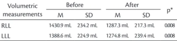
The results of volumetric measurements revealed that the mean volume of water varied from one side to the other. The initial assessments of right lower limbs (RLL) resulted in a mean volume of 1,430.9 mL (±234.2), which reduced to 1,287.3 mL (±217.3) after vascular physiotherapy, which is a difference of 143.6 mL. For the left lower limb (LLL), means were 1,388.6 mL (±224.9) before and 1,274.8 mL (±239.4) after treatment, which is a difference of 113.8 mL, as shown in Table 1.
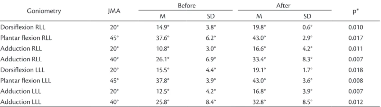
The goniometry assessments demonstrated signiicant
gains in JMA for all movement variables after vascular physiotherapy. Results for degrees of ankle movement are expressed as means with standard deviation and are shown in Table 2 together with their p values.
All of the mean scores for the AVVQ-Brazil domains reduced from their initial values after the vascular physiotherapy sessions, indicating improvement in
the patients’ disease‑speciic QoL, as can observed
in Table 3.
Figure 1. Distribution of patients by primary and secondary symptoms.
Table 1. Means, standard deviations and p values for volumetric measurement of lower limbs, before and after treatment.
Volumetric measurements
Before After
p*
M SD M SD
RLL 1430.9 mL 234.2 mL 1287.3 mL 217.3 mL 0.008
LLL 1388.6 mL 224.9 mL 1274.8 mL 239.4 mL 0.008
Mean scores for all of the domains of the SF-36 questionnaire increased after vascular physiotherapy treatment, indicating improvements in general QoL for all aspects assessed, as shown in Table 4.
DISCUSSION
All of the ten patients studied were female. There are statistical data showing that CVD patients are predominantly women,17,27,28 due to hormonal factors
and also aesthetic reasons that mean that women seek treatment three times more often than men.29
Comparison of the characteristics of the sample assessed in this study with the results of certain prevalence studies shows that both the risk factors for CVD and the age range are in line with the literature. The average age was 43.1 years, and it has been observed that the prevalence of CVD increases progressively with age from the third decade of life onwards and, according to Evans et al.,30 there is a
progressive increase in the frequency of varicose veins from puberty onwards.
There is no consensus in the literature on predominant physical posture as a risk factor for CVD. Activities that require people to remain standing or sitting for
long periods have a signiicant link to development
and maintenance of the disease and also to the emergence of ulcers and to whether they become chronic.31 This study found that the majority of
patients remained standing for prolonged periods, which can provoke muscle fatigue and deterioration of the capillaries, compromising blood and lymphatic circulation and making the emergence of circulation disorders more likely.
Studies demonstrate a predominance of CVD among people with low educational levels, correlating this factor with their working conditions, understanding of how to care for their health and a lack of economic planning, interfering with access to treatments and making it more likely that the condition will become chronic.32 This was borne out in the present study,
in which the majority of patients had not completed primary education.
None of the patients in this study were classiied
as obese, but 80% of them had high BMI and were
classiied as overweight. It is probable that both obese
and overweight people have increased compression of abdominal veins caused by increased abdominal volume and also have sedentary lifestyle habits, which can be correlated with worsening of varicose veins in the LL, since the calf muscle pump is compromised.2,29
Table 4. Mean scores for SF-36 domains before and after treatment and p values.
SF-36 Before After
p*
Domain M M
Functional capacity 39.0 80.0 0.005
Role physical 15.0 90.0 0.005
Pain 70.0 93.8 0.005
General health status 42.0 86.8 0.005
Vitality 37.0 77.5 0.005
Role social 45.0 88.8 0.005
Role emotional 30.0 90.0 0.014
Mental health 51.6 73.2 0.005
*Statistical signiicance (p<0.05). Wilcoxon test; M: Mean score. SF-36: Short Form-36.
Table 2. Reference values for normal JMA, and means, standard deviations and p values for goniometry of ankles, before and after treatment.
Goniometry JMA Before After p*
M SD M SD
Dorsilexion RLL 20° 14.9° 3.8° 19.8° 0.6° 0.010
Plantar lexion RLL 45° 37.6° 6.2° 43.0° 2.9° 0.017
Adduction RLL 20° 10.8° 3.0° 16.6° 4.2° 0.011
Adduction RLL 40° 26.1° 6.9° 33.4° 8.3° 0.007
Dorsilexion LLL 20° 15.5° 4.4° 19.1° 1.7° 0.018
Plantar lexion LLL 45° 37.8° 3.9° 43.0° 3.6° 0.008
Adduction LLL 20° 12.5° 4.2° 16.8° 3.9° 0.007
Adduction LLL 40° 25.8° 8.4° 32.8° 8.5° 0.012
*Statistical signiicance (p<0.05). Wilcoxon test; JMA: Amplitude of movement; M: Mean; SD: Standard deviation; RLL: Right lower limb; LLL: Left lower limb.
Table 3. Mean total score and domain scores for AVVQ-Brazil before and after treatment with p values.
AVVQ-Brazil Before After
p*
Domain M M
Total score 26.4 14.2 0.005
Pain and dysfunction 38.5 4.7 0.008
Aesthetic appearance 77.4 55.8 0.018
Extent of varicosity 22.8 17.6 0.005
Complications 12.4 4.7 0.017
The CEAP classiication results in this study showed
that there was a predominance of patients with C3, and edema is present in classes from C3 onwards. It is worth pointing out that, according Timi et al.,33
this can contribute to reducing the amplitude of ankle joint movement, which in turn reduces in proportion to disease progression.
Volumetric measurements of the LL were conducted using the water displacement technique, which
demonstrated a signiicant difference in volume after
physiotherapy treatment. This is considered, by many authors, to be 100% reliable for precise measurement of the volume of the limb and for estimating variations
in volume in relation to other speciic factors.21
The results of this study demonstrate the reliability of goniometry measurements, since by using them it proved possible to verify improvements in the amplitude of all movements of the ankle after the intervention with vascular physiotherapy. This is in agreement with Nolasco et al.34 who state that goniometers are
reliable and easy-to-use instruments that can be used to monitor the progress of patients and, primarily,
verify the eficacy of procedures employed to improve
the JMA of the ankle.
The SF-36 and AVVQ-Brazil were used as instruments to assess the QoL of patients with CVD. Some authors state that these assessment methods are capable of monitoring patient progress and response to treatment, assessing the quality of healthcare,35
which was the case in the present study, in which these instruments demonstrated improvements in the QoL of the patients in all of the aspects assessed.
This study demonstrated that the patients’ clinical
status improved signiicantly after vascular physiotherapy,
eliminating their complaints of tiredness and pain, reducing swelling and increasing amplitude of ankle movements and, consequently, improving their QoL. According to some studies, physical exercises increase
blood low and muscle strength and resistance, in
addition to improving venous hemodynamics and joint mobility, which improves the symptoms listed above, thereby improving the QoL of people with CVD4,26,36.
In addition to the physical exercises, MLD is another physiotherapy resource that can be used for treatment of CVD, since it can ameliorate the changes caused by the disease through a global approach to the patient, resulting in improved mobility and reduced edema and pain, and better performance in functional activities,26 in conirmation of the results
demonstrated in this study.
As such, this study provides evidence, in clinical
practice, of signiicant improvement in the clinical
status of patients after intervention with vascular physiotherapy, and these improvements are proven by the very satisfactory statistical results.
It is therefore suggested that further research into the subject be conducted with larger numbers of participants in order to obtain additional information
on the beneits of physiotherapy treatment for CVD.
CONCLUSIONS
Vascular physiotherapy is capable of modifying clinical status in CVD, promoting a positive change to the health status of patients and, consequently, a real improvement to their quality of life.
REFERENCES
1. Silva RMV, Costa LS, Carlos AG, Machini MG. Perfil clínico de pacientes atendidos na clínica de fisioterapia angiovascular na universidade Potiguar. Caderno da Escola de Saúde. 2013;2(10):118-27. 2. Alberti LR, Petroianu A, França DC, Silva TMF. Relação entre
exercício físico e insuficiência venosa crônica. Rev Med Minas Gerais. 2010;20(1):30-5.
3. Silva GCC, Medeiros RJD, Oliveira LS, et al. Treinamento de sobrecarga muscular não afeta o diâmetro das principais veias dos membros inferiores em mulheres adultas com insuficiência venosa. Rev Med Esporte. 2010;16(6):413-7. http://dx.doi.org/10.1590/ S1517-86922010000600003.
4. Lima RCM, Santiago L, Moura RMF, et al. Efeitos do fortalecimento muscular da panturrilha na hemodinâmica venosa e na qualidade de vida em um portador de insuficiência venosa crônica. J Vasc Bras. 2002;1(3):219-26.
5. Rosa AC, Montandon I. Efeitos do aquecimento sobre a amplitude de movimento: uma revisão crítica. R Bras Ci e Mov. 2006;14(2):103-10. 6. Sales JV, Morais HCR, Araújo FCS. Respostas cardiovasculares
a partir da imersão na fase de recuperação do protocolo de reabilitação cardíaca. Rev. Bras. Promoc. Saúde. 2011;24(2):123-8. 7. Azoubel R, Torres GV, Silva LW, Gomes FV, Reis LA. Efeitos da
terapia física descongestiva na cicatrização de úlceras venosas. Rev Esc Enferm USP. 2010;44(4):1085-92. http://dx.doi.org/10.1590/ S0080-62342010000400033. PMid:21337794.
8. Silva DK, Nahas MV. Prescrição de exercícios físicos para pessoas com doença vascular periférica. Rev Bras Ciên e Mov. 2002;10(1):55-61. 9. Fonseca FM, Pires JLVR, Magalhães MG, Paiva FA, Sousa CT, Bastos VPD. Estudo comparativo entre a drenagem linfática manual e atividade física em mulheres no terceiro trimestre de gestação. Fisioterapia Ser. 2009;4(4):225-33.
10. Steins A, Jünger M. Physical therapy in patients with chronic venous insufficiency. Steins Phlebologie. 2000;29(2):48-53. 11. Silva RH. Drenagem linfática manual no tratamento de pacientes
portadores de feridas venosas crônicas em membros inferiores em uso de curativos bioativos [tese]. Botucatu: Universidade Estadual Paulista, Faculdade de Medicina de Botucatu; 2010.
12 . Nakamura CM, Vanini TM, Chingui LJ, Silva CA. Avaliação das repercussões cardiovasculares da drenagem linfática manual em mulheres idosas. Anuário da Produção de Iniciação Científica Discente. 2010;13(17):43-51.
14. Alberti LR, Petroianu A, Corrêa D, Franco Silva T. Efeito da actividade física na insuficiência venosa crónica dos membros inferiores. Acta Med Port. 2008;21(3):215-20. PMid:18674413.
15. França LHG, Tavares V. Insuficiência venosa crônica: uma atualização. J Vasc Br. 2003;2(4):318-28.
16. Castro e Silva M, Cabral ALS, Barros JRN, Castro AA, Santos MERC. Diagnóstico e tratamento da Doença Venosa Crônica. J Vasc Br. 2005;4(3 Supl 2):185-94.
17. Santos RFFN, Porfírio GJM, Pitta GBB. A diferença na qualidade de vida de pacientes com doença venosa crônica leve e grave. J Vasc Bras. 2009;8(2):143-7. http://dx.doi.org/10.1590/ S1677-54492009000200008.
18. Ciconelli RM, Ferraz MB, Santos W, Meinão I, Quaresma MR. Tradução para a língua portuguesa e validação do questionário genérico de avaliação de qualidade de vida SF-36 (Brasil SF-36). Rev Bras Reumatol. 1999;39(3):143-50.
19. Leal FJ, Couto RC, Pitta GBB, et al. Tradução e adaptação cultural do Questionário Aberdeen para Veias Varicosas. Porto Alegre. J Vasc Bras. 2012;11(1):34-42. http://dx.doi.org/10.1590/ S1677-54492012000100007.
20. Leal FJ. Validação no Brasil de questionário de qualidade de vida na doença venosa (AVVQ – Brasil) [dissertação]. São Paulo: Programa de Pós-graduação em Saúde Coletiva, Escola Paulista de Medicina, Universidade Federal de São Paulo; 2012. 21. Belczac CEQ, Godoy JMP, Seidel AC, Silva JÁ, Cavalheri G Jr, Belczak
SQ. Influência da atividade diária na volumetria dos membros inferiores medida por perimetria e pela pletismografia de água. J Vasc Bras. 2004;3(4):304-10.
22. Belczak CE, Godoy JMP, Ramos R, Oliveira MA, Belczak SQ, Caffaro RA. Influência do turno laboral na formação de edema dos membros inferiores em indivíduos normais. J Vasc Bras. 2008;7(3):225-30. http://dx.doi.org/10.1590/S1677-54492008000300007. 23. Silva TA, Justo IRG, Valente FM, Godoy MFG, Godoy JMP. Efeitos
da imersão e da hidrocinesioterapia na reabilitação do linfedema. Rev Inst Ciênc Saúde. 2008;26(1):51-3.
24. Vianna DL, Greve JMD. Relação entre mobilidade do tornozelo e pé e a magnitude da força vertical de reação do solo. Rev Bras Fisioter. 2006;10(3):339-45. http://dx.doi.org/10.1590/ S1413-35552006000300014.
25. Venturini C, Ituassú NT, Teixeira LM, Deus CVO. Confiabilidade intra e interexaminadores de dois métodos de medida da amplitude ativa de dorsiflexão do tornozelo em indivíduos saudáveis. Rev Bras Fisiter. 2006;10(4):407-11. http://dx.doi.org/10.1590/ S1413-35552006000400008.
26. Meyer PF, Chacon DA, Lima ACM. Estudo piloto dos efeitos da pressoterapia, drenagem linfática manual e cinesioterapia na insuficiência venosa crônica. Reabilitar. 2006;31(8):11-7. 27. Costa LM, Higino WJF, Leal FJ, Couto RC. Clinical and
socio-demographic profile of patients with venous disease treated in health centers of Maceió (AL), Brazil. J Vasc Bras. 2012;11(2):108-13. http://dx.doi.org/10.1590/S1677-54492012000200007. 28. Krijnen RMA, de Boer EM, Bruynzeel DP. Epidemiology of venous
disorders in the general and occupational populations. Epidemiol Rev. 1997;19(2):294-309. http://dx.doi.org/10.1093/oxfordjournals. epirev.a017959. PMid:9494789.
29. Iannuzzi A, Panico S, Ciardullo AV, et al. Varicose veins of the lower limbs and venous capacitance in postmenopausal women:
relationship with obesity. J Vasc Surg. 2002;36(5):965-8. http:// dx.doi.org/10.1067/mva.2002.128315. PMid:12422106.
30. Evans CJ, Fowkes FGR, Hajivassiliou CA, Harper DR, Ruckley CV. Epidemiology of varicose veins: a review. Int Angiol. 1994;13(3):263-70. PMid:7822904.
31. Proença RPC, Bertoldi CML. Doença venosa e sua relação com as condições de trabalho no setor de produção de refeições. Rev Nutr. 2008;21(4):447-54.
32. Costa IKF. Qualidade de vida de pessoas com úlcera venosa: associação dos aspectos sociodemográficos, de saúde, assistência e clínicos da lesão [tese]. Natal: Universidade Federal do Rio Grande do Norte; 2011.
33. Timi JR, Belczak SQ, Futigami AY, Pradella FM. Anquilose tíbio- társica e sua importância na insuficiência venosa crônica. J Vasc Bras. 2009;8(3):214-8. http://dx.doi.org/10.1590/S1677-54492009000300005.
34. Nolasco CS, Reis FA, Figueiredo AM, Laraia EMS. Confiabilidade e aplicabilidade de dois métodos de avaliação da amplitude de movimento de dorsiflexão do tornozelo. ConScientiae Saúde. 2011;10(1):83-92. http://dx.doi.org/10.5585/ConScientiaeSaude/2011/ v10n1/2439.
35. Mansilha A, Leal J. Como avaliar o impacto da doença venosa crônica na qualidade de vida. Angiol Cir Vasc. 2010; 6(4):173-184. 36. Prado RA, Teixeira ALC, Langa CJSO, Egydio PRM, Izzo P. A
influência dos exercícios resistidos no equilíbrio, mobilidade funcional e na qualidade de vida de idosas. O Mundo da Saúde. 2010; 34(2):183-191.
*
Correspondence
Vanessa de Oliveira Tenório Condomínio Residencial Dom Adelmo Machado, Bloco 4, apto 405 - Cruz das Almas CEP 57038-050 - Maceió (AL), Brazil Tel.: +55 (82) 9940-6602 E-mail: [email protected]
Author information
FJL and RCC - Physical herapists, MScs in Sciences from Universidade Federal de São Paulo (UNIFESP); Assistant Professors, Universidade Estadual de Ciências da Saúde de Alagoas (UNCISAL). TPS and VOT - Physical therapists from Universidade Estadual de Ciências da Saúde de Alagoas (UNCISAL).
Author contributions
Conception and design: VOT, TPS, FJL, RCC Analysis and interpretation: VOT, TPS, FJL, RCC Data collection: VOT, TPS Writing the article: VOT, TPS Critical revision of the article: VOT, TPS, FJL, RCC Final approval of the article*: VOT, TPS, FJL, RCC Statistical analysis: VOT, TPS, FJL, RCC Overall responsibility: VOT, TPS, FJL, RCC