RBCCV 44205-1549 DOI: 10.5935/1678-9741.20140029
Vacuum-assisted drainage in cardiopulmonary
bypass: advantages and disadvantages
Sistema a vácuo na circulação extracorpórea: vantagens e desvantagens
Élio Barreto de Carvalho Filho
1, Fernando Augusto de Lima Marson
1, MD; Loredana Nilkenes
Gomes da Costa
1, Nilson Antunes
1, PhD
1Faculdade de Ciências Médicas da Universidade Estadual de Campinas
(FCM-Unicamp), Campinas, SP, Brazil.
This study was carried out at Hospital das Clínicas da Faculdade de Ciên-cias Médicas da Universidade Estadual de Campinas (HC-FCM-Unicamp), Campinas, SP, Brazil.
No inancial support.
Correspondence address:
Fernando Augusto de Lima Marson
Unicamp - Universidade Estadual de Campinas
Tessália Vieira de Camargo, 126 - Cidade Universitária “Zeferino Vaz” Campinas, SP, Brazil - Zip code: 13083-887
E-mail: [email protected]
Article received on April 6th, 2013
Article accepted on September 2nd, 2013
Abstract
Systematic review of vacuum assisted drainage in car-diopulmonary bypass, demonstrating its advantages and disadvantages, by case reports and evidence about its effects on microcirculation. We conducted a systematic search on the period 1997-2012, in the databases PubMed, Medline, Lilacs and SciELO. Of the 70 selected articles, 26 were included in the review. Although the vacuum assisted drainage has sig-nificant potential for complications and requires appropriate technology and professionalism, prevailed in literature re-viewed the concept that vacuum assisted drainage contributed in reducing the rate of transfusions, hemodilutions, better operative field, no significant increase in hemolysis, reduced complications surgical, use of lower prime and of smaller diameter cannulas.
Descriptors: Cardiopulmonary Bypass. Review. Thoracic Surgery.
Resumo
Revisão sistemática sobre drenagem assistida a vácuo na circu-lação extracorpórea, demonstrando seus benefícios, desvantagens, por relatos de casos e evidências sobre seus efeitos na microcircu-lação. Realizou-se pesquisa sistemática, no período de 1997-2012, nas bases de dados do PubMed-Medline, Lilacs e SciELO. Termos: “circulação extracorpórea”, “vácuo”, “drenagem”, “cirurgia car-díaca” e suas correspondentes traduções, em condições variadas. Dos 70 artigos selecionados, 26 foram incluídos na revisão. Embora
a drenagem assistida a vácuo possua potencial signiicante de com
-plicações e exija tecnologia e proissionalismo respectivo adequado,
prevaleceu na literatura revisada o conceito de que a drenagem assistida a vácuo contribuiu na redução no índice de transfusões, hemodiluições, melhor campo operatório, não aumento de hemólise
signiicativa, redução de complicações pós-cirúrgicas, uso de menor
prime e uso de cânulas de menor calibre.
Descritores: Circulação Extracorpórea. Revisão. Cirurgia Torácica.
The CPB circuit has two reservoirs, and the venous res-ervoir receives blood from the venous drainage and the
car-diotomy reservoir receives blood from the operative ield, re -covered by aspiration. In addition, it has oxygenator coupled to a heat exchanger. Between the oxygenator and the arterial
cannula an arterial line ilter is installed. Some equipments
are used according to the method of the team, as the pre-by-INTRODUCTION
Cardiopulmonary bypass (CPB) is a set of machines,
devices, tubes and techniques that temporarily replace the
Abbreviations, acronyms and symbols
CPB cardiopulmonary bypass VAD vacuum assisted drainage TNP topical negative pressure CVP central venous pressure
After this step, the process of selecting studies for
ex-amining titles and abstracts was initiated. The irst inclusion criterion used was identiication of relevant studies, whereas
those in which the drainage system was the main subject of the article (Table 1). At this moment, all studies that only cited vacuum as a method used in surgeries and/or described events were excluded.
Summary of Data
47 articles were found using the keywords, in the irst search
in the PubMed-Medline and 23 articles in SciELO-Lilacs. Through analysis of the abstracts included in this phase,
we deined as criteria for recovery of full articles: case report,
systematic reviews, meta-analyzes, randomized controlled trials, whose results dealt with the direct effect of the use of vacuum in the patient (positively or negative). The review was concluded with the reading of 27 full articles.
The 27 studies analyzed were divided into three groups: (I) reported cases with the use of vacuum drainage (n=3); (II)
use of vacuum in surgical processes and their inluence on
hemodynamic (n=5); (III) use of vacuum pump procedures and their evaluation (n=19).
In group I, there were three case reports, two of which describe methods that were successful and one with accident
using the vacuum technique. Shin et al.[7] used the vacuum
drainage in surgery for tumor in the right atrium. At the end
of the procedure the technique was assessed as effective, al -lowing the removal of the tumor in a safe and simple way. Similarly, Fukuda et al.[8] describe the surgery of the tricuspid valve in a patient with severe congestion caused by insufi
-cient heart valve with calciied tissue. At the end of the study,
the authors report a successful procedure citing that
hemo-lysis caused by the method was insigniicant and the patient
remained stable postoperatively, with minimal bleeding and improvement in edema of the lungs and kidneys.
Gregory et al.[9] in a case report, mentioned massive air
embolism in patient who underwent the Fontan operation. The problem occurred in the venous reservoir pressurization
caused by suction pad that blocked the air low from within
the reservoir. Therefore they recommend the deep knowledge of the perfusionist on the material used in order to avoid this type of accident.
In group II, ive studies using vacuum as treatment of sur -gical wounds and/or abscess have been described. Petzina et al.[10] in their study showed that the use of vacuum system
generated immediate reduction in cardiac output and systolic volume, as well as the left ventricular end-diastolic volume. Kadohama et al.[11] in a case report, also demonstrated the eficacy of using vacuum to post sternotomy, even in pedi -atric patients, as well as Anne et al.[12] who mentioned the eficiency of the vacuum in wound closure in mediastinum.
Lindstedt et al.[13] showed the beneits of vacuum treatment
on myocardial microcirculation.
pass ilter, cardioplegia system as well as the use of the vac -uum system.
The use of vacuum-assisted drainage system (VAD) emerged in an attempt to reduce the deleterious effects of hemodilution and with the advent of minimally invasive surgery has gained prominence, in addition to be used in heart[2-5] surgeries.
The vacuum technique consisted of using negative pres -sure in the venous reservoir, allowing the active drainage vein, eliminating the principles of gravity and siphoning[6].
This way it is possible to shorten the circuit, reducing the prime volume, hemodilution and possibly the incidence of blood transfusion[2-5].
The systematic review aimed to examine the use of VAD in CPB citing its advantages and disadvantages through case reports and evidence about its effects on microcirculation.
METHODS
Systematic research was performed, including articles pub-lished in Portuguese and English, in the period 1997 to 2012.
The reference search was performed in the databases of the following databases: PubMed-Medline (http://www.ncbi.nlm. nih.gov/pubmed), Lilacs (http://lilacs.bvsalud.org/), SciELO (http://www.scielo.org/php/index.php) using the keywords: “by-pass”, “vacuum”, “drain”, “heart surgery” and its corresponding translations and synonyms, in varied conditions.
The reviews on the subject and reference lists of included articles were consulted in search of new items for inclusion in this study.
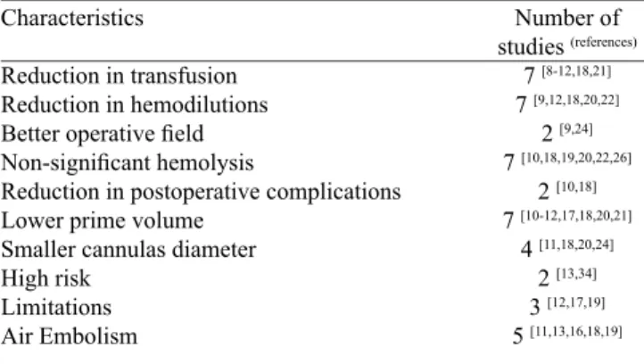
Table 1. Overview of randomized studies that examined the use of vacuum assisted drainage.
Characteristics
Reduction in transfusion Reduction in hemodilutions
Better operative ield Non-signiicant hemolysis
Reduction in postoperative complications Lower prime volume
Smaller cannulas diameter High risk
Limitations Air Embolism
Number of studies (references)
7 [8-12,18,21] 7 [9,12,18,20,22]
2 [9,24] 7 [10,18,19,20,22,26]
2 [10,18] 7 [10-12,17,18,20,21]
Finally, Chen et al.[14] demonstrated that a vacuum assist to aid in wound scarring can be used for its beneits such as
to restore the integrity of the basement membrane, reducing the endothelial space and edema, blood vessel patency, in-creasing their diameter and capillary volume and stimulate angiogenesis.
In group III were analyzed the following
characteris-tics: blood transfusion, hemodilution, surgical ield, he -molysis, postoperative complications, prime volume, size of the cannulas used, air embolism, limitations and risks
of the technique.
DISCUSSION
The development of research in CPB has helped to
clarify doubts of some procedures, often for non-scientiic
reasons, but from experience in the surgical routine. This systematic review shows that the use of a vacuum drainage
system can be effective, safe, providing new techniques to
be performed.
Analyzing three case reports found (group I), we ob-served that in one case there was an accident with massive air embolism. Gregory et al.[9] reported that the accident
was not caused solely by the vacuum system used, but suction pad not suitable for the procedure, which gener-ated the pressurization system. Replacing the suction pad
for another that could maintain and facilitate the low of
air with the environment, the procedure has become safe. Thus, the author calls attention to the materials used in the procedure, which can cause accidents when unknown by professionals.
On analysis, it was observed that 79% (n=15) of the
au-thors consider that the technique of vacuum assist provides beneit to the procedure and/or patient. The reduction in the
number of transfusions[5,6,15-19] contributes to not overload
in blood banks. The reduction occurs by improving venous
drainage and, consequently, no need for volume increase in
the venous reservoir to maintain levels of security against ingress of air into the system.
The lower use of blood products contributes to reduction of postoperative[16,20] complications, and the technique offers
reduced total prime[5,16-21] reducing hemodilution[5,6,18-21] and
maintaining hematocrit and hemoglobin levels at acceptable levels. There was disagreement with respect to generation of hemolysis by the use of a vacuum. Most authors[16,18,20,22-25]
considers that hemolysis caused in procedures with negative pressure procedures were similar to hemolysis in gravitation-al drainage. However, when comparing vacuum drainage with drainage by centrifugal pump, Cirri et al.[26] showed that
the vacuum drainage causes higher degree of hemolysis,
ac-cording conclusions conirmed by Gregoretti et al.[27].
How-ever, Lau et al.[28] and Shin et al.[24] disagree, showing similar
levels of hemolysis.
Another reported benefit was the improvement in vi-sualization of the operative field with the reduction of blood, resulting in greater security and convenience to the procedure[6,24]. The limiting venous drainage promotes
more congested surgical field and imposes difficulties in viewing by the surgeon. The possibility of using smaller diameter tubes is another important factor[17,18,20,24]. This
possibility is explained because the vacuum system im-proves the flow rate through the cannula, allowing great-er flow.
With this, one can use smaller caliber of venous cannulas, which improves visualization during surgery, without compromising venous drainage. It also allows cannulation of smaller vessels. The vacuum drainage is closely related to minimally invasive surgeries advo-cating small surgical incisions and optimization of the operative field. However, due to the benefits this type of drainage can be used in normal infusions, provided that safety measures are taken: (i) the use of own modern
equipment, (ii) the use of filters and (iii) knowledge of the technique by the perfusionist[29].
Only two authors considered the type of drainage ana-lyzed as being at high risk[29,30], being a procedure with high
probability of accidents due to the rapid rate at which the volume changes in the reservoir. Even with such statement, the studies conclude that the procedure is effective and safe, because even with the condition of rapid change in volume in
the reservoir, the experience in the described cases conirms the viability of an eficient procedure together with technical
mastery.
Davila et al.[30] stated that the procedure has risk because the technique is different from the usually practiced, forcing
the perfusionist to have knowledge of the system. In a vac-uum pump system, the system must be built to allow
alter-natives in cases of accidents. In the speciic situation of use
of negative pressure, blood circulation methods and depres-surization of the tanks must be designed to provide safe pro-cedure. Exemplifying the fact, Davila et al.[30] demonstrated
that simple change in the valve position of the circuit assem-bly can generate pressurization system, preventing or hinder-ing the correction of the accident.
In our survey, air embolism was reported in ive stud -ies[17,18,22,29,31] showing that the venous vacuum assistance
produced almost 10 times more embolism in the arterial line compared to the gravitational line, despite the use of
in the use of prime reduced, as with lowered volume levels in the reservoir the turbulence generated in CPB can allow air embolism.
The other authors cited conirm the possibility of embo -lism, however claim to be controllable the risks of this in-volvement, since the perfusionist has knowledge of the
ap-plied technique, knowing the limitations and risks involved.
Even with increased chances of accidents, Carrier et al.[32]
and Murai et al.[25] argue that the use of vacuum does not
in-crease the chance of neurological and general complications,
conirming the theory that the risks can be controlled.
In addition to the risks mentioned above, some
limita-tions on vacuum technique are taking into account by some
authors as Colangelo et al.[22], who reported the technique
as costly, while mentioning the procedure using centrifugal pump to perform the drainage. However, the statement be-comes fragile. Ii is of global knowledge that there are other methods of lower cost for the procedure, such as: (i) use of the monitor to control the vacuum from the vacuum system installed in operating rooms, (ii) vacuum pumps and (iii) roll-er pump.
Another limitation is cited by Taketani et al.[5], afirming
that the vacuum procedure presents instability due to impre-cise control of negative pressure. However, this limitation is overcome when using valves or monitors that control levels of negative pressure.
The survey also included studies that deal with the
in-luence of vacuum in hemodynamic parameters, especially
on the microcirculation. Currently, there is little knowlege-ment about the effects of the vacuum in the microcirculation, a place that really cares perfusionists, because in this area
it is dificult to achieve blood perfusion. There are studies
reporting the effectiveness of vacuum treatment[10-12] as that
detailed by Gustafsson et al.[33] on surgical wounds such as
post sternotomy mediastinitis. The mechanism by which the topical negative pressure (TNP) promotes wound healing is
by stimulating blood low in the periphery of the wound and
skeletal muscle. The mechanical stress and pressure gradient through the tissue cause increased blood volume in the area.
Both mechanical stress or increased blood low are known
to stimulate endothelial proliferation, capillary budding and angiogenesis[34].
Seeking to use knowledge of TNP for improved myocar-dial microcirculation, Lindstedt et al.[13] performed a study
with 6 pigs simulating myocardial ischemia by occluding the left posterior descending artery and using the TNP as a way to improve the microcirculation. The results proved that the use of 50 mmHg of topical negative pressure stimulated a 25 mm depth perfusion in the skeletal muscle and there was a
doubling of the blood low in the myocardium detected using
Laser Doppler.
Petzina et al.[10] performed a sternotomy in 6 pigs and
treated the surgical wound using vacuum, and
demonstrat-ed that the use of negative pressure rdemonstrat-educdemonstrat-ed cardiac output and stroke volume of the animals undergoing the surgical process. This procedure would be used in patients with deep sternal infections that have ischemic heart failure. Concom-itant to this, Chen et al.[14] show the effects of the vacuum in
the microcirculation. The author studied wounds in rabbits,
analyzing the speed of capillary blood low, as well as its
size, capillary density, the structure of the endothelium and the healing process.
The best rated pressure level was -10 kPa, which achieved maximum speed of blood in the capillaries in 4 minutes and
remained with this low for a longer time. After the comple -tion of the wounds on the rabbits’ ears, it was observed in the tissue: turgid mitochondria, membrane-targeting, large endothelial spaces, few cell junctions and many pinocytic vesicles. After 2 minutes of use of vacuum, the capillaries become more round, the endothelium cubic and the basal membrane were almost completely recovered. In 10 minutes, the capillaries become elliptical and dilated. In 30 minutes capillary sprouts emerged as villous processes, meaning an-giogenesis. In 2 hours new vessels were found, the
endothe-lial spaces have been reduced and cell junctions were irmer.
In 24 hours we observed reduction in pinocytic vesicles. In the control group, in 3 days was still observed fragmented cell membrane and diverse cell membrane. We attribute to these facts the increased blood volume caused by stimulation of negative pressure gradient, favoring membrane integrity,
resulting in reduced permeability, which consequently will reduce edema formation and thus heal the site quickly.
Transposing this study for the cardiopulmonary bypass, even in different conditions, we think that the use of vacuum
can beneit the microcirculation, promoting better tissue per
-fusion and minimizing interstitial edema caused by inlam
-mation and by changing low regime generated by cardio -pulmonary bypass. A study that still needs to be performed.
Munster et al.[35] reported the perfusion procedure with
the aid of vacuum in 54 patients. The system described in the study is similar to that normally used in cardiopulmonary by-pass with addition of negative pressure monitor, disposable pressure transducer and “Y” connector to attach the tank to the vacuum source. The relief valve of the venous reservoir already composed the system and therefore it did not have to be added. By using this system, the author claims to have enabled the heart to be always empty, as well as allowed the use of smaller cannulas facilitating the surgical procedure. Other factors were reported as the maintainability of patient’s central venous pressure (CVP) close to zero, reducing the
ad-dition of 250 ml of prime luid on average and no haemolysis
was observed postoperatively.
With these data, Munster et al.[35] reiterate the authors pre-viously cited of some beneits of using negative pressure in
CPB, adding that the procedure used is low cost and
CVP, thereby improving venous drainage and reducing pre-load momentarily until the CPB. The effect of better venous drainage allows the perfusionist maneuvers, depending on the hematocrit (greater than 25%) and hemoconcentration.
This procedure can reduce the excess liquid, even reducing
– after CPB – the preload on the myocardium, as well as reducing the formation of edema in the patient, resulting in
signiicant improvement in clinical outcome.
CONCLUSION
In conclusion, although the VAVD has signiicant poten
-tial for complications and requires technology and appropri -ate professionalism, it prevailed in the reviewed literature the concept that the VAVD contributed in reducing the rate of
transfusions, hemodilutions, better operative ield, no signif -icant increase in hemolysis, reduced postoperative compli-cations, smaller use of prime and smaller cannulas diameter.
REFERENCES
1. Souza MHL, Elias DO. Fundamentos da circulação extracorpórea 2ª ed. Rio de Janeiro: Centro editorial Alfa Rio; 2006. 828p.
2. Canêo LF, Lourenço Filho DD, Rocha e Silva R, Jatene FB, Turri F, Leirner AA. Drenagem venosa assistida através da utilização controlada de vácuo no reservatório venoso do oxigenador. Rev Bras Cir Cardiovasc. 1999;14(2):135-8.
3 . S o u z a D D , B r a i l e D M . Av a l i a ç ã o d e n o v a t é c n i c a de hemoconcentração e da necessidade de transfusão de hemoderivados em pacientes submetidos à cirurgia cardíaca com circulação extracorpórea. Rev Bras Cir Cardiovasc. 2004;19(3):287-94.
4. Souza HJB, Moitinho RF. Estratégias para redução do uso de hemoderivados em cirurgia cardiovascular. Rev Bras Cir Cardiovasc. 2008;23(1):53-9.
5. Taketani S, Sawa Y, Masai T, Ichikawa H, Kagisaki K, Yamaguchi
T, et al. A novel technique for cardiopulmonary bypass using
vacuum system for venous drainage with pressure relief valve: an experimental study. Artif Organs. 1998;22(4):337-41.
6. Chalegre ST, Salerno PR, Salerno LMVO, Melo ARS, Pinheiro AC, Frazão CS, et al. Drenagem venosa assistida a vácuo na circulação EBCF Data Survey, review of articles and textual construction FALM Reviewer
LNGC Reviewer
IN Reviewer and author responsible for publishing Authors’ roles & responsibilities
extracorpórea e necessidade de hemotransfusão: experiência de serviço. Rev Bras Cir Cardiovasc. 2011;26(1):122-7.
7. Shin H, Mori M, Matayoshi T, Suzuki R, Yozu R. Resection of giant right atrial lymphoma using vacuum-assisted cardiopulmonary bypass without snaring the inferior vena cava. Ann Thorac Cardiovasc Surg. 2004;10(4):249-51.
8. Fukuda W, Aoki C, Daitoku K, Fukuda I. Vacuum-assisted venous drainage in tricuspid valve re-replacement. Interact Cardiovasc Thoracic Surg. 2011;13(1)101-3.
9. Gregory SM, Kussman BD, Wagner JW, Boyle SL, Howe RJ, Pigula FA et al. Massive Air Embolism in a Fontan patient. J Extracorp Technol. 2011;43(2):79-83.
10. Petzina R, Ugander M, Gustafsson L, Engblom H, Sjögren J, Hetzer R, et al. Hemodynamic effects of vacuum-assisted closure therapy in cardiac surgery: Assessment using magnetic resonance imaging. J Thorac Cardiovasc Surg. 2007;133(5):1154-62.
11. Kadohama T, Akasaka N, Nagamine A, Nakanishi K, Kiyokawa K, Goh K, et al. Vacuum-assisted closure for pediatric
post-sernotomy mediastinitis: are low negative pressures suficient?
An Thorac Surg. 2008;85(3):1094-6.
12. Conquest AM, Garofalo JH, Maziarz DM, Mendelson KG, Su
Sun Y, Wooden WA, et al. Hemodynamic effects of the vacuum-assisted closure device on open mediastinal wounds. J Surg Res. 2003;115(2):209-13.
13. Lindstedt S, Malmsjö M, Ingemansson R. Blood low changes
in normal and ischemic myocardium during topically applied negative pressure. Ann Thorac Surg. 2007;84(2):568-73.
14. Chen SZ, Li J, Li XY, Xu LS. Effects of vacuum-assisted Closure on Wound Microcirculation: An experimental Study. Asian J Surg. 2005;28(3):211-7.
15. Zangrillo A, Garozzo FA, Biondi-Zoccai G, Pappalardo F, Monaco F, Crivellari M, et al. Miniaturized cardiopulmonary bypass improves short-term outcome in cardiac surgery: a meta-analysis of randomized controlled studies. J Thorac Cardiovasc Surg. 2010;139(5):1162-9.
16. Nasso G, Costantini C, Petralia A, Del Prete A, Lopriore V, Fattouch K, et al. A new extracorporeal vacuum-assited device to optimize cardiopulmonary bypass. Comparison with the conventional system. Interact Cardiovasc Thorac Surg. 2011;12(4):591-5.
17. Banbury MK, White JA, Blackstone EH, Cosgrove DM 3rd. Vacuum-assisted venous return reduces blood usage. J Thorac Cardiovasc Surg. 2003;126(3):680-7.
18. Bevilacqua S, Matteucci S, Ferrarini M, Kacila M, Ripoli A,
19. Nakanishi K, Shichijo T, Shinkawa Y, Takeuchi S, Nakai M, Kato G, et al. Usefulness of vacuum-assisted cardiopulmonary bypass circuit for pediatric open-heart surgery in reducing homologous blood transfusion. Eur J Cardiothorac Surg. 2001;20(2):233-8.
20. Hayashi Y, Kagisaki K, Yamaguchi T, Sakaguchi T, Naka Y, Sawa Y, et al. Clinical application of vacuum-assisted cardiopulmonary bypass with a pressure relief valve. Eur J Cardiothorac Surg. 2001;20(3):621-6.
21. Pappalardo F, Corno C, Franco A, GiardinaG, Scandroglio AM, Landoni G, et al. Reduction of hemodilution in small adults undergoing open heart surgery: a prospective randomized trial. Perfusion. 2007;22(5):317-22.
22. Colangelo N, Torracca L, Lapenna E, Moriggia S, Crescenzi
G, Alieri O. Vacuum-assisted venous drainage in extrathoracic
cardiopulmonary bypass management during minimally invasive cardiac surgery. Perfusion. 2006;21(6):361-5.
23. Mueller XM, Tevaeara HT, Horisberger J, Augstburger M, Burki M, von Segesser LK. Vacuum assisted venous drainage does not increase trauma to blood cells. ASAIO J. 2001;47(6):651-4.
24. Shin H, Yozu R, Maehara T, Matayoshi T, Morita M, Kawai Y, et al. Vacuum assisted cardiopulmonary bypass in minimally invasive cardiac surgery: its feasibility and effects on hemolysis. J Artif Organs 2000;24(6):450-3.
25. Murai N, Cho M, Okada S, Chiba T, Saito M, Shioguchi S, et al. Venous drainage method for cardiopulmonary bypass in single-access minimally invasive cardiac surgery: siphon and vacuum-assisted drainage. J Artif Organs. 2005;8(2):91-4.
26. Cirri S, Negri L. Babbini M, Latis G, Khlat B, Tarelli G, et al. Haemolysis due to active venous drainage during
cardiopulmonary bypass: comparison of two different techniques.
Perfusion. 2001;16(4):313-8.
27. Gregoretti S. Suction-induced hemolysis at various vacuum pressures: implications for intraoperative blood salvage. Transfusion. 1996;36(1):57-60.
28. Lau CL, Posther KE, Stephenson GR, Lodge A, Lawson JH, Darling EM, et al. Mini-circuit cardiopulmonary bypass with vacuum assisted venous drainage. Feasibility of an asanguineous prime in the neonate. Perfusion. 1999;14(5):389-96.
29. Kiyama H, Imazeki T, Katayama Y, Murai N, Mukouyama M, Yamauti N. Vacuum-assisted venous drainage in single-access minimally invasive cardiac surgery J Artif Organs. 2003;6(1):20-4.
30. Davila RM, Rawles T, Mach MJ. Venoarterial air embolus: a complication of vacuum-assisted venous drainage. Ann Thorac Surg. 2001;71(4):1369-71.
31. Willcox TW, Mitchell SJ, Gorman DF. Venous air in the bypass circuit: a source of arterial line emboli exacerbated by vacuum-assisted drainage. Ann Thorac Surg. 1999;68(4):1285-9.
32. Carrier M, Cyr A, Voisine P, Pellerin M, Perrault LP, Cartier R, et al. Vacuum-assisted venous drainage does not increase the neurological risk. Heart Surg Forum. 2002;5(3):285-8.
33. Gustafsson RI, Sjogren J, Ingemansson R. Deep sternal wound
infection: a sternal-sparing technique with vacuum-assisted
closure therapy. Ann Thorac Surg. 2003;76(6):2048-53.
34. Vandenburgh HH. Mechanical forces and their second messengers in stimulating cell growth in vitro. Am J Physiol. 1992;262(3 Pt 2):R350-5.