www.jcol.org.br
Journal of
Coloproctology
Original article
Risk factors for recurrence of stage I/II (TNM) colorectal
adenocarcinoma in patients undergoing surgery with
curative intent
☆Marssoni Deconto Rossoni
a,*, José Ederaldo Queiroz Telles
b,
Andrea Maciel de Oliveira Rossoni
c, Jorge Eduardo Fouto Matias
da Postgraduate Program in Clinical Surgery, Universidade Federal do Paraná (UFPR), Curitiba, PR, Brazil b Department of Clinical Pathology, Division of Health Sciences, UFPR, Curitiba, PR, Brazil
c Department of Internal Medicine, UFPR, Curitiba, PR, Brazil
d Department of Surgery and Postgraduate Program in Clinical Surgery, UFPR, Curitiba, PR, Brazil
a r t i c l e i n f o
Article history:
Received 6 January 2013 Accepted 19 February 2013
Keywords:
Neoplastic invasiveness Adenocarcinoma Prognosis
Colorectal neoplasms
a b s t r a c t
Objective: Evaluate risk factors for colorectal cancer recurrence after surgical treatment. Methods: Sixty-fi ve patients with colorectal adenocarcinoma, stage I and II (TNM), undergoing curative-intent surgery and followed for fi ve years were studied. Presence of adjuvant/neo-adjuvant therapy, tumor differentiation degree, lymphatic and venous vascular infi ltration, depth of tumor invasion, and disease staging was analyzed, using recurrence relative risk ratios for each parameter calculated at two years, after two years and fi ve years of follow up. Results: At fi ve years, recurrence was 21.4% (14/65), with equal incidence (10.7%) for the separated periods. Only lymphatic and venous vascular infi ltration showed statistically signifi cant association with recurrence during times analyzed. Relative risk (RR) of recur-rence was signifi cantly related to the presence of lymphatic infi ltration [RR = 6 (1.3 – 28.5) p = 0.01] and venous infi ltration [RR = 9.5 (2.6 – 34.9) p < 0.001] after two years of follow-up. At fi ve years follow-up, only venous infi ltration remained with signifi cant relative risk for recurrence [RR = 3.9 (1.8 – 8.8) p < 0.001]. In a multivariate analysis, only venous vascular infi ltration was associated with recurrence [accuracy 81.5% (p < 0.001)].
Conclusion: In this series, the factors associated with risk of colorectal cancer recurrence were the presence of lymphatic and venous vascular infi ltration.
© 2013 Elsevier Editora Ltda. All rights reserved.
☆ This work is part of the doctoral thesis of the student Marssoni Deconto Rossoni, Postgraduate Program in Clinical Surgery of UFPR.
* Corresponding author.
E-mail: [email protected] (M.D. Rossoni)
Palavras-chave:
Invasividade neoplásica Adenocarcinoma Prognóstico
Neoplasias colorretais
r e s u m o
Fatores de risco para recidiva em pacientes com adenocarcinoma colorretal estádio I e II (TNM) submetidos à cirurgia com intenção curativa
Objetivo: Analisar fatores de risco para recidiva de câncer colorretal após tratamento ci-rúrgico.
Método: Avaliou-se 65 pacientes com adenocarcinoma colorretal, estadio I e II (TNM), sub-metidos à cirurgia com intenção curativa, acompanhados por cinco anos após a operação. Analisou-se presença de tratamento adjuvante/neoadjuvante, grau de diferenciação do tumor, ini ltração vascular linfática e venosa, profundidade de invasão tumoral e estadia-mento da doença, estabelecendo-se para cada um o risco relativo de recidiva aos dois anos, após dois anos e aos cinco anos de seguimento.
Resultados: Recidiva global em cinco anos foi 21,4% (14/65), com idêntica incidência (10,7%) nos períodos separados. Somente as ini ltrações vasculares linfáticas e venosas apresen-taram associação estatisticamente signii cativa com a recidiva nos períodos de análise. Encontrou-se risco relativo (RR) estatisticamente signii cativo após dois anos relacionados à presença de ini ltração linfática [RR = 6 (1,3 – 28,5) p = 0,01] e ini ltração venosa [RR = 9,5 (2,6 – 34,9) p < 0,001]. Após cinco anos, apenas a ini ltração venosa manteve a signii cância estatística, com risco relativo elevado para ocorrência de recidiva [RR = 3,9 (1,8 – 8,8) p < 0.001]. Na análise multivariada apenas a presença de ini ltração vascular venosa com 81,5% de acerto foi associada à recidiva (p < 0.001).
Conclusão: Nesta série, os únicos fatores associados com risco de recidiva do câncer color-retal foram a presença de ini ltração vascular linfática e venosa.
© 2013 Elsevier Editora Ltda. Todos os direitos reservados.
Introduction
Colorectal cancer (CRC) ranks second in mortality in the U.S. and in Brazil, second only to lung cancer. The National Insti-tute of Health reported 141,210 new cases and 49,380 deaths
in the year 2011.1 In Brazil, according to the National Cancer
Institute (INCA) data, 30,140 new cases of colon and rectum cancer are expected for the year 2012, with a slightly higher
incidence in women.2
Although there has been progress in understanding the genesis of colorectal tumors, CRC-related deaths are still
high, with great impact on public health programs.2 The
overall median survival at 5 years is around 55% in devel-oped countries and 40% in developing countries, and can
reach higher rates.2
Currently, colorectal cancer staging is based on clinico-pathological staging proposed by the International Union Against Cancer (TNM staging system), i.e., depth of tumor invasion into the colon wall, presence of lymph node and distant metastases. However, patients with the same stage may have different clinical outcomes, indicating that the currently used staging may not rel ect the actual
aggressive-ness of each individual tumor.3,4
Several efforts have been made regarding the identii ca-tion of the colorectal cancer prognostic factors. This study objective was to analyze in a case series of colorectal cancer (TNM staging I-II) the relationship between the specii c clin-icopathological factors and recurrence after surgical treat-ment with curative intent.
Methods
This study was approved by the Human Research Ethics Com-mittee of the Hospital de Clínicas (HC), Universidade Federal do Paraná (UFPR), and registered under the No 82.001/2003-01.
Patients: After reviewing 550 cases of colorectal cancer treat-ed at the General Surgery and Digestive Surgery centers (HC– UFPR), from September 1995 to January 2003, 65 patients with neoplasia, classii ed as TNN clinical stage I and II and under-wenting curative surgical treatment were selected. Exclusion criteria were familial adenomatous polyposis, inl ammatory bowel disease, and postoperative death.
Follow-up: After surgery, patients were followed-up for at least i ve years, according to the following protocol: clinical history, physical examination, measurement of blood carci-noembryonic antigen (CEA), chest X-ray, abdominal ultra-sound, chest and abdominal CT scans, and colonoscopy. In the i rst year after surgery, follow-up visits were quarterly and colonoscopy semiannually; from the second year onward, the visits were semiannual and colonoscopy annual. CEA blood test, chest X-ray, and abdominal ultrasound were performed every three months for the i rst year and every six months after the second year. Chest and abdominal CT scan was per-formed every six months in the i rst year and annually from the second year onwards.
therapy used were correlated with the incidence of relapse when detected up to two years, after two years, and at i ve years of follow-up.
Statistical Analysis: The measures of central tendency con-sidered were average, standard deviation, and 95% coni dence interval for continuous variables and absolute frequencies and percentages for categorical variables. The estimated risk was performed by calculating the relative risk for variables with three levels of classii cation, always comparing the second lev-el with the i rst, then the third levlev-el with the i rst, and i nally both levels with the i rst. Multivariate logistic regression and discriminant analysis models were used to assess the predic-tive power for disease recurrence. To estimate the difference between variables, Student’s t-test was used for continuous variables and chi-square test and Fisher’s exact test for cat-egorical variables; p values < 0.05 were considered statistically signii cant.
Results
Patient’s mean age was 58.5 ± 12.6 years (24-79 years), with a slight predominance of male (53.3%). Rectal neoplasm ac-counted for 40% of cases (26), with 40% represented by other lesions in the left colon and the remaining 20% (13) located in the right colon. Surgical treatment consisted of anterior recto-sigmoidectomy or recto-sigmoidectomy in 29 cases (44.7%). Abdom-inoperineal excision of rectum was performed in 17 patients (26.1%). Fifteen patients (23.1%) underwent right colectomy and four patients (6.1%) were treated for left colectomy.
Table 1 shows the frequency of the analyzed parameters and recurrence occurred during the follow-up times. Adju-vant/neoadjuvant therapy was used in 44.6% of cases. Grade 1 histology was predominant (69.3%). Lymphatic ini ltration overcame venous ini ltration (29.2% vs 12.3%), while T3 tu-mor depth and TNM stage II predominated (64.6%).
Recurrence was observed in 14 patients over the i ve year of follow-up (21.4%), divided equitably (seven cases, 10.7%) be-tween times up to two years and after two years. Topographi-cally, the locations with the highest frequency of recurrence were the liver and pelvic region, with six cases each (9.2%). Recurrence was also seen in the lung and anastomotic line in one case each (1.5%) (Table 1).
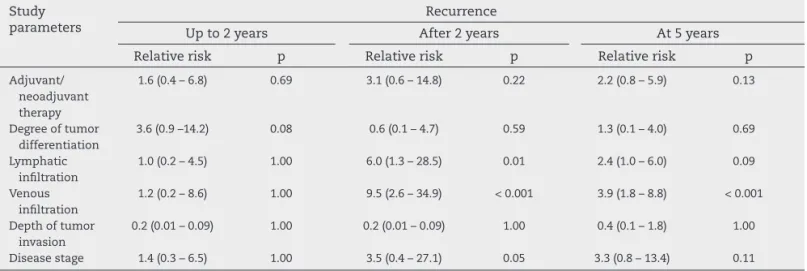
Regarding relative risk of relapse according to the studied parameters, there were no statistically signii cant differences between any of the times studied and the adjuvant/neoadju-vant therapy used, tumor differentiation degree, depth of tu-mor invasion, and disease stage (Table 2).
Presence of lymphatic and venous ini ltration was associ-ated with a statistically signii cant relative risk of recurrence after two years of follow up. The risk of recurrence after two years of follow-up was six times higher with lymphatic ini l-tration (95% CI = 1.3 – 28.5) than without lymphatic involve-ment. Similarly, the risk of tumor recurrence after two years of follow-up was 9.5 times higher (95% CI = 2.6 – 34.9) with venous ini ltration than without venous vascular invasion. The latter persisted with statistical signii cance for increased risk of recurrence also when analyzing the total follow-up time (i ve years), with a risk of recurrence 3.9 times higher (95% CI = 1.8 – 8.8) (Table 2).
Table 1 – Frequency of presented parameters and recurrence in the study patients.
Characteristics Values
Adjuvant/neoadjuvant therapy
Chemotherapy and/or radiotherapy: 29 cases (44.6%)
Grade of histological differentiation
Grade I: 45 cases (69.3%) Grade II: 9 cases (13.8%) Grade III: 11 cases (16.9%) Vascular ini ltration Lymphatic: 19 cases (29.2%)
Venous: 8 cases (12.3%)
Lymphatic and venous: 6 cases (9.2%) Depth of tumor invasion T1: 1 case (1.5%)
T2: 22 cases (33.9%) T3: 42 cases (64.6%) TNM staging Stage I: 23 cases (35.4%)
Stage II: 42 cases (64.6%) Recurrence Present: 14 cases (21.4%) Liver: 6 cases (9.2%) Pelvic region: 6 cases (9.2%) Anastomotic line: 1 case (1.5%) Lung: 1 case (1.5%)
Time of recurrence:
Up to two years: 7 cases (10.7%) After 2 years: 7 cases (10.7%) At 5 years: 14 cases (21.4%)
Table 2 – Relative risk of relapse according to the study parameters. Study
parameters
Recurrence
Up to 2 years After 2 years At 5 years
Relative risk p Relative risk p Relative risk p
Adjuvant/ neoadjuvant therapy
1.6 (0.4 – 6.8) 0.69 3.1 (0.6 – 14.8) 0.22 2.2 (0.8 – 5.9) 0.13
Degree of tumor differentiation
3.6 (0.9 –14.2) 0.08 0.6 (0.1 – 4.7) 0.59 1.3 (0.1 – 4.0) 0.69
Lymphatic ini ltration
1.0 (0.2 – 4.5) 1.00 6.0 (1.3 – 28.5) 0.01 2.4 (1.0 – 6.0) 0.09
Venous ini ltration
1.2 (0.2 – 8.6) 1.00 9.5 (2.6 – 34.9) < 0.001 3.9 (1.8 – 8.8) < 0.001
Depth of tumor invasion
0.2 (0.01 – 0.09) 1.00 0.2 (0.01 – 0.09) 1.00 0.4 (0.1 – 1.8) 1.00
Considering relapse as dependent variable, discriminant analysis showed that the studied parameter selected with the highest discrimination power was the presence of ve-nous vascular ini ltration, with 81.5% of correct classii cation (p < 0.001).
Discussion
In the present study, as often observed in the literature,1,5
the most frequent tumor location was in the left colon and rectum; histological type was adenocarcinoma; and grade of histological differentiation was Grade I.
There was no recurrence in 78% of patients, which can be explained by the early clinical stage of tumors (I-II). Re-currence was more frequent in the liver and pelvic region,
which is consistent with literature reports.6,7
Diagnosis and treatment of colorectal cancer have evolved considerably in recent years, particularly with regard to de-cisions on limited or complete resection and the type and indication of the adjuvant therapy used. In both situations, decisions are mainly based on the histopathological i ndings
of the resected specimen.8-10 The indication of adjuvant
ther-apy in clinical stages I and II patients remains controversial in the literature because, besides the possibility of not in-creasing the survival of these individuals, it has a high cost
and may expose them to the adverse effects of QT and RT.8,11
In our series, the evaluation of adjuvant or neoadjuvant therapy as a prognostic factor for recurrence showed no sig-nii cant association. This may be evidence that in patients with early clinical stage disease the use of adjuvant or neo-adjuvant treatment does not change the course of disease.
Although adjuvant chemotherapy is indicated for pa-tients with colorectal cancer with lymph nodes positive for malignancy, a small proportion of patients with negative nodes have an unfavorable clinical course, and thus adju-vant therapy could be benei cial for these patients,
justify-ing costs and adverse effects.8,12 Therefore, identii cation of
patients at higher risk for local, lymph node, and distance metastasis recurrence is critical, regardless of the informa-tion obtained by the applicainforma-tion of the current pathological TNM staging system.
Contrary to what literature often reports, in this series we found no association between frequency of tumor re-currence and degree of differentiation, depth of tumor
in-vasion, and staging.13,14 Coincidentally, the classii cation of
these parameters evaluated in this study whose classii ca-tion could not be categorized in a dichotomous (present/ absent), requiring two (TNM staging) or three classes (grade of histological differentiation and depth of tumor invasion), subdividing the number of patients prior to statistical analy-sis. Unfortunately, we cannot discard dei nitively that the analysis of these parameters for the associations estimated as non-signii cant may have been a type II error due to the sample size.
The presence of lymphatic and venous vascular ini ltra-tion by neoplastic colorectal cells is described in literature as a prognostic factor indicative of more aggressive
neo-plasm.7,15-18 In this study, 29% of patients had lymphatic
vas-cular ini ltration and 12% had venous ini ltration. Moreover,
there was a statistically signii cant association between venous or lymphatic ini ltration and tumor recurrence rate measured at follow-up.
When we stratii ed the time of global recurrence, as-sessed at i ve years follow-up in two different times (up to two years and after two years recurrence), we found similar behavior of lymphatic and venous ini ltration according to the sub-period analyzed (Table 2). Both parameters (venous and lymphatic ini ltration) did not reach statistical signii -cance when studied for association with early relapse within two years. On the other hand, both parameters were signii -cantly associated with tumor recurrence after the third year of follow up. We believe that this fact has important clini-cal implications relevant to current and future strategies of postoperative follow-up of patients undergoing colorectal cancer surgery. We could ask, for example, to patients at follow-up (with known high risk of recurrence) the reasons for the infrequency, except from the third year of follow up. Are frequency and the means currently available to detect recurrences suitable for the earlier periods of follow-up? Is there a different clinical proi le among patients with early recurrence compared to others?
In this series, the predictive factors of recurrence in mul-tivariate analysis revealed vascular venous ini ltration as the only factor capable of predicting recurrence alone with sta-tistical signii cance (p < 0.001).
Currently, the biological behavior of malignant colorectal neoplasm still surpasses the ability to predict recurrence pa-rameters of known value and used for evaluation and char-acterization of clinical placements.
Conclusion
In this work, it was possible to concluded that among the clinicopathological factors analyzed, the ones associated with risk of recurrence were lymphatic and venous vascular ini l-tration by tumor cells; nevertheless, only after two years of follow-up.
Further studies with new criteria to predict early relapse are still needed in order to have an even better performance in colorectal cancer follow-up.
R E F E R E N C E S
1. Colorectal cancer 2011 [cited 2011 14 Dezembro]. Disponível em: www.cancer.org.
2. Câncer colorretal 2011 [cited 2011 14 Dezembro]. Disponível em: www.inca.gov.br.
3. Okuyama T, Oya M, Ishikawa H. Budding as a useful prognostic marker in pT3 well- or
moderately-differentiated rectal adenocarcinoma. J Surg Oncol. 2003 May;83(1):42-7.
4. Okuyama T, Nakamura T, Yamaguchi M. Budding is useful to select high-risk patients in stage II well-differentiated or moderately differentiated colon adenocarcinoma. Dis Colon Rectum. 2003 Oct;46(10):1400-6.
6. Di Gregorio C, Benatti P, Losi L, Roncucci L, Rossi G, Ponti G, et al. Incidence and survival of patients with Dukes’ A (stages T1 and T2) colorectal carcinoma: a 15-year population-based study. Int J Colorectal Dis. 2005 Mar;20(2):147-54.
7. Losi L, Ponti G, Gregorio CD, Marino M, Rossi G, Pedroni M, et al. Prognostic signii cance of histological features and biological parameters in stage I (pT1 and pT2) colorectal adenocarcinoma. Pathol Res Pract. 2006;202(9):663-70. 8. Chun P, Wainberg ZA. Adjuvant Chemotherapy for Stage II
Colon Cancer: The Role of Molecular Markers in Choosing Therapy. Gastrointest Cancer Res. 2009 Sep;3(5):191-6. 9. Porschen R, Arkenau HT, Kubicka S, Greil R, Seufferlein T,
Freier W, et al. Phase III study of capecitabine plus oxaliplatin compared with l uorouracil and leucovorin plus oxaliplatin in metastatic colorectal cancer: a i nal report of the AIO Colorectal Study Group. J Clin Oncol. 2007 Sep;25(27):4217-23. 10. Northover JM. Staging and management of colorectal cancer.
World J Surg. 1997 Sep;21(7):672-7.
11. Gill S, Loprinzi CL, Sargent DJ, Thomé SD, Alberts SR, Haller DG, et al. Pooled analysis of l uorouracil-based adjuvant therapy for stage II and III colon cancer: who benei ts and by how much? J Clin Oncol. 2004 May;22(10):1797-806.
12. Zlobec I, Molinari F, Martin V, Mazzucchelli L, Saletti P, Trezzi R, et al. Tumor budding predicts response to anti-EGFR therapies in metastatic colorectal cancer patients. World J Gastroenterol. 2010 Oct;16(38):4823-31.
13. Kajiwara Y, Ueno H, Hashiguchi Y, Mochizuki H, Hase K. Risk factors of nodal involvement in T2 colorectal cancer. Dis Colon Rectum. 2010 Oct;53(10):1393-9.
14. Tateishi Y, Nakanishi Y, Taniguchi H, Shimoda T, Umemura S. Pathological prognostic factors predicting lymph node metastasis in submucosal invasive (T1) colorectal carcinoma. Mod Pathol. 2010 Aug;23(8):1068-72.
15. Gabbert H. Mechanisms of tumor invasion: evidence from in vivo observations. Cancer Metastasis Rev. 1985;4(4): 293-309.
16. Gabbert H, Wagner R, Moll R, Gerharz CD. Tumor dedifferentiation: an important step in tumor invasion. Clin Exp Metastasis. 1985 1985 Oct-Dec;3(4):257-79. 17. Merkel S, Wein A, Günther K, Papadopoulos T, Hohenberger
W, Hermanek P. High-risk groups of patients with Stage II colon carcinoma. Cancer. 2001 Sep;92(6):1435-43.