Evolution of hepatitis B serological markers
in HIV coinfected patients: a case study
Ana Luiza de Castro Conde ToscanoI,II, Maria Cássia Mendes CorrêaII,III
I Instituto de Infectologia Emílio Ribas. São Paulo, SP, Brasil
II Departamento de Doenças Infecciosas. Faculdade de Medicina. Universidade de São Paulo. São Paulo, SP, Brasil III Instituto de Medicina Tropical de São Paulo. Laboratório de Investigação Médica 52. São Paulo, SP, Brasil
ABSTRACT
OBJECTIVE: To describe the evolution of serological markers among HIV and hepatitis B coinfected patients, with emphasis on evaluating the reactivation or seroreversion of these markers.
METHODS: he study population consisted of patients met in an AIDS Outpatient Clinic in São Paulo State, Brazil. We included in the analysis all HIV-infected and who underwent at least two positive hepatitis B surface antigen serological testing during clinical follow up, with tests taken six months apart. Patients were tested with commercial kits available for hepatitis B serological markers by microparticle enzyme immunoassay. Clinical variables were collected: age, sex, CD4+ T-cell count, HIV viral load, alanine aminotransferase level, exposure to antiretroviral drugs including lamivudine and/or tenofovir.
RESULTS: Among 2,242 HIV positive patients, we identiied 105 (4.7%) patients with chronic hepatitis B. Follow up time for these patients varied from six months to 20.5 years. All patients underwent antiretroviral therapy during follow-up. Among patients with chronic hepatitis B, 58% were hepatitis B “e” antigen positive at the irst assessment. Clearance of hepatitis B surface antigen occurred in 15% (16/105) of patients with chronic hepatitis B, and 50% (8/16) of these patients presented subsequent reactivation or seroreversion of hepatitis B surface antigen. Among hepatitis B “e” antigen positive patients, 57% (35/61) presented clearance of this serologic marker. During clinical follow up, 28.5% (10/35) of those who initially cleared hepatitis B “e” antigen presented seroreversion or reactivation of this marker.
CONCLUSIONS: Among HIV coinfected patients under antiretroviral therapy, changes of HBV serological markers were frequently observed. hese results suggest that frequent monitoring of these serum markers should be recommended.
DESCRIPTORS: HIV infection, Hepatitis B, Chronic, immunology. Coinfection. Biomarkers. Seroepidemiologic Studies.
Correspondence:
Ana Luiza de Castro Conde Toscano Av. Dr. Arnaldo, 165
01246-900 São Paulo, SP, Brasil E-mail: ana.toscano@emilioribas. sp.gov.br
Received: 16 Sep 2015 Approved: 17 Jan 2016
How to cite: Toscano ALCC, Mendes-Corrêa MC. Evolution of hepatitis B serological markers in HIV coinfected patients: a case study. Rev Saude Publica. 2017;51:24.
Copyright: This is an open-access article distributed under the terms of the Creative Commons Attribution License, which permits unrestricted use, distribution, and reproduction in any medium, provided that the original author and source are credited.
INTRODUCTION
Among the estimated 40 million people living with human immunodeiciency virus (HIV) worldwide, it is believed that approximately four million (10%) are coinfected with hepatitis B virus (HBV)1. his is mainly because both viruses share the same routes of transmission.
he presence of HIV seems to alter the natural evolution of hepatitis B infection. It is believed that the presence of HIV could decrease the chance of HBV viral clearance after an acute infection and increase the risk for HBV chronic infection. Individuals with HIV-HBV coinfection have higher HBV DNA levels, rapid liver disease progression, and increased liver disease-related mortality, when compared to HBV monoinfected patients2.
Immunosuppression associated with HIV infection or interruption of anti-HBV treatment may lead to HBV reactivation, even in individuals with evidence of prior positive antibody to the hepatitis B surface antigen (anti-HBs)3,4. Furthermore, immunosuppression associated with HIV infection may be related to the inability to produce antibodies in the presence of an HBV infection. herefore, atypical serological patterns have been described in this population. he presence of HBV variants with speciic mutations has also been frequently described in the same population, and has been associated with some atypical serological patterns found in coinfected patients5.
Evolution of hepatitis B serological markers among HIV coinfected patients has been evaluated by some authors, and these studies reveal great variation of these markers during clinical follow up6,7. In Brazil, however, data regarding this aspect of HBV infection in this population is scarce. his study aimed to describe the evolution of hepatitis B surface antigen (HBsAg) and hepatitis B “e” antigen (HBeAg) among HIV coinfected patients, with emphasis on the reactivation or seroreversion of these markers in this group of patients.
METHODS
he enrolled patients were selected from those regularly followed up at an HIV Outpatient Clinic in São Paulo, Brazil, between May 2006 and July 2011. All HIV-infected patients with serum hepatitis B surface antigen HBsAg were identiied. HBsAg-positive patients who underwent at least two repeated HBV serological testing, during clinical follow up, with tests taken six months apart, were included in the analysis. he study was conducted from June 2011 to July 2012.
he search for information about HBsAg-reactive patients started with an electronic and written database query. he serological proile analysis relative to hepatitis B in all patients with regular treatments during the determined time frame (between May 2006 and July 2011) identiied HBsAg positive patients. We included all serological tests found for each patient, containing serological markers of HBV infection.
Medical records were reviewed retrospectively to ascertain demographic and clinical characteristics and laboratory indings during the six months preceding the serological test of interest (we considered the data sample in which serological reactivation of HBsAg and/or HBeAg markers was irst detected).
he following clinical and demographic variables were considered for analysis: sex, age, HIV or HBV risk factors, treatment adherence; CD4+ T-cell count; HIV viral load, alanine aminotransferase (ALT) level; use and duration of exposure to antiretroviral drugs (ARV); use and duration of use of HBV-active drugs (lamivudine, tenofovir, entecavir, or interferon). Epidemiological, clinical, and laboratory features were described based on the date of serological test of interest.
the serological test of interest. he absolute values of HIV viral load will not be described, since the long period of follow-up did not allow the analysis using a single method.
For the purpose of this study, we considered HBsAg or HBeAg seroclearance when HBsAg or HBeAg, respectively, became non-reagent after being reagent, during clinical follow up. HBsAg or HBeAg seroreversion was considered whenever these markers were detected reagent after being previously detected as non-reagent (with no appearance of anti-HBs or anti-HBe, respectively) during clinical follow up.
HBsAg or HBeAg reactivationwere considered whenever these markers were detected reagent after been previously detected as non-reagent (with appearance of anti-HBs or anti-HBe, respectively) during clinical follow-up. Follow up time was considered as the period of time between the irst and last serologic sample containing HBsAg or HBeAg markers.
HIV results were obtained using two commercial enzyme immunosorbent assays (Organon Technika, Tournault, Belgium and Embrabio, São Paulo, Brazil) and conirmed using GS-HIV-1 Western Blot (Bio-Rad, Hercules, CA). HIV RNA was quantiied using the Versant HIV-1 RNA 3.0 bDNA assay (Siemens Healthcare, Erlangen, Germany) and Nucleic Acid Sequence-Based-Ampliication (NASBA-NUCLISENS). CD4 T-lymphocyte counts were determined using the BD Multitest/Trucount (BD Biosciences, San Jose, CA).
A microparticle enzyme immunoassay (MEIA, AxSYM, Abbott Laboratories, Abbott Park, IL) was used to detect serological markers (HBsAg, anti-Hepatitis B core total antibodies, anti-HBs, HBeAg, and anti-HBe) at the Central Laboratory of the Hospital das Clínicas, São Paulo, Southeastern Brazil.
his project was approved by the Research Ethics Committee of the Department of Infectious and Parasitic Diseases of the Faculdade de Medicina da Universidade de São Paulo (CAPPesq Protocol 0197/11, from November 5, 2011). It was exempted of the need for an informed consent form for patients. Secrecy and conidentiality were guaranteed in this study. he analyses were performed from a database entered into Microsoft Excel. A descriptive
analysis of the study variables was carried out.
Due to the small number of patients included in the study, we were not able to compare the possible associations between the diferent patterns of serological evolution observed and the clinical or epidemiological variables described.
RESULTS
Initially, 2,242 patients were enrolled in the AIDS Outpatient Clinic with conirmed HIV infection and serological markers for hepatitis B. Of those, 105 (4.7%) were patients with chronic hepatitis B.
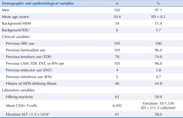
he clinical and epidemiological featuresof the 105 included HBsAg-positive patients are shown in Table 1.
Most patients were male (97%), with the main risk factor for exposure to both infections (HIV and HBV) being risky sexual behavior (including same-sex relations). All patients underwent antiretroviral therapy during the monitoring phase. A total of 44% (46/105) of patients had a history of one or more previous AIDS deining illness. CD4+ T-cell counts varied from 10 to 1,536 cells/mm3, with the average count being 449.2 cells/mm3 (standard deviation [SD] = 211.5 cells/mm3). A total of 101 (96%) and 78 (74%) patients used lamivudine or tenofovir, respectively, in their therapeutic regimen during follow up.
Follow up time for these 105 patients varied from six months to 20.5 years.
Among those with HBsAg positive, 58% (61/105) were also HBeAg positive.
Clearance of HBsAg occurred in 15% (16/105) of patients with chronic hepatitis B (1.7 cases per 100 person-years), and 50% of them (8/16) presented subsequent seroreversion or reactivation of HBsAg during clinical follow up. Two patients (25%) presented anti-HBs before HBsAg reactivation.
Follow up time for patients who underwent HBsAg reappearance varied from 1,775 to 6,051 days (4.9 years to 16.8 years).
Clinical characteristics of patients who presented HBsAg reactivation are summarized in Table 2.
Among HBeAg positive patients at the irst serologic test, 57% (35/61) underwent clearance of this serologic marker (5.8 cases per 100 person-years). During clinical follow up, 28.5%
Table 1. Epidemiological, clinical, and laboratory features of 105 HBsAg-reactive patients coinfected with HIV.
Demographic and epidemiological variables n %
Men 102 97.1
Mean age (years) 50.4 SD = 8.2
Background MSM 54 51.4
Background IDU 6 5.7
Clinical variables
Previous ARV use 105 100
Previous lamivudine use 101 96.0
Previous tenofovir use (TDF) 78 74.0
Previous LAM, TDF, ENT, or IFN use 101 96.0
Previous entecavir use (ENT) 4 3.8
Previous interferon use (IFN) 5 4.7
History of AIDS-defining illness 46 43.8
Laboratory variables
HBeAg reactivity 61 58.0
Mean CD4+ T-cells 4,492 Variation: 10-1,536
SD = 211.5 cells/mm3
Elevation ALT >1.5 × ULN† 61 58.0
MSM: men who have sex with men; IDU: injection drug use; ARV: antiretroviral drug; TDF: Tenofovir; LAM: Lamivudine; ENT: Entecavir; IFN: Interferon; ALT: alanine aminotransferase; ULN: upper limit of normality
Table 2. Clinical characteristics of patients who underwent HBsAg reappearance.
ID HBeAg at first
assessment
Mean CD4 count at seroreversion
detection
History of AIDS-defining
illness
ARV exposure
Detectable HIV viremia at
seroreversion detection
Exposure to anti-hepatitis B drugs before
seroreversion detection
History of anti-hepatitis drugs interruption
before seroreversion
detection
ALT level at seroreversion
detection
1 Non reagent 380 No Yes No No NA 1.5 to 2.5 ULN
2 Non reagent 462 No No Yes No NA < 1.5 ULN
3 Reagent 448 No Yes Yes LAM No 2.5 to 3.5 ULN
4 Reagent 618 No Yes Yes No NA 3.5 to 5 ULN
5 Non reagent 546 No Yes No LAM No < 1.5 ULN
6 Reagent 422 No Yes No No NA < 1.5 ULN
7 Non reagent 478 Kaposi Sarcoma Yes Yes LAM No < 1.5 ULN
8 Non reagent 365 No Yes No LAM+TDF No < 1.5 ULN
(10/35) of those who initially cleared HBeAg presented HBeAg reactivation or seroreversion. Five patients (50%) presented anti-HBe before HBeAg reactivation.
Follow up time for patients who underwent HBeAg reappearance varied from 1,296 to 5,087 days (3.6 years to 14.1years).
Clinical characteristics of these patients are described in Table 3. All patients were male, with average age of 56 years (SD = 11). he average CD4+ T-cell count was 588 cells/mm3.
DISCUSSION
In this study, changes of HBV serological markers were frequent among HIV-HBV coinfected patients during their clinical follow up. We identiied high initial HBsAg and HBeAg clearance rates, at 15.2% and 57%, respectively. hen, we observed HBsAg and HBeAg reappearance in 50% and 28.6% of patients, respectively, throughout their clinical monitoring.
HBsAg and HBeAg serological clearance has been analyzed and extensively described among monoinfected HBV patients. his is a phenomenon observed in the natural course of this infection and depends on diferent factors. During hepatitis B treatment, the type of drug and period of exposure to a speciic regimen are associated with diferent clearance rates observed throughout clinical monitoring for both antigens; however, the clearance rates increase with monitoring time7,8. Few studies have evaluated this issue among HIV coinfected patients, yet our study conirms what was previously evaluated by some authors. After ive years of monitoring, Sheng et al.9 found an HBsAg clearance rate of 14.4% in HIV coinfected patients. On the other hand, Nunez et al.10 and Maylin et al.11 observed a loss of HBsAg and HBeAg, varying from 2.8 to 13% and 17.7 to 27.7%, respectively.
Overall, the adequate use of ARV may have an important impact on the natural course of HIV infection, restoring speciic adaptive and nonspeciic innate immune responses2,12. It is important to emphasize that this impact is independent of the use of drugs with anti-HBV action in this medication regimen. he use of lamivudine, emtricitabine, or tenofovir enhances this impact, as HBV infection replication decreases, thus contributing concurrently and signiicantly to the patient’s overall immune response restoration. Coinfected patients with no treatment exhibit a lower average CD4+ count and greater HIV viral loads when compared to HBV monoinfected individuals13,14.
Table 3. Clinical characteristics of patients who underwent HBeAg reappearance.
ID HBeAg at first
assessment
Mean CD4 count at seroreversion
detection
History of AIDS-defining
illness
ARV exposure
Detectable HIV viremia at
seroreversion detection
Exposure to anti hepatitis B drugs before
seroreversion detection
History of anti-hepatitis drugs interruption
before seroreversion
detection
ALT level at seroreversion
detection
10 reagent 682 No No Yes No NA < 1.5 ULN
17 reagent 871 No Yes Yes LAM+TDF Yes 1.5 to 2.5 ULN
18 reagent 613 No Yes No LAM No < 1.5 ULN
19 reagent 494 No Yes Yes LAM+TDF Yes < 1.5 ULN
20 reagent 431 No Yes No LAM No 1.5 to 2.5 ULN
21 reagent 292 Lung Tb No No No NA > 5 ULN
22 reagent 498 No Yes No LAM No 1.5 to 3.5 ULN
23 reagent 739 No Yes No LAM+TDF No 1.5 to 2.5 ULN
24 reagent 858 No Yes No LAM+TDF No < 1.5 ULN
25 reagent 402 No No Yes No NA < 1.5 ULN
herefore, it is possible to suppose that the high HBsAg and HBeAg clearance rates observed among coinfected patients may be partly due not only to the antiviral action of anti-HBV drugs used in most patients, but also to the immunomodulatory action of the medication therapy as a whole. In accordance to this, HBsAg seroconversion to anti-HB has been frequently described after introduction of antiretroviral treatment in this population15. High rates of HBsAg and HBeAg reappearance were observed in our study. Matthews et al.16 identiied HBsAg and HBeAg reactivation, respectively, in one and two patients, among the 47 monitored over 42 months. Di Martino et al.17 observed HBeAg reversion in ive patients, among the 14 monitored over 24 months, who were subjected to interferon therapy for chronic hepatitis B treatment.
It is possible that diferent causes might be associated with the frequent serological variations observed in our study. First, to the best of our knowledge, until now, no other research group has monitored patients’ HBV serological markers, among HIV infected patients using ARV, for such a long time. Second, aspects related to the sensitivity of the serological methods involved could also be associated with some of the serological alterations observed among our patients. Small titer oscillations of those markers above or below the detection limit could eventually have generated some of the alterations observed in the patients included in our study; as such, the HBsAg or HBeAg reemergence may not have represented real reactivations, but instead titer oscillations of these markers. It is also possible that intercurrent plasmatic factors associated with other concomitant infections in this group of patients could have, in some way, interfered with some of the results observed. In this sense, Rabenau et al.18, using the AxSYMTM method for HBsAg detection, described signiicant statistical diferences between samples of anti-HB-positive and anti-HB-negative plasma saturated with HBsAg.
hird, it is possible to suppose that some of the included patients could have selected mutations in the surface antigen genes or in the precore/core genes. he presence of these mutations could have been associated with the absence of HBsAg or HBeAg in some of the included patients. In this regard, a recent study evaluated alterations in the HBV genome among coinfected patients over time, and identiied important evolutionary alterations in the patients’ viral genome19. Numerous amino acid variations were found; however, the mutants speciically selected for defects in diagnostic tests were not observed in that particular study.
Also, all patients included in this study were under ARV. Among them, 96% had used lamivudine and 74% had used tenofovir, according to medical records. herefore, nearly 26% of patients were submitted to lamivudine monotherapy for some time. Lamivudine monotherapy is frequently associated with the selection of mutants in the polymerase region20. In this case, mutations in the S gene region could eventually have been selected among the analyzed patients21. Unfortunately, HBV DNA quantiication or the identiication of mutations in the serum of these patients was not available for analyses.
he presence of the virus genotype G among the patients could also have contributed to the HBeAg variations observed. Genotype G has been associated with the presence of two codons responsible for the translation termination in the HBV precore region, which seem to inhibit the synthesis of HBeAg22. It is interesting to note that the presence of genotype G was previously described by other authors in a few patients from the same study population23. he presence of the HBV genotypes C and F could also have been associated with a greater chance of HBeAg seroreversion in that population24.
severity, should not be discarded. Studies on HBV monoinfection have clearly associated HBsAg reactivation and HBeAg seroreversion with a worse prognosis of liver disease25. Also, the small sample of patients included did not allow us to analyze possible associations between the evolution of serological markers and the clinical and laboratory variables described.
In conclusion, changes of HBV serological markers were frequently observed in our study. High HBsAg and HBeAg clearance rates, as well as reemergence of these markers, were also observed in this population. Our data suggest that periodic measurements of HBV serological markers should be recommended.
REFERENCES
1. Alter M. Epidemiology of viral hepatitis and HIV co-infection. J Hepatol.2006;44 Suppl 1:S6-9. https://doi.org/10.1016/j.jhep.2005.11.004
2. Puoti M, Torti C, Bruno R, Filice G, Carosi G. Natural history of chronic hepatitis B in co-infected patients. J Hepatol. 2006;44 Suppl 1:S65-70. https://doi.org/10.1016/j.jhep.2005.11.015
3. Manegold C, Hannoun C, Wywiol A, Dietrich M, Polywka S, Chiwakata CB, et al. Reactivation of hepatitis B virus replication accompanied by acute hepatitis in patients receiving highly active antiretroviral therapy. Clin Infect Dis. 2001;32(1):144-8. https://doi.org/10.1086/317535
4. Idoko J, Meloni S, Muazu M, Nimzing L, Badung B, Hawkins C, et al. Impact of hepatitis B virus infection on human immunodeficiency virus response to antiretroviral therapy in Nigeria.
Clin Infect Dis. 2009;49(8):1268-73. https://doi.org/10.1086/605675
5. Avelino-Silva VI, Miraglia JL, Gomes-Gouvêa MS, Pinho JR, Mendes-Corrêa MC. Absence of anti-hepatitis B virus (HBV) core in HIV/HBV coinfection with advanced immunosuppression.
HIV Med. 2013;14(7):453-4. https://doi.org/10.1111/hiv.12040
6. Piroth L, Binquet C, Vergne M, Minello A, Livry C, Bour JB, et al. The evolution of hepatitis B virus serological patterns and the clinical relevance of isolated antibodies to hepatitis B core antigen in HIV infected patients. J Hepatol. 2002;36(5):681-6. https://doi.org/10.1016/S0168-8278(02)00019-3
7. Lin SM, Yu ML, Lee CM, Chien RN, Sheen IS, Chu CM, et al. Interferon therapy in HBeAg positive chronic hepatitis reduces progression to cirrhosis and hepatocellular carcinoma.
J Hepatol. 2007;46(1):45-52. https://doi.org/10.1016/j.jhep.2006.08.021
8. Lau GK, Piratvisuth T, Luo KX, Marcellin P, Thongsawat S, Cooksley G, et al. Peginterferon Alfa-2a, lamivudine, and the combination for HBeAg-positive chronic hepatitis B. N Engl J Med. 2005;352(26):2682-95. https://doi.org/10.1056/NEJMoa043470
9. Sheng WH, Kao JH, Chen PJ, Huang LM, Chang SY, Sun HY, et al. Evolution of hepatitis B serological markers in HIV-infected patients receiving highly active antiretroviral therapy. Clin
Infect Dis. 2007;45(9):1221-9. https://doi.org/10.1086/522173
10. Núñez M, Ramos B, Díaz-Pollán B, Camino N, Martín-Carbonero L, Barreiro P, et al.
Virological outcome of chronic hepatitis B virus infection in HIV-coinfected patients receiving anti-VHB active antiretroviral therapy. AIDS Res Hum Retroviruses. 2006;22(9):842-8. https://doi.org/10.1089/aid.2006.22.842
11. Maylin S, Boyd A, Lavocat F, Gozlan J, Lascoux-Combe C, Miailhes P, et al. Kinetics of hepatitis B surface and envelope antigen and prediction of treatment response to tenofovir in antiretroviral-experienced HIV-hepatitis B virus-infected patients. AIDS. 2012;26(8):939-49. https://doi.org/10.1097/QAD.0b013e328352224d
12. Kosi L, Reiberger T, Payer BA, Grabmeier-Pfistershammer K, Strassl R, Rieger A, et al.
Five-year on-treatment efficacy of lamivudine-, tenofovir- and tenofovir + emtricitabine-based HAART in HBV-HIV-coinfected patients. J Viral Hepat. 2012;19(11):801-10.
https://doi.org/10.1111/j.1365-2893.2012.01601.x
13. Gilson RJC, Hawkins AE, Beecham MR, Ross E, Waite J, Briggs M, et al. Interactions between HIV and hepatitis B virus in homosexual men: effects on the natural history of infection. AIDS. 1997;11(5):597-606. https://doi.org/10.1097/00002030-199705000-00007
15. Velasco M, Morán A, Téllez MJ. Resolution of chronic hepatitis B after ritonavir treatment in an HIV-infected patient. N Engl J Med. 1999;340(22):1765-6. https://doi.org/10.1056/NEJM199906033402215
16. Matthews GV, Ali RJ, Avihingsanon A, Amin J, Hammond R, Bowden S, et al. Quantitative HBsAg and HBeAg predict hepatitis B seroconversion after initiation of HAART in HIV-HBV coinfected individuals. PLoS One.2013;8(4):e61297. https://doi.org/10.1371/journal.pone.0061297
17. Di Martino V, Thevenot T, Colin JF, Boyer N, Martinot M, Degos F, et al. Influence of HIV infection on the response to interferon therapy and the long-term outcome of chronic hepatitis B.
Gastroenterology. 2002;123(6):1812-22. https://doi.org/10.1053/gast.2002.37061
18. Rabenau H, Schütz R, Berger A, Doerr HW, Weber B. How accurate is serologic testing of plasma pools for hepatitis B virus surface antigen, anti-human immunodeficiency virus 1 and 2, and anti-hepatitis C virus? Infusionsther Transfusionsmed. 1996;23(3):124-30. https://doi.org/10.1159/000223281
19. Taffon S, Genovese D, Blasi M, Pierotti P, Degli Esposti A, Catone S, et al. HBV whole-genome mutation profile in HIV-1/HBV coinfected patients in a long-term follow-up study. Infection. 2014;42(4):675-87. https://doi.org/10.1007/s15010-014-0616-2
20. Benhamou Y, Bochet M, Thibault V, Di Martino V, Caumes E, Bricaire F, et al. Long-term incidence of hepatitis B virus resistance to lamivudine in human immunodeficiency
virus-infected patients. Hepatology. 1999;30(5):1302-6. https://doi.org/10.1002/hep.510300525
21. Matthews GV, Bartholomeusz A, Locarnini S, Ayres A, Sasaduesz J, Seaberg E, et al. Characteristics of drug resistant HBV in an international collaborative study of
HIV-HBV-infected individuals on extended lamivudine therapy. AIDS. 2006;20(6):863-70. https://doi.org/10.1097/01.aids.0000218550.85081.59
22. Kato H, Orito E, Gish RG, Sugauchi F, Suzuki S, Ueda R, et al. Characteristics of hepatitis B virus isolates of genotype G and their phylogenetic differences from the other six genotypes (A through F). J Virol.2002;76(12):6131-7. https://doi.org/10.1128/JVI.76.12.6131-6137.2002
23. Silva AC, Spina AMM, Lemos MF, Oba IT, Guastini CF, Gomes-Gouvêa MS, et al. Hepatitis B genotype G and high frequency of lamivudine-resistance mutations among human
immunodeficiency virus/hepatitis B virus co-infected patients in Brazil. Mem Inst Oswaldo Cruz. 2010;105(6):770-8. https://doi.org/10.1590/S0074-02762010000600007
24. Livingston SE, Simonetti JP, Bulkow LR, Homan CE, Snowball MM, Cagle HH, et al. Clearance of hepatitis B e antigen in patients with chronic hepatitis B and genotypes A, B, C, D, and F.
Gastroenterology. 2007;133(5):1452-7. https://doi.org/10.1053/j.gastro.2007.08.010
25. Lu ZH, Chen W, Ju ZC, Pei H, Yang XJ, Gu XB, et al. Pathological features and prognosis in chronic hepatitis B virus carriers. J Int Med Res. 2011;39(1):71-7. https://doi.org/10.1177/147323001103900109
Authors’ Contribution: Conception and planning of the study, data collection, data analysis and interpretation: ALCCT, MCMC. Preparation and writing of the manuscript: ALCCT. Critical review of the manuscript: MCMC. Both authors have approved the inal version to be published.