○ ○ ○ ○ ○ ○ ○ ○ ○ ○ ○ ○ABSTRACT○ ○ ○ ○ ○ ○ ○ ○ ○ ○ ○ ○ ○ ○ ○ ○ ○ ○ ○ ○ ○ ○ ○ ○ ○ ○
INTRODUCTION
Since the 1980s, sputum induction by in-halation of hypertonic saline has been success-fully used for diagnosing Pneumocystis carinii pneumonia in patients infected with HIV. Pitchenik et al. (1986) showed that, with 5% hypertonic saline administered via an ultrasonic nebulizer for 10 or 20 minutes, sputum could be induced in the majority of patients with aids and in patients with Pneumocystis carinii pneu-monia.1 Pin et al. (1992) adapted the method for use in asthmatic subjects, and this was the first study to attempt to use induced sputum for examining the inflammatory response in asthma.2 In recent years, sputum induction by hypertonic saline and its subsequent process-ing has been refined as a noninvasive research tool providing important information about in-flammatory events in the lower airways. In con-clusion, the ability to study inflammation has changed considerably with the development of this technique as a research tool and increas-ingly as a clinical tool.3 Induced sputum has been used for studying various illnesses: asthma, chronic obstructive pulmonary disease, tuber-culosis, Pneumocystis carinii pneumonia, cystic fibrosis, lung cancer and chronic cough.
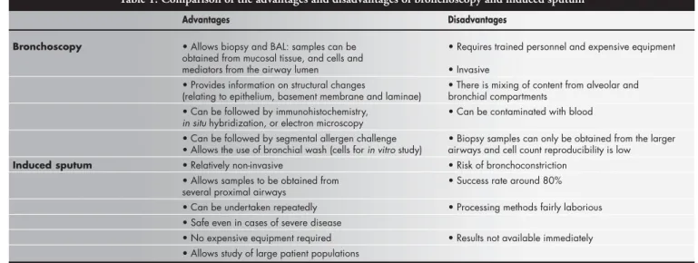
Induced sputum has several advantages over other techniques. Bronchoscopy is an in-vasive procedure and is not easily applicable on a large scale in follow-up studies. Sputum analy-sis might be an alternative to this, for obtain-ing airway secretions that may potentially be used for monitoring airway inflammation.4 Although fiberoptic bronchoscopy with trans-bronchial biopsy, trans-bronchial brushing and bronchoalveolar lavage are relatively safe
pro-cedures, they still entail some morbidity and are relatively unpleasant and expensive proce-dures compared with sputum induction.1 With sputum induction, samples can be obtained from the lower airways with minimal discom-fort to the patient. Bronchoscopy allows sam-pling of the cells and mediators in the airway lumens by means of bronchoalveolar lavage and enables biopsy of the mucosal tissue. The com-bined information thus obtained is therefore superior to that of sputum alone. However, bronchoalveolar lavage samples only distinguish lung segments that are distal to the bronchus into which the bronchoscope is wedged. Fur-thermore, significant mixing of distal, alveolar and proximal bronchial compartments occurs. The mediators are usually diluted in the large volumes of physiological saline solution used in the washing, and some exchange with the blood compartment is inevitable. In contrast, induced sputum probably provides a more rep-resentative sample of several proximal airways, although with prolonged induction the distal parts can also be sampled, as evident from in-creased numbers of macrophages from the al-veolar compartment. The comparison between bronchoscopy and induced sputum can be seen in Table 1.
CLINICAL APPLICATIONS
○ ○ ○ ○ ○ ○ ○ ○ ○ ○ ○ ○ ○ ○ ○ ○ ○ ○ ○ ○
OF INDUCED SPUTUM
Asthma
In clinical practice, it is difficult to assess airway inflammation and the effects of medi-cation on such inflammation. Subjective as-sessment of symptoms is always difficult and has often been found to be unsatisfactory for
• Elcio Oliveira Vianna
review of literature and
proposal for a protocol
Pulmonary Division, Department of Medicine, Faculdade de Medicina de
Ribeirão Preto, Universidade de São Paulo, Ribeirão Preto, São Paulo,
Brazil
Since the 1980s, sputum induction by inhala-tion of hypertonic saline has been successfully used for diagnosing Pneumocystis carinii pneu-monia in patients infected with HIV. In recent years, sputum induction and its subsequent processing has been refined as a noninvasive research tool providing important information about inflammatory events in the lower airways, and it has been used for studying various ill-nesses. In asthma, one application is to use spu-tum inflammatory indices to increase our under-standing of complex relationships between in-flammatory cells, mediators, and cytokine mecha-nisms. In chronic obstructive pulmonary disease, sputum assessment could be used as a screen-ing test before decidscreen-ing on long-term corticoster-oid treatment. In tuberculosis, sputum induction is a valuable diagnostic tool for HIV-seropositive patients who do not produce sputum. Sputum induction appears to be a relatively safe, noninvasive means of obtaining airway secre-tions from subjectswith cystic fibrosis, especially from those who do not normally produce spu-tum. Moreover, sputum induction can also be used in chronic cough and lung cancer. Gener-ally, induction is performed through ultrasonic nebulizers, using hypertonic saline. It is recom-mended that sputum be processed as soon as possible, with complete homogenization by the use of dithiothreitol. We have also shown in this article an example of a protocol for inducing and processing sputum employing a nebulizer produced in Brazil.
KEY WORDS: Sputum. Asthma. Cytological tech-niques. Tuberculosis. Cystic fibrosis.
R
eview A
monitoring asthma severity.5 Nonetheless, the regular use of peak flow measurements has been shown to improve asthma control, peak flow rates and diurnal peak flow relapse. Meas-urements of the levels of exhaled gases such as nitric oxide may be useful, but more data are needed to fully evaluate the importance of such markers in assessing airway inflamma-tion in asthma, especially since nitric oxide can be produced in large amounts in paranasal sinuses and the stomach.5
Asthma is commonly associated with spu-tum eosinophilia. Up to 80% of corticoster-oid-naive subjects and more than 50% of cor-ticosteroid-treated subjects with currently symptomatic asthma have a sputum eosinophil count that is outside of the normal range. The validity of a high sputum eosinophil count for the identification of asthma is better than peak expiratory flow measurement.6
The short-term response to inhaled corticosteroids differs markedly according to the sputum eosinophil count, with little evi-dence of improvement in symptoms and air-way responsiveness in subjects with a baseline sputum eosinophil count of less than 3%. These findings suggest that measuring the underlying airway inflammation might pro-vide a better guide as to the need for corticos-teroid treatment than assessment of functional abnormality.6
Occupational asthma is associated with an increase in sputum eosinophilia. There is some evidence that sputum eosinophil counts in-crease during workplace exposure in subjects with occupational asthma.6
One obvious application of sputum induc-tion is to use sputum inflammatory indices to increase our understanding of complex
relation-ships between inflammatory cells, mediators and cytokine mechanisms in asthma. The spu-tum fluid phase seems to be suitable for meas-uring eosinophil cationic protein, some cytokines and histamine. Assessment of airway inflammation using sputum could also be used for evaluating the effects of drugs on asthmatic airway inflammation and relating their anti-inflammatory effect to the effects on symptoms and disordered airway function.
Chronic obstructive pulmonary disease
Chronic obstructive pulmonary disease is a clinical entity that is characterized by the presence of blockage or chronic limitation of the airflow that presents slow and irreversible progression. The origin of such alterations is the pulmonary combination of chronic bron-chitis and emphysema. The pathophysiology of chronic obstructive pulmonary disease in-volves an inflammatory disorder characterized by neutrophilicinflammation in airway secre-tions, with the presence of macrophagesand lymphocytes on airway tissue.Bronchoscopic investigations areoften not possible due to disease severity.7 Thus, sputum induction is a valuable tool for pathophysiology studies. The sputum neutrophil count is usually high, and the neutrophil count can be correlated with a reduction in forcedexpiratory volume in one second (FEV1) and the rate of decline in FEV1, thus suggesting that neutrophilic airway in-flammation is functionally important. Peleman et al. (1999) studied the cellular com-position of induced sputum in chronic ob-structive pulmonary disease and found marked sputum neutrophilia.8
Despite its nonspecific nature, the early inflammatory responseto cigarette smoke is
probably crucial to the development of subse-quent tissue damage and disease in suscepti-ble individuals.Neutrophils and macrophages can potentially produce large quantitiesof proteases, of which the various elastase en-zymes have attractedthe most attention as likely causes of the loss of elastic recoiland destruction of elastic fibers in the lung paren-chyma.Indeed, lung specimens from patients with panlobular emphysemahave a signifi-cantly decreased elastin content.9
Confalonieri et al. (1998)studied the ef-fects of two months of treatment with inhaled beclomethasonedipropionate (1,500 µg/day) on bronchial inflammation in patients with stable, mild to moderate chronic obstructive pulmonary disease, by using sputum induc-tion. They found that the number of neu-trophils present in inducedsputum samples decreased after treatment.10 Moreover, a short course of oral glucocorticoid therapyhas been demonstrated to improve pulmonary function in somepatients with chronic obstructive pul-monary disease, but not all.11
In a recent prospective trial, Borbeau et al. (1998) showed that, in a group of 140 chronic obstructive pulmonary disease patients, 19 (13.5%) responded to the two-week treatment with 40 mg prednisone daily. Response to treat-ment was defined as a 15% improvetreat-ment in FEV1.12 Also employing FEV
1 to assess response, Mendella et al. (1982) showed that 17% of chronic obstructive pulmonary disease cases were considered responsive to a course of 32 mg/d methylprednisolone for two weeks.13 Furthermore, Brazilian authors studying spu-tum eosinophilia in smokers have found that eosinophilic inflammation can occur in smok-ers with or without chronic airflow limitation
Table 1. Comparison of the advantages and disadvantages of bronchoscopy and induced sputum
Advantages Disadvantages
Bronchoscopy • Allows biopsy and BAL: samples can be • Requires trained personnel and expensive equipment obtained from mucosal tissue, and cells and
mediators from the airway lumen • Invasive
• Provides information on structural changes • There is mixing of content from alveolar and (relating to epithelium, basement membrane and laminae) bronchial compartments
• Can be followed by immunohistochemistry, • Can be contaminated with blood in situ hybridization, or electron microscopy
• Can be followed by segmental allergen challenge • Biopsy samples can only be obtained from the larger • Allows the use of bronchial wash (cells for in vitro study) airways and cell count reproducibility is low
Induced sputum • Relatively non-invasive • Risk of bronchoconstriction
• Allows samples to be obtained from • Success rate around 80% several proximal airways
• Can be undertaken repeatedly • Processing methods fairly laborious • Safe even in cases of severe disease
• No expensive equipment required • Results not available immediately • Allows study of large patient populations
(chronic obstructive pulmonary disease) and that sputum eosinophilia may predict those patients who will benefit from steroid therapy.14 Fujimoto et al. (1999) investigated the influ-ence of glucocorticoid in the reversibility of eosinophilic inflammation in patients with pulmonary emphysema. They found that the reversibility ofairway obstruction following the treatment could be correlated with the eosi-nophil count in the induced sputum, and that the treatment significantly reduced eosinophil count and eosinophil mediators.In addition, patients who did not show improvement in FEV1, had lower baselineeosinophil counts.11 In conclusion, sputum assessment could be used as a screening test before deciding on long-term corticosteroid treatment in chronic obstructive pulmonary disease.
Cough
Chronic cough is associated with pre-dominant sputum neutrophilia, but up to 40% of subjects with cough have a sputum eosinophil count of more than 3%. Patients with cough and sputum eosinophilia exhibit an objective response to corticosteroid treat-ment that occurs in parallel with a treattreat-ment- treatment-associated fall in the sputum eosinophil count. In contrast, patients without sputum eosi-nophilia do not respond.6
Tuberculosis
Pulmonary tuberculosis remains one of the most important health problems in the world.15 The World Health Organization rec-ommends the detection of acid-fast bacilli in respiratory specimens as the initial approach to the diagnosis of tuberculosis.16 However, this method has low sensitivity and has little value in patients who cannot produce sputum spontaneously. In Brazil, with an estimated annual prevalence of 129,000 cases, approxi-mately 22% of adult HIV-seronegative pa-tients with suspected tuberculosis do not pro-duce sputum spontaneously, or have negative acid-fast bacilli smears.17 Thus, the diagnosis of tuberculosis in these patients is difficult, and in most cases they are treated empirically on the basis of clinical and chest radiographic findings. However, empirical therapy may re-sult in unnecessary cost and toxicity. Moreo-ver, HIV-seropositive patients who do not produce sputum often undergo expensive and more invasive procedures.17 Thus, sputum induction is a valuable tool for diagnosing pulmonary tuberculosis.
Conde et al. (2000) compared sputum in-duction with fiberoptic bronchoscopy in the diagnosis of tuberculosis in a reference center
in Rio de Janeiro, Brazil. They found that spu-tum induction is a safe procedure with high diagnostic yield and high agreement with the results from fiberoptic bronchoscopy, for the diagnosis of tuberculosis in HIV-seronegative and HIV-seropositive patients. In localities where fiberoptic bronchoscopy is not readily available, and as part of the work-up of sus-pected tuberculosis prior to bronchoscopy, in-duced sputum offers an alternative or addi-tional approach to the diagnosis of sputum smear-negative tuberculosis, and would en-hance diagnostic sensitivity in resource-poor areas.17 Anderson et al. (1995) compared spu-tum induction to fiberoptic bronchoscopy in the diagnosis of pulmonary tuberculosis in immunocompromised patients and found that sputum induction was well-tolerated, low-cost and provided the same, if not bet-ter, diagnostic yield compared with bron-choscopy in the diagnosis of smear-negative pulmonary tuberculosis.18
Bacteriological confirmation of pulmo-nary tuberculosis in infants and children re-mains difficult. Older children can produce or be induced to producesputum. However, there are no reports of its use in infants or childrenyounger than 3years of age.
Gastric lavage is regarded as the standard procedure for obtaining specimens for stain-ing and culture of Mycobacterium tuberculosis in younger children, because they swallow their sputum and do notexpectorate. But spu-tum induction can be effectively performed and is well tolerated and safe, even in infants. Zar et al. (2000) compared induced sputum and gastric lavage for the isolation of M. tu-berculosis in bothHIV-infected and uninfected infants and children and found that induced sputum is better than gastric lavage.19 The bacteriologicalyield from sputum or gastric lavage in cases of pulmonary tuberculosis does not differaccording to HIV status. The use of induced sputum should be considered as a first-line investigation in children suspected of having pulmonarytuberculosis, especially in circumstances in which a culture-confirmed diagnosis needs to be vigorously sought (such as when the sourceof the case is unknown, drug resistance is suspected, or cutaneous al-lergyoccurs).19
Cystic fibrosis
Cystic fibrosis is a hereditary disease of autosomal recessive transmission also known as mucoviscidosis or cystic fibrosis of the pan-creas. The fundamental abnormality consists of the production of abnormal secretions from a variety of exocrine glands. It is chiefly a
dis-ease of infants and children, although adult cases are being recognized with greater fre-quency. There is no sex predominance. In-volvement of the lungs usually is manifested clinically by recurrent chest infections ( Pseu-domonas aeruginosa, Staphylococcus aureus, Haemophilusinfluenzae) that are associated with wheezing, dyspnea, productive cough, and hemoptysis, as a result of bronchiectasis. Respiratory insufficiency and cor pulmonale develop frequently in the later stages of the disease. The lack of pancreatic enzymes results in poor digestion, particularly of fat.20 Inflam-mation begins at an early age, even in the ab-sence of concomitant infection, and persists and progresses throughout life, ultimately leading to lung destruction. Quantitative measurements of infection and inflammation aretherefore important in disease staging and new treatment evaluation.21
Each of the current techniquesused for defining the microbiology and inflammatory response of thecystic fibrosis airway has nota-ble limitations. Expectorated sputum provides an accurate measure of infection and inflam-mation in the lowerairways, but many chil-dren with cystic fibrosis are unable to sponta-neously expectoratesputum.21 Fiberoptic bron-choscopy with bronchoalveolar lavageis inva-sive, risky and costly. Serial bronchoalveolar lavages are particularly difficultto perform. Furthermore, lavage generally samples only one or twosegments of the lung, thereby possibly limiting the detection of infection. Oropharyn-geal cultures, commonly used in young chil-dren with cystic fibrosiswho are not capable of expectorating, do not reliably predictthe pres-ence of lower airway pathogens, lack sensitiv-ityfor identifying Pseudomonas aeruginosa and Staphylococcus aureus, and provide no informa-tion aboutinflammation.21
is suspected.25 Kirsch et al. (1990), studying 62 patients with possible aids-associated Pneumocystis carinii pneumonia to determine the diagnostic usefulness of sputum analysis, found that sputum analysis is a sensitive, spe-cific, rapid and low-cost technique for the di-agnosis of Pneumocystis carinii pneumonia.24
METHODS FOR SPUTUM
○ ○ ○ ○ ○ ○ ○ ○ ○ ○ ○ ○ ○ ○ ○ ○ ○ ○ ○ ○
INDUCTION AND PROCESSING
Induction
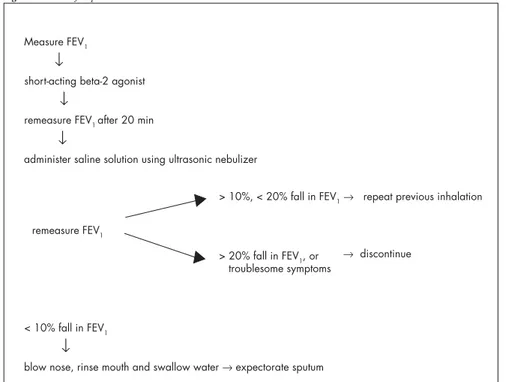
Ultrasonic nebulizers are recommended for sputum inducing since other nebulizers do not usually have sufficient saline aerosol output. Spirometry is necessary to assess the baseline airway caliber and avoid excessive bronchoconstriction during saline inhalation. Spirometers are preferable to peak flow me-ters because of the greater sensitivity of FEV1 in detecting induced bronchoconstriction. Sputum induction requires a high degree of cooperation from the patient. The procedure should be conducted by an experienced tech-nician under the supervision of an experi-enced physician.26
Because hypertonic saline causes bron-choconstriction in asthmatic subjects,27 pre-treatment with a short-acting beta-2 agonist is recommended as the standard procedure.2,28 Salbutamol, usually 200-400 mmg, i.e. 2-4 puffs from a standard metered-dose inhaler, has generally been used for pretreatment.26
The concentration of the saline used for sputum induction has ranged from 0.9 to 7%.28-30 The concentration may be changed during the procedure, starting with 3% and subsequently increasing to 4 or 5%.2,31 Hyper-tonic saline solution is reportedly more effec-tive than isotonic saline in inducing sputum.32 Different compartments of the respiratory tract are sampled at different time-points dur-ing induction, i.e. central airways are sampled early, whereas peripheral airways and alveoli are sampled later. Shorter inhalation times (15-20 min) appear to have feasibility and success rates that are similar to those of longer inha-lation times (30 min). The consensus is to use cumulative nebulization duration of 15-20 minutes.26 The procedures for induction can be seen in Figure 1.
Sputum Processing
It is recommended that sputum be proc-essed as soon as possible or within two hours, in order to ensure optimum cell counting and staining.33,34 Complete homogenization is im-portant and can be achieved by the use of dithiothreitol (DTT). Cells that are incom-Airway inflammation and infection are
sig-nificantly increasedin both non-expectorat-ing and expectoratnon-expectorat-ing children with cystic fi-brosis, incomparison with healthy children.21 Also, induced sputum samples appearto be comparable to spontaneously expectorated samples in describingboth inflammation and infection in the cystic fibrosisairway.21 Induced sputumdiffers from spontaneous sputum by having a higher number of viablecells and less squamous cell contamination.22
Lung Cancer
With regard to lung cancer, identification of early (or pre-symptomatic) lung cancer in smokers is considered the best strategy for preventing this disease.5 But cytological exami-nation of sputum has been shown to lead to lung cancer detection at an earlier stage, thereby resulting in an improved five-year sur-vival rate. Recent studies of sputum specimens and clinical data linking specimens to lung cancer outcomes may make it possible to de-termine molecular diagnoses of cancer several years before its clinical presentation. This has become possible through the use of tests to evaluate altered gene expression, including specific oncogene activation and tumor sup-pressor gene detection, as well as genomic in-stability and abnormal methylation. Such studies clearly indicate that good sputum sam-ples ought to allow complicated genetic
analy-sis to be performed, thus providing further impetus for considering the induced sputum technique as a tool for lung cancer screening.5
PneumocystisCarinii Pneumonia
Pneumocystis carinii pneumonia remains a significant cause of morbidity and mortal-ity in HIV-infected individuals, causing clini-cally apparent pneumonia virtually exclu-sively in immunosuppressed patients. The clinical presentation is characterized by fe-ver, shortness of breath, substernal tightness, and nonproductive cough. Especially in HIV-infected patients, the symptoms can be rela-tively mild and slowly progressive, which may delay diagnosis.23 Transbronchial biopsy and bronchoalveolar lavage have been shown to have 98 to 100% yield and 92 to 100% nega-tive predicnega-tive value for the diagnosis of Pneumocystis carinii pneumonia. Although these are considered to be the gold standard, they still entail some morbidity and are rela-tively expensive procedures.24 Thus, induced sputum may have a role in diagnosing Pneumocystis carinii pneumonia.
It was reported in the mid-1980s that the examination of sputum induced by the inha-lation of hypertonic saline solution was fre-quently diagnostic for Pneumocystis carinii pneumonia.1 Since then, this diagnostic method has generally become the first em-ployed when Pneumocystis carinii pneumonia
FEV1 = forced expiratory volume in one second. Measure FEV1
↓
short-acting beta-2 agonist
↓
remeasure FEV1 after 20 min
↓
administer saline solution using ultrasonic nebulizer
< 10% fall in FEV1
↓
blow nose, rinse mouth and swallow water → expectorate sputum
> 10%, < 20% fall in FEV1 → repeat previous inhalation
> 20% fall in FEV1, or troublesome symptoms
→ discontinue remeasure FEV1
pletely released from mucus tend to stain darkly, making correct identification difficult. DTT (0.1%) has been shown to be more effective for dispersing cells than phosphate-buffered saline (PBS), and has no adverse effects on cell counts. The volume of mucolytic agents used during the processing of all the expectorated sputum, although fixed at 1:1, is variable in relation to the sputum/saliva ratio, which is an unknown.34,35 Filtration through a 48-µm ny-lon mesh is commonly used to remove mucus and debris, and is strongly recommended.36 Centrifugation is necessary to separate sputum cells from the fluid phase. Centrifugal forces used in studies to date have ranged from 300 to 1,500 xg and the duration of centrifuga-tion from 5 to 10 minutes. The total cell count is performed manually using a hemo-cytometer, and cell viability is determined by the trypan blue exclusion method.34,37 Fluid phase storage temperatures used have ranged from –20 to –70º C.36
Preparation of cytospins with an optimum number of cells (40-60 x 103 cells) provides a more accurate estimate of cell distribution than smears.Cytocentrifugation speeds range from 10 to 51 xg (using a cytocentrifuge), with the most common conditions being 22 xg for 6 minutes.37,38 Cytospin staining for differen-tial cell counts can be achieved using either Wright or Giemsa staining. The differential cell count is determined by counting a mini-mum of 400 non-squamous cells, and is re-ported as the relative numbers of eosinophils,
neutrophils, macrophages, lymphocytes and bronchial epithelial cells, expressed as a per-centage of total non-squamous cells. The sq-uamous cell percentage should always be re-ported separately.36 The processing procedures can seen be in Figure 2.
Objective quantitative analysis of cells in sputum
Manual differential counting of sputum cytospins is tedious to perform and, although based on objective morphological criteria, observer variability makes more objective as-saying desirable. The laser scanning cytom-eter is a novel microscope-linked and compu-ter-operated instrument that measures fluo-rescence and optically scans cells labeled with fluorescent probes on a microscope slide.39
EXAMPLE OF PROTOCOL
○ ○ ○ ○ ○ ○ ○ ○ ○ ○ ○ ○ ○ ○ ○ ○ ○ ○ ○ ○
USED IN BRAZIL
The protocol for inducing and process-ing sputum used by the Pulmonary Division, Medical School of Ribeirão Preto,
Univer-sidade de São Paulo, is as follows.
Nebulizer
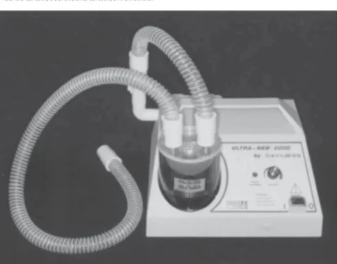
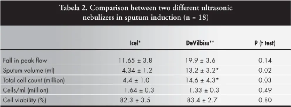
We compared two ultrasonic nebulizers for sputum induction, one Brazilian and the other imported, in terms of safety (bronchos-pasm risk) and yield (sputum volume and cell numbers).40 Patients with mild or moderate asthma formed two groups that underwent sputum induction by inhalation of NaCl 4.5% for 20 minutes. Peak flow measurements were done before induction and every five minutes during induction. Group 1 used the Icel nebulizer (Figure 3) (Evolusonic 1000BR, Icel, São Paulo, SP, Brazil), a low-output nebulizer whose measured output rate was 1 ml/min (n = 9). Group 2 used the DeVilbiss nebulizer (Figure 4) (Ultra-Neb 2000, DeVilbiss-Sun-rise Medical, Somerset, Pennsylvania, United States of America), a high-output nebulizer with a rate of 2.5 ml/min (n = 9).
The results can be seen in Table 2. We concluded that the much greater spu-tum induction seen in Group 2 might be a consequence of the higher output rate pro-vided by the nebulizer used in that group
Figure 3. Icel ultrasonic nebulizer (model Evolusonic
1000BR).
Figure 4. DeVilbiss ultrasonic nebulizer (model Ultra-Neb 2000).
TCC: total cell count; DCC: differential cell count; DTT: dithiothreitol.
Figure 2. Sputum processing method
1. Pitchenik AE, Ganjei P, Torres A, Evans DA, Rubin E, Baier H. Sputum examination for the diagnosis of Pneumocystis carinii pneumonia in the acquired immunodeficiency syndrome. Am Rev Respir Dis 1986;133(2):226-9.
2. Pin I, Gibson PG, Kolendowicz R, et al. Use of induced spu-tum cell counts to investigate airway inflammation in asthma. Thorax 1992;47(1):25-9.
3. Djukanovic R, Sterk PJ, Fahy JV, Hargreave FE. Standardised methodology of sputum induction and processing. Eur Respir J 2002; 37(Suppl):1s-2s.
4. Grootendorst DC, Sont JK, Willems LN, et al. Comparison of inflammatory cell counts in asthma: induced sputum vs bronchoalveolar lavage and bronchial biopsies. Clin Exp Al-lergy 1997;27(7):769-79.
5. Vignola AM, Rennard SI, Hargrave FE, et al. Future directions. Eur Respir J 2002;20(Suppl 37):51s-55s.
6. Pavord ID, Sterk PJ, Hargreave FE, et al. Clinical applications of assessment of airway inflammation using induced sputum. Eur Respir J 2002;37(Suppl):40s-3s.
7. Kelly MG, Brown V, Martin SL, Ennis M, Elborn JS. Com-parison of sputum induction using high-output and low-out-put ultrasonic nebulizers in normal subjects and patients with COPD. Chest 2002;122(3):955-9.
8. Peleman RA, Rytilâ PH, Kips JC, Joos GF, Pauwels RA. The cellular composition of induced sputum in chronic obstructive pulmonary disease. Eur Respir J 1999;13(4):839-43. 9. Cosio MG, Majo J, Cosio MG. Inflammation of the airways
○ ○ ○ ○ ○ ○ ○ ○ ○ ○ ○ ○ ○ ○ ○ ○ ○ ○ ○ ○ ○ ○ ○ ○ ○ ○ ○ ○ ○ ○ ○ ○ ○ ○ ○ ○ ○ ○ ○ ○ ○ ○ ○ ○ ○ ○ ○ ○ ○ ○ ○ ○ ○ ○ ○ ○ ○ ○ ○ ○ ○ ○ ○ ○
REFERENCES
and lung parenchyma in COPD: role of T cells. Chest 2002;121(5 Suppl):160S-5S.
10. Confalonieri M, Mainardi E, Della Porta R, et al. Inhaled corticosteroids reduce neutrophilic bronchial inflammation in patients with chronic obstructive pulmonary disease. Thorax 1998;53(7):583-5.
11. Fujimoto K, Kubo K, Yamamoto H, Yamaguchi S, Matsuzawa Y. Eosinophilic inflammation in the airway is related to gluco-corticoid reversibility in patients with pulmonary emphysema. Chest. 1999;115(3):697-702.
12. Bourbeau J, Rouleau MY, Boucher S. Randomised controlled trial of inhaled corticosteroids in patients with chronic obstruc-tive pulmonary disease. Thorax 1998;53(6):477-82. 13. Mendella LA, Manfreda J, Warren CP, Anthonisen NR.
Ster-oid response in stable chronic obstructive pulmonary disease. Ann Intern Med 1982;96(1):17-21.
14. Pizzichini E, Pizzichini MM, Gibson P, et al. Sputum eosi-nophilia predicts benefit from prednisone in smokers with chronic obstructive bronchitis. Am J Respir Crit Care Med 1998;158(5 Pt 1):1511-7.
15. Dye C, Scheele S, Dolin P, Pathania V, Raviglione MC. Con-sensus statement. Global burden of tuberculosis: estimated in-cidence, prevalence, and mortality by country. JAMA 1999;282(7):677-86.
16. World Health Organization. Treatment of tuberculosis: guide-lines for national programs. Geneva: World Health Organiza-tion; 1993.
17. Conde MB, Soares SL, Mello FC, et al. Comparison of sputum induction with fiberoptic bronchoscopy in the diagnosis of tu-berculosis: experience at an acquired immune deficiency syn-drome reference center in Rio de Janeiro, Brazil. Am J Respir Crit Care Med 2000;162(6):2238-40.
18. Anderson C, Inhaber N, Menzies D. Comparison of sputum in-duction with fiber-optic bronchoscopy in the diagnosis of tuber-culosis. Am J Respir Crit Care Med 1995;152(5 Pt 1):1570-4. 19. Zar HJ, Tannenbaum E, Apolles P, Roux P, Hanslo D, Hussey
G. Sputum induction for the diagnosis of pulmonary tubercu-losis in infants and young children in an urban setting in South Africa. Arch Dis Child 2000;82(4):305-8.
20. Fraser RS, Paré JAP, Fraser RG, Paré PD. Diseases of the air-way. In: Fraser RS, Paré JAP, Fraser RG, Paré PD, eds. Synop-sis of diseases of the chest. Philadelphia: WB Saunders; 1996:683-87.
21. Sagel SD, Kapsner R, Osberg I, Sontag MK, Accurso FJ. Air-way inflammation in children with cystic fibrosis and healthy children assessed by sputum induction. Am J Respir Crit Care Med 2001;164(8 Pt 1):1425-31.
22. HenigNR, Tonelli MR, Pier MV, Burns JL, Aitken ML. Spu-tum induction as a research tool for sampling the airways of subjects with cystic fibrosis. Thorax 2001;56(4):306-11. 23. Kovacs JA, Gill VJ, Meshnick S, Masur H. New insights into
transmission, diagnosis, and drug treatment of Pneumocystis carinii pneumonia. JAMA 2001;286(19):2450-60. 24. Kirsch CM, Azzi RL, Yenokida GG, Jensen WA. Analysis of
(DeVilbiss). On the basis of this conclusion, we have been utilizing the high-output nebulizer (DeVilbiss) for routine induction. However, in cases of severe asthma, we may prefer low output so as to decrease the risk of bronchospasm. Moreover, the high cost of the imported equipment may lead us to use the Brazilian nebulizer, which is also effective in inducing sputum.
Induction Method
Measure peak flow. Apply 2 to 4 puffs of salbutamol (200-400 µg). After 20 minutes, measure peak flow and calculate the critical peak flow (fall of 10%). Administer NaCl 4.5%, using a ultrasonic nebulizer like DeVilbiss. Interrupt this every 5 minutes, to discard saliva and collect sputum in a specific tube. Total induction time is 20 minutes, or
Tabela 2. Comparison between two different ultrasonic nebulizers in sputum induction (n = 18)
Icel* DeVilbiss** P (t test)
Fall in peak flow 11.65 ± 3.8 19.9 ± 3.6 0.14
Sputum volume (ml) 4.34 ± 1.2 13.2 ± 3.2* 0.02
Total cell count (million) 4.4 ± 1.0 14.6 ± 4.3* 0.03
Cells/ml (million) 1.64 ± 0.3 1.33 ± 0.3 0.49
Cell viability (%) 82.3 ± 3.5 83.4 ± 2.7 0.80
* Evolusonic 1000BR, Icel, Brazil; ** Ultra-Neb 2000, DeVilbiss-Sunrise Medical, United States of America.
PBS. Prepare cytospins: 75 µl/cups to spin for 60 seconds at 1,000 rpm (in cytocentrifuge). Stain the slides using the May-Grunwald and Giemsa methods.
○ ○ ○ ○ ○ ○ ○ ○ ○ ○ ○ ○ ○ ○ ○ ○ ○ ○ ○ ○
FUTURE DIRECTIONS
The identification of biomarkers that al-low early diagnosis, monitoring and optimi-zation of lung disease therapy is one of the most ambitious goals in respiratory medicine. The induced sputum technique allows sam-pling of the airways in a noninvasive fashion and thus offers a unique opportunity for iden-tifying biomarkers of potential clinical use in respiratory medicine. It is hoped that, in the future, induced sputum will provide clinicians with useful markers that can be used routinely for performing more accurate and, ideally, more rapid determination of disease pheno-types in many lung diseases.
The hope is that the induced sputum tech-nique will provide a simple and cost-effective tool for monitoring airway inflammation in the clinical setting, an approach that was pre-cluded by previous techniques such as bron-chial biopsy and bronchoalveolar lavage. In addition, since the induced sputum technique enables regular monitoring of inflammation, it will be of great help in assessing the anti-inflammatory potential of new treatments. less if peak flow falls to the critical value.
Processing Method
induced sputum in the diagnosis of Pneumocystis carinii pneu-monia. Am J Med Sci 1990;299(6):386-91.
25. Metersky ML, Aslenzadeh J, Stelmach P. A comparison of in-duced and expectorated sputum for the diagnosis of Pneumocystis carinii pneumonia. CHEST 1998;113(6):1555-9. 26. Paggiaro PL, Chanz P, Holtz O, et al. Sputum induction. Eur
Respir J 2002;20(Suppl 37):3s-8s.
27. Smith CM, Anderson SD. Inhalation provocation tests using nonisotonic aerosols. J Allergy Clin Immunol 1989;84(5 Pt 1):781-90.
28. Wong HH, Fahy JV. Safety of one method of sputum induc-tion in asthmatic subjects. Am J Respir Crit Care Med 1997;156(1):299-303.
29. Pavia D, Thomson ML, Clarke SW. Enhanced clearance of se-cretions from the human lung after the administration of hyper-tonic saline aerosol. Am Rev Respir Dis 1978;117(2):199-203. 30. Bacci E, Cianchetti S, Paggiaro PL, et al. Comparison between hypertonic and isotonic saline-induced sputum in the evalua-tion of airway inflammaevalua-tion in subjects with moderate asthma.
Clin Exp Allergy 1996;26(12):1395-400.
31. Bacci E, Cianchetti S, Ruocco L, et al. Comparison between eosinophilic markers in induced sputum and blood in asthmatic patients. Clin Exp Allergy 1998;28(10):1237-43. 32. Bartoli ML, Bacci E, Carnevali S, et al. Quality evaluation of
samples obtained by spontaneous or induced sputum: compari-son between two methods of processing and relationship with clinical and functional findings. J Asthma 2002;39(6):479-86. 33. Pizzichini E, Pizzichini MM, Efthimiadis A, Hargreave FE, Dolovich J. Measurement of inflammatory indices in induced sputum: effects of selection of sputum to minimize salivary con-tamination. Eur Respir J 1996;9(6):1174-80.
34. Fahy JV, Liu J, Wong H, Boushey HA. Cellular and biochemi-cal analysis of induced sputum from asthmatic and from healthy subjects. Am Rev Respir Dis 1993;147(5):1126-31. 35. Gershman NH, Wong HH, Liu JT, Mahlmeister MJ, Fahy JV.
Comparison of two methods of collecting induced sputum in asthmatic subjects. Eur Respir J 1996;9(12):2448-53. 36. Efthimiadis A, Spanevello A, Hamid Q, et al. Methods of
spu-tum processing for cell counts, immunocytochemistry and in situ hybridisation. Eur Respir J 2002; 37(Suppl):19s-23s. 37. Pizzichini E, Pizzichini MM, Efthimiadis A, et al. Indices of
airway inflammation in induced sputum: reproducibility and validity of cell and fluid-phase measurements. Am J Respir Crit Care Med 1996;154(2 Pt 1):308-17.
38. Popov T, Gottschalk R, Kolendowicz R, Dolovich J, Powers P, Hargreave FE. The evaluation of a cell dispersion method of sputum examination. Clin Exp Allergy 1994;24(8):778-83. 39. Woltmann G, Ward RJ, Symon FA, Rew DA, Pavord ID,
Wardlaw AJ. Objective quantitative analysis of eosinophils and bronchial epithelial cells in induced sputum by laser scanning cytometry. Thorax 1999;54(2):124-30.
40. Vianna EO, Matos FL, Martinez JAB, Terra-Filho J. Sputum induction in asthma. Comparison of two ultrasonic nebulizers [abstract]. ATS 2001, 2002 & 2003. 3 Year Abstracts 2 ViewTM.
Available from: URL: http://www.abstracts2view.com/atsall/ search.php?search=do&intMaxHits=10&where%5B%5D =&andornot%5B%5D=&query=Vianna. Accessed on 22/07/03.
Indução de escarro: revisão de literatura e pro-posta de protocolo
Desde a década de 80, a indução de escarro pela inalação de solução salina hipertônica tem sido usada com sucesso para
diagnosti-car pneumonia por Pneumocystis carinii em
pacientes infectados pelo HIV. Em anos re-centes, a indução de escarro e seu subseqüente processamento foram refinados como uma ferramenta de pesquisa não invasiva que for-nece informações importantes sobre eventos inflamatórios nas pequenas vias aéreas, sen-do usada para estudar várias sen-doenças. Na asma, uma aplicação é para o uso de índices inflamatórios do escarro para aumentar nos-sa compreensão de complexos relacionamen-tos entre célula inflamatória, mediador e mecanismos das citocinas. Na doença pulmo-nar obstrutiva crônica, a avaliação do escarro pode ser usada como um teste de seleção an-tes de se decidir pelo tratamento a longo
pra-○ ○ ○ ○ ○ ○ ○ ○ ○ ○ ○ ○ ○ ○ ○ ○ ○ ○ ○ ○ ○ ○ ○ ○ ○ ○ ○ ○ ○ ○ ○ ○ ○ ○ ○ ○ ○ ○ ○ ○ ○ ○
RESUMO
Marcos Eduardo Scheicher, BSc (PT), MSc. Faculdade de Medicina de Ribeirão Preto, Universidade de São Paulo, Ribeirão Preto, São Paulo, Brazil.
João Terra Filho, MD, PhD. Faculdade de Medicina de Ribeirão Preto, Universidade de São Paulo, Ribeirão Preto, São Paulo, Brazil.
Elcio Oliveira Vianna, MD, PhD. Faculdade de Medicina de Ribeirão Preto, Universidade de São Paulo, Ribeirão Preto, São Paulo, Brazil.
Sources of funding: Fapesp — Process nº. 02/02194-2
Conflict of interest: Not declared Date of first submission: April 16, 2003 Last received: April 16, 2003
Accepted: May 30, 2003
Address for correspondence Marcos Eduardo Scheicher
Divisão de Pneumologia, Hospital das Clínicas Faculdade de Medicina de Ribeirão Preto — USP Av. Bandeirantes, 3900
Ribeirão Preto/SP — Brasil — CEP 14048-900 Tel. (+55 16) 602-2631 — Fax (+55 16) 633-6695 E-mail: [email protected]
COPYRIGHT © 2003, Associação Paulista de Medicina ○ ○ ○ ○ ○ ○ ○ ○ ○ ○ ○ ○ ○ ○ ○ ○ ○ ○ ○ ○
Publishing information
zo com corticosteróide. Na tuberculose, a indução do escarro é uma ferramenta valiosa para o diagnóstico nos pacientes HIV-soropositivos que não produzem escarro. A indução de escarro parece ser um meio rela-tivamente seguro, não-invasivo de se obter secreções das vias aéreas de sujeitos com fibrose cística, especialmente aqueles que não produzem normalmente o escarro. Além dis-so, a indução do escarro também pode ser usada na tosse crônica e no câncer de pul-mão. Geralmente, a indução é executada atra-vés dos nebulizadores ultra-sônicos, usando solução salina hipertônica. Recomenda-se que o escarro seja processado o mais cedo possí-vel, com homogeneização completa pelo uso de ditiotreitol. Mostramos também neste ar-tigo um exemplo de protocolo que emprega um nebulizador de fabricação nacional para induzir o escarro.
PALAVRAS-CHAVE: Escarro. Asma. Técnicas