○ ○ ○ ○ ○ ○ ○ ○ ○ ○ ○ ○ ABSTRACT○ ○ ○ ○ ○ ○ ○ ○ ○ ○ ○ ○ ○ ○ ○ ○ ○ ○ ○ ○ ○ ○ ○ ○ ○ ○ ○
INTRODUCTION
Tuberculosis is the most common infec-tious disease afflicting the human species, af-fecting 8.4 million people throughout the world in 1999. It has been estimated that this number will rise to 10.2 million by 2005, with the majority of cases occurring in emerging countries.1 In Brazil, 90,000 new cases are re-ported each year, with an estimated 130,000 active at present.2
The diagnosis of pulmonary tuberculosis in Brazil is based upon two positive direct bacilloscopy findings in the sputum, or a posi-tive culture for Mycobacterium tuberculosis. In the absence of these discoveries, suggestive radiological images or other complementary associated tests based upon clinical discover-ies are indicative of active disease.3 Adequate knowledge of images consistent with tuber-culosis activity is therefore an important re-source for its diagnosis and treatment, par-ticularly in those cases in which it is not pos-sible to achieve bacteriological confirmation. Chest radiography remains the primary imaging technique for the diagnosis and fol-low-up of pulmonary tuberculosis. However, computed tomography can help identify or confirm the presence of findings that may be used to suggest a tuberculosis diagnosis when the radiographic findings are inconsistent but tuberculosis is suspected clinically.
Computed tomography of the thorax is used in cases of clinical suspicion of pulmo-nary tuberculosis, particularly in those cases in which the initial thoracic radiography ap-pears normal, as well as in differentiating this from other thoracic diseases and aids or fever
of unknown origin.2,4 In a study of 42 patients with tuberculosis confirmed by bacteriologi-cal tests, Campos et al.5 concluded that high resolution computed tomography can be strongly suggestive of pulmonary disease ac-tivity. This is particularly helpful in patients with negative smear and/or indeterminate ra-diograms and allows proper treatment to be established, even before mycobacteria are iden-tified via culturing.
In a study performed by Lee et al.,6 the diagnosis of tuberculosis through high reso-lution computed tomography was accurate in 88% of the patients (165/188), for ruling out or confirming the pulmonary disease. Other studies have confirmed that computed tom-ography is superior to thoracic raditom-ography in the initial evaluation of tuberculosis.7-9
The objective of this study was to utilize conventional computed tomography for evaluating the structural alterations in the pulmonary parenchyma caused by tuberculo-sis, during the active phase of the disease and also after concluding the treatment.
○ ○ ○ ○ ○ ○ ○ ○ ○ ○ ○ ○ ○ ○ ○ ○ ○ ○ ○ ○
METHODS
Twenty patients with pulmonary tuber-culosis and serologically negative for the HIV virus were prospectively studied. Tuberculo-sis was confirmed by positive culturing for Mycobacterium tuberculosis in sputum (19 pa-tients) or via a lung fragment obtained through transbronchial biopsy (one patient).
All patients were informed of the proce-dures to be performed, and were subsequently submitted to conventional computed tomog-raphy at Hospital das Clínicas of the Univer-• Márcia Seiscento
• Mário Terra Filho
Tomographic evaluation
in the active and
post-treatment phases
Pulmonary Division, Hospital das Clínicas, Faculdade de Medicina da
Universidade de São Paulo, Brazil
Original A
rticle
CONTEXT: Adequate knowledge of images consistent with tuberculosis activity is an important resource for tuberculosis diagnosis and treatment.
OBJECTIVE: To evaluate the structural alterations caused by tuberculosis in the pulmonary parenchyma, both during the active phase of the disease and after the end of the treatment, through computerized tomography of the thorax.
TYPE OF STUDY: Prospective study.
SETTING: Pulmonary Division, Hospital das Clínicas, Faculdade de Medicina da Universidade de São Paulo.
PARTICIPANTS: 20 patients, carriers of pulmonary tu-berculosis, confirmed by Mycobacterium tubercu-losis culture.
PROCEDURES: Conventional tomography scans of the patients were obtained at two times: upon diag-nosis and after the end of the treatment. The fol-lowing were considered suggestive signs of tuber-culosis activity: centrilobular nodules with seg-mented distribution, confluent micronodules, con-solidations, thick-walled cavities, nodules, masses, thickening of the bronchial walls, tree-in-bud ap-pearance and cylindrical bronchiectasis.
MAIN MEASUREMENTS: The presence of sugges-tive signs of tuberculosis activity was compared between the start and the end of treatment by means of the signs test (z).
RESULTS: All patients (20/20) presented suggestive signs of tuberculosis activity at the start of treat-ment. After the end of treatment, 13 patients (13/ 20) still presented some suggestive signs consist-ent with activity. A reduction in the extconsist-ent of lung attack was seen post-treatment, in relation to its start (z = 10.10). This change was statistically sig-nificant (p < 0.001).
CONCLUSION: Signs suggestive of tuberculosis activ-ity are present in the active disease and are seen via computed tomography. The extent of parenchymal attack significantly decreases follow-ing treatment. Such signs may be useful in the di-agnosis of pulmonary tuberculosis.
sidade de São Paulo. Tomographic images were obtained using a conventional Phillips Tomoscan LX tomographic scanner (axial cuts of 10 mm thickness, in 10-mm increments from the apical area to the base of the lungs). The study protocol was approved by the Ethics Committee for Research Project Assess-ment at Hospital das Clínicas of the Universidade de São Paulo.
The tomographic evaluation performed was based on two separate time periods: the first, from the diagnosis of tuberculosis until 30 days after the start of treatment with ri-fampicin, isoniazid and pyrazinamide; and the second, until 30 days after the completion of the proposed treatment (six months), when all the patients were considered to be cured, according to clinical criteria. The images were analyzed by three observers.
Suggestive signs of tuberculosis activity included: centrilobular nodules with segmen-tal distribution, confluent micronodules, consolidations, thick-walled cavities, nod-ules, masses, thickening of the bronchial walls, tree-in-bud appearance and cylindri-cal bronchiectasis.3,5,9-13
For the analyses of the images obtained, the lungs were divided into three sections: up-per, middle and lower. Furthermore, each sec-tion was divided into two parts: anterior and posterior. Analyses of 240 fields were performed (12 fields each patient) at the two study times. The tomographic findings were classified into three grades in accordance with the ob-served extent of one or more of the signs of tuberculosis activity within each field analyzed: Grade 0: absence of suggestive signs of tuberculosis activity.
Grade 1: presence of suggestive signs of tuberculosis activity in up to 50% of the analyzed field.
Grade 2: presence of suggestive signs of tuberculosis activity in more than 50% of the analyzed field.
The sensitivity and specificity of this method were calculated. The presence of signs of tuber-culosis activity, as shown by computed tomog-raphy, was compared between the start and com-pletion of the treatment using the signs test (z). The significance level of 5% was adopted.
○ ○ ○ ○ ○ ○ ○ ○ ○ ○ ○ ○ ○ ○ ○ ○ ○ ○ ○ ○
RESULTS
The patients’ mean age was 34.3 years (range: 16 to 71 years old) with a standard deviation of 13.8 years. Of the 20 individuals studied, eleven were male (55%) and nine were female (45%). All the patients presented symp-toms clinically consistent with tuberculosis.
From computed tomography, all patients (20/20) presented signs consistent with tuber-culosis activity at the start of the treatment. After completion of the treatment, seven pa-tients did not present any of the previously noted signs (7/20), while 13 patients (13/20) still presented some suggestive signs of activ-ity. The sensitivity of the computed tomogra-phy was 100%, while the specificity was 35%.
Compromised parenchyma (disease ex-tent of grade 1 or 2) was observed in 117 fields of the 240 analyzed at the start of treat-ment, and in only 30 fields after the end of treatment (Figure 1).
Upon the completion of treatment, it was observed that in the fields still presenting signs of tuberculosis activity, as seen via computed tomography, the extent of the lesions was
simi-Figure 1. Histogram of extent of parenchyma attack at the beginning and post-treatment.
Table 1. Tomographic findings from 20 patients with pulmonary tuberculosis with suggestive signs at the start of treatment and post-treatment
Computed tomography findings Start of treatment Post-treatment Post-treatment/start of treatment
N % N % N %
Thick-walled cavities 16 80.0 1 5.0 1/16 6.2
Centrilobular nodules 19 95.0 5 25.0 5/19 26.3
Confluent micronodules 16 80.0 0 - 0/16
-Nodules 14 70.0 7 35.0 7/14 50.0
Consolidations 9 45.0 3 15.0 3/9 33.3
Masses 12 60.0 9 45.0 9/12 75.0
Thickening of bronchial walls 13 65.0 4 20.0 4/13 30.7
Tree-in-bud appearance 12 60.0 1 5.0 1/12 8.3
Cylindrical bronchiectasis 4 20.0 0 - 0/4
-Thin-walled cavities 0 - 5 25.0 5/0
-Traction bronchiectasis 0 - 7 35.0 7/0
-Bands 2 10.0 14 70.0 14/2
-lar in 17 of the analyzed fields (grades 2/2 or 1/1). In 13 fields, the extent was less than was observed at the start of the treatment (grade 2/1). In 87 fields, there was an absence of sug-gestive signs of tuberculosis activity, when such signs had been present at the start of the treat-ment (grades 2/0 or 1/0) (Figure 2).
There was a decrease in the extent of sug-gestive signs of tuberculosis activity observed via computed tomography after the end of the treatment, in comparison with what had been observed at the start of the treatment (z = 10.10). This change was statistically signifi-cant (p < 0.001).
The tomographic suggestive signs of tu-berculosis found in the 20 patients are de-scribed in Table 1.
○ ○ ○ ○ ○ ○ ○ ○ ○ ○ ○ ○ ○ ○ ○ ○ ○ ○ ○ ○
DISCUSSION
In the initial phase of Mycobacterium tu-berculosis infection, the inhaled bacilli reach the alveoli, where a process of nonspecific in-flammation takes place, mediated by alveolar neutrophils and macrophages. The release of oxidizing substances and elastin creates a core of pointed alveolar exudate, characterized by necrosis of the alveoli, fibrin exudation, de-generate neutrophils and a great number of viable bacilli.12-14 This, the host’s first line of defense, determines the formation of exuda-tive nodules. The filling of the alveoli by a material that is denser than air is defined in radiology as consolidation.15,16 As such, this first stage of the infectious process can be radiologically identified as small nodules and consolidations in the pulmonary parenchyma. During the present study, an appearance of segmental or lobular consolidation was ob-served via computed tomography in 45% of the cases, as presented in Table 1.
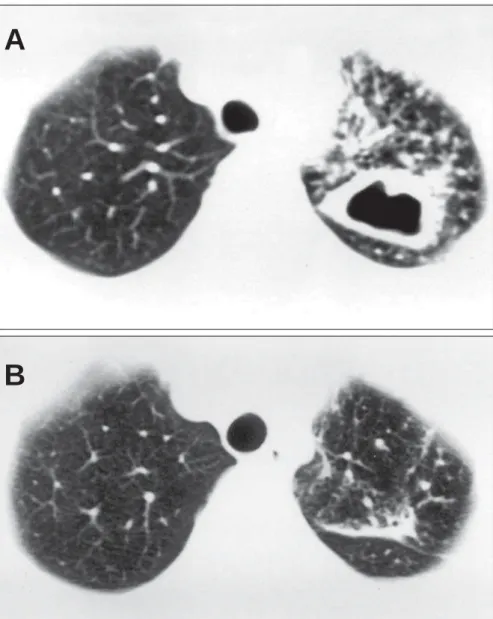
Figure 3. (A) Computed tomography at the start of treatment: thick-walled cavity in the left lung. (B) Computed tomography post-treatment, for the same patient: there is a band in the place where a cavity had been observed at the start of treatment.
A
If the exudative response is not sufficient to contain the progress of the bacilli, macrophages are activated and cause phago-cytosis of the bacilli. This introduces antigens of the microorganism to the lymphoid tissue associated with the bronchus. The activated macrophages in the alveoli crowd around the bacilli and become epithelium cells, which themselves group together to form the gigan-tic multinuclear cells that form the granuloma of the tuberculosis.14
Groupings of formed granulomas are re-ferred to as Ghon nodules. Groups of Ghon nodules constitute the primary tuberculosis complexes, which are known as lymphangi-tis and lymphadenilymphangi-tis. These may develop into the cure or the disease itself, depending upon the number and virulence of the ba-cilli, as well as the degree of host hypersensi-tivity and resistance.17
The disease begins from the initial paren-chymatous or ganglionic lesion. After necro-sis of the lesion center, liquefaction follows (the elimination of liquid material by bron-chial drainage and subsequent formation of cavities). These thick-walled cavities appear during the active phase of the tuberculosis. During the present study, thick-walled cavi-ties were observed upon diagnosis in 80% of the patients (Figure 3).
Centrilobular nodules with segmental distribution, representative of the broncho-genic spread of tuberculosis, are most fre-quently discovered through tomographic scan-ning in the active phase of the disease, and are present in up to 82% of the cases.5,6 In the present study, they were seen on 95% of the tomographic scans analyzed in the start of treatment (Figure 4).
These nodules tend to converge or form larger nodules and masses in 62% to 71% of the cases.6 In our sample, 80% and 70% of the patients presented confluent micronodules and larger nodules, respectively. Massive ap-pearance was observed in 60% of the patients. Thickening of the bronchial walls and cy-lindrical bronchiectasis are described in the literature in 62% and 23% of the patients with active pulmonary tuberculosis, respectively. Tree-in-bud appearance is present in up to 57% of the cases.2,5,6 During the present study, 65% of the patients showed thickened bron-chial walls. Bronchiectasis was present in 20% and the tree-in-bud appearance was observed in 60% of the cases.
A variety of sequelae and complications can occur in pulmonary tuberculosis in treated or untreated patients. These can be catego-rized as parenchymal or airway lesions, which
include thin-walled cavities, bands and bron-chiectasis.18 The cavities are formed by scar-ring. The residual characteristics of such cavi-ties include grooving, calcification and retrac-tion of the attacked parenchyma. The cavities may also remain after curing, with their walls thinned, which represents the succession or inactivation of that specific process.19,20 Some authors suggest that, in radiology, these find-ings should be described as “stable” rather than “inactive”, because of the possibility of future recrudescence of latent bacilli.21
Such residual cavities and bronchiectasis may be colonized by Aspergillus species, nontu-berculous mycobacteria or other microorgan-isms. Hemoptysis may be the clinically most important consequence of these sequelae. Af-ter the completion of treatment, thin-walled cavities were present in 25%, traction bron-chiectasis in 35% and bands in 70% of the
pa-tients appraised in our study. These post-treat-ment findings, in relation to the initial condi-tions (Table 1), provide important evidence for the follow-up of patients with pulmonary tu-berculosis. Therefore, recognition of radiologi-cal manifestations of the pulmonary sequelae is important for facilitating the understanding of complications due to the disease.18,22
○ ○ ○ ○ ○ ○ ○ ○ ○ ○ ○ ○ ○ ○ ○ ○ ○ ○ ○ ○
CONCLUSIONS
We have concluded that signs suggestive of tuberculosis activity are present in the ac-tive disease, and are seen via computed tom-ography. The extent of the parenchyma attack decreases significantly upon completion of the treatment. Such findings may be useful in the diagnosis of pulmonary tuberculosis, particu-larly when it is not possible to achieve bacte-riological confirmation.
Figure 4. (A) Computed tomography at the start of treatment: centrolobular nodules with segmental distribution in the anterior parts of both lungs. (B) Computed tomography post-treatment, for the same patient: absence of the alterations observed at the start of treatment.
A
Tuberculose pulmonar: avaliação tomográfica na fase ativa e pós-tratamento
CONTEXTO: O adequado conhecimento das
imagens compatíveis com atividade da tuber-culose é um importante recurso para o seu diagnóstico e acompanhamento.
OBJETIVO: Avaliar as alterações estruturais da
tuberculose no parênquima pulmonar, duran-te a fase ativa da doença e após o término do tratamento, através da tomografia computa-dorizada do tórax.
TIPO DE ESTUDO: Estudo prospectivo.
LOCAL: Disciplina de Pneumologia da Faculdade
de Medicina da Universidade de São Paulo.
PARTICIPANTES: 20 pacientes portadores de
tuberculose pulmonar confirmadas por cul-tura de Mycobacterium tuberculosis.
PROCEDIMENTOS: Foram obtidas
tomogra-fias convencionais desses pacientes em dois momentos: quando do diagnóstico e após o término do tratamento. Foram considerados sinais compatíveis com atividade de tuber-culose: nódulos centrolobulares de distribui-ção segmentar, micronódulos confluentes, consolidações, cavidades de paredes espessas,
○ ○ ○ ○ ○ ○ ○ ○ ○ ○ ○ ○ ○ ○ ○ ○ ○ ○ ○ ○ ○ ○ ○ ○ ○ ○ ○ ○ ○ ○ ○ ○ ○ ○ ○ ○ ○ ○ ○ ○ ○ ○
RESUMO
This data forms part of research protocol no. 228/00, as approved by the Ethics Committee for Research Project Assessment at Hospital das Clínicas, Universidade de São Paulo, on July 3, 2000.
Sidney Bombarda, MD, PhD. Attending physician, Pul-monary Division, Faculdade de Medicina da Universidade de São Paulo, São Paulo, Brazil.
Cláudia Maria Figueiredo, MD, PhD. Attending physi-cian, Radiology Division, Faculdade de Medicina da Universidade de São Paulo, São Paulo, Brazil.
Márcia Seiscento, MD, PhD. Attending physician, Pul-monary Division, Faculdade de Medicina da Universidade de São Paulo, São Paulo, Brazil.
Mário Terra Filho, MD, PhD. Associate professor, Pul-monary Division, Faculdade de Medicina da Universidade de São Paulo, São Paulo, Brazil.
Sources of funding: Not declared
Conflict of interest: Not declared
Date of first submission: February 19, 2003
Last received: May 12, 2003
Accepted: May 30, 2003
Address for correspondence
Sidney Bombarda
Rua Ezequiel Freire, 35 — Sala 33 — Santana São Paulo/SP — Brasil — CEP 02415-001 Tel./Fax (+55 11) 6977-5227 E-mail: [email protected]
COPYRIGHT © 2003, Associação Paulista de Medicina
○ ○ ○ ○ ○ ○ ○ ○ ○ ○ ○ ○ ○ ○ ○ ○ ○ ○ ○ ○
Publishing information
nódulos, massas, espessamento de paredes brônquicas, aspecto de árvore em florescência e bronquiectasias cilíndricas.
VARIÁVEIS ESTUDADAS: Comparou-se a
pre-sença de sinais compatíveis com atividade de tuberculose no início do tratamento e após o término do tratamento através do teste dos sinais (z).
RESULTADOS: Todos os pacientes (20/20)
apre-sentavam sinais compatíveis com atividade de tuberculose no início do tratamento. Após o término do tratamento, 13 pacientes (13/20) ainda apresentavam algum sinal sugestivo de atividade. Houve diminuição da extensão do acometimento pulmonar no pós-tratamento em relação ao início do tratamento (z = 10,10). Essa mudança foi estatisticamente significativa (p < 0,001).
CONCLUSÃO: Os sinais sugestivos de
ativida-de da tuberculose, na tomografia computa-dorizada do tórax, estão presentes na doença ativa e a extensão do parênquima acometido diminui significativamente após o término do tratamento.
PALAVRAS-CHAVE: Tuberculose. Tomografia
computadorizada por raios x. Diagnóstico.
1. World Health Organization. Global Tuberculosis Control: Sur-veillance, Planning, Financing. WHO Report 2002. Available from: URL: http://www.who.int/gtb/publications/globrep02/ contents.html. Accessed on 10/07/03
2. Bombarda S, Figueiredo CM, Funari MBG, Soares-Júnior J, Seiscento M, Terra-Filho M. Imagem em tuberculose pulmonar. J Pneumol 2001;27(6):329-40.
3. Brasil. Ministério da Saúde. Plano Nacional de Controle da Tuberculose. Manual de normas para o controle da tuberculose. Brasília: Ministério da Saúde; 2000.
4. Goo JM, Im JG. CT of tuberculosis and nontuberculous myco-bacterial infections. Radiol Clin North Am 2002;40(1):73-87. 5. Campos CA, Marchiori E, Rodrigues R. Tuberculose pulmonar:
achados na tomografia computadorizada de alta resolução do tórax em pacientes com doença em atividade comprovada bacteriologicamente. J Pneumol 2002;28(1):23-9. 6. Lee KS, Hwang JW, Chung MP, Kim H, Kwon OJ. Utility of
CT in the evaluation of pulmonary tuberculosis in patients with-out AIDS. Chest 1996;110(4):977-84.
7. Hatipoglu ON, Osma E, Manisali M, et al. High resolution computed tomographic findings in pulmonary tuberculosis. Thorax 1996;51(4):397-402.
8. Lee KS, Im JG. CT in adults with tuberculosis of the chest:
○ ○ ○ ○ ○ ○ ○ ○ ○ ○ ○ ○ ○ ○ ○ ○ ○ ○ ○ ○ ○ ○ ○ ○ ○ ○ ○ ○ ○ ○ ○ ○ ○ ○ ○ ○ ○ ○ ○ ○ ○ ○ ○ ○ ○ ○ ○ ○ ○ ○ ○ ○ ○ ○ ○ ○ ○ ○ ○ ○ ○ ○ ○ ○
REFERENCES
characteristic findings and role in management. AJR Am J Roentgenol 1995;164(6):1361-7.
9. Im JG, Itoh H, Shim YS, et al. Pulmonary tuberculosis: CT findings - early active disease and sequential change with an-tituberculous therapy. Radiology 1993;186(3):653-60. 10. Leung AN. Pulmonary tuberculosis: the essentials. Radiology
1999;210(2):307-22.
11. McAdams HP, Erasmus J, Winter JA. Radiologic manifesta-tions of pulmonary tuberculosis. Radiol Clin North Am 1995;33(4):655-78.
12. Lee KS, Song KS, Lim TH, Kim PN, Kim IY, Lee BH. Adult-onset pulmonary tuberculosis: findings on chest radiographs and CT scans. AJR Am J Roentgenol 1993;160(4):753-8. 13. Bombarda S, Figueiredo CM, Seiscento M, Terra-Filho M.
Estudo comparativo entre a radiografia e a tomografia compu-tadorizada de tórax na forma ativa da tuberculose pulmonar. J Pneumol 2000;26(Suppl 3):S18.
14. Capellozi, VL. Tuberculose. In: Brasileiro-Filho G, Bogliolo L, editors. Patologia. 6ª ed. Rio de Janeiro: Guanabara Koogan; 2000. p.320-2.
15. Tuddenham WJ. Glossary of terms for thoracic radiology: rec-ommendations of the Nomenclature Committee of the Fleischner Society. AJR Am J Roentgenol 1984;143(3):509-17.
16. Souza-Júnior AS, Araujo CN, Jasinovodolinsky D, et al. Terminologia para a descrição de tomografia computadorizada de tórax: sugestões iniciais para um consenso brasileiro. [Ter-minology for the description of the thoracic computed tomog-raphy: first suggestions for a brazilian consensus]. Radiol Bras 2002;35(2):125-8.
17. Rook GW, Zumla A. Advances in the immunopathogenesis of pulmonar y tuberculosis. Curr Opin Pulm Med 2001;7(3):116-23.
18. Kim HY, Song KS, Goo JM, Lee JS, Lee KS, Lim TH. Tho-racic sequelae and complications of tuberculosis. Radiographics 2001;21(4):839-58; discussion 859-60.
19. Collins J. CT signs and patterns of lung disease. Radiol Clin North Am 2001;39(6):1115-35.
20. Lee JY, Lee KS, Jung KJ, et al. Pulmonary tuberculosis: CT and pathologic correlation. J Comput Assist Tomogr 2000;24(5):691-8.
21. Miller WT, MacGregor RR. Tuberculosis: frequency of unu-sual radiographic findings. AJR Am J Roentgenol 1978;130(5):867-75.