○ ○ ○ ○ ○ ○ ○ ○ ○ ○ ○ ○ABSTRACT○ ○ ○ ○ ○ ○ ○ ○ ○ ○ ○ ○ ○ ○ ○ ○ ○ ○ ○ ○ ○ ○ ○ ○ ○ ○
INTRODUCTION
Childhood cancer can be distinguished from other cancers by its short latency period, rapid growth and invasive aggressiveness, as well as by an apparent reduced influence from environmental carcinogens. These peculiari-ties apply to the most frequent type of child-hood cancer, i.e. acute lymphoblastic leukemia.1 Studies carried out in the 1960s showing that the fusion of normal and neoplastic cells can suppress tumorigenicity led to the isolation of the Rb tumor suppressor gene in 1986.2
The cell cycle consists of an alternation between periods of rest and periods of cell di-vision until cell death. Although the duration of the different phases varies according to cell type, at least two great barriers, in the G1→S and G2→M transitions, seem to operate in all eukaryotic cells. These cells only divide af-ter receiving instructions or extracellular stimulation, either in the form of stimulating circulating mitogenic substances, or blockers of antiproliferative cytokines, or even through contact with adjacent cells or substrates. The transition from the early/intermediate phase to the late G1 phase is called the restriction point (R point), while transitions that occur at other points of the cell cycle are called check-points, with the most important at the G2 to M transition. Cancerous cells abandon their control mechanisms and continue to divide without evolving to programmed death.2 The decision to divide occurs as soon as the cell passes the R point, with the cell following its own program thereafter until division. The passage of a cell through the R point and
checkpoints is regulated by a family of kinase proteins that include a regulatory subunit, cyclin, and a catalytic subunit, cyclin-depend-ent kinase (CDK). Cyclin D activates CDK4, and also CDK6 in some cells, thus leading the cell to go beyond the R point.3
The genes p16, p15 and ARF1/p19 of the
INK4 family are a group of cyclin inhibitors located on the 9p21 band, a frequent site of abnormalities in human tumors, with p15 and p16 forming the multiple tumor suppressor (MTS) locus.4 The products of these genes bind to CDK 4 and 6, thus inhibiting the CDK-cyclin D complex, which results in the blockade of the phosphorylation of protein Rb, thereby preventing cell cycle progression. Genes with this mode of action are called tumor suppressor genes and have been stud-ied as risk indicators for poor therapeutic re-sponse. Some studies5,6 have found an in-creased rate of p15 inactivation in children with acute lymphoblastic leukemia, which was also correlated with disease prognosis, while others7 have not.
The objective of the present study was to analyze p15 gene inactivation by homologous deletion and point mutations in Brazilian chil-dren with acute lymphoblastic leukemia.
○ ○ ○ ○ ○ ○ ○ ○ ○ ○ ○ ○ ○ ○ ○ ○ ○ ○ ○ ○
SERIES AND METHODS
A total of 106 pediatric patients with acute lymphoblastic leukemia were admitted for treatment to the Pediatric Clinic of the Faculdade de Medicina de Ribeirão Preto, Universidade de São Paulo, between January 1991 and June 1999. Eighty-three patients were eligible for the study and 23 were
ex-Original A
rticle
• Rosana Cipolotti
• José Alexandre Rodrigues Lemos
• Ricardo Defavery
• Carlos Alberto Scrideli
• Amaury Lellis Dal Fabbro
• Luiz Gonzaga Tone
Inactivation of the p15
gene in children with acute
lymphoblastic leukemia
Laboratory of Molecular Biology, Department of Pediatrics, Faculdade de
Medicina de Ribeirão Preto, Universidade de São Paulo, Ribeirão Preto,
São Paulo, Brazil
CONTEXT: Tumor suppressor genes act on the control of cell cycle progression. In pediatric neoplasias, some of these genes may be considered to be markers for diagnosis or relapse, thus probably representing prognostic indicators.
OBJECTIVE: To study the inactivation of the p15 gene in children with acute lymphoblastic leukemia.
TYPE OF STUDY: Retrospective study.
SETTING: Laboratory of Molecular Biology, Department of Pediatrics, Faculdade de Medicina de Ribeirão Preto, Universidade de São Paulo.
PARTICIPANTS: Eighty-three children and adolescents with acute lymphoblastic leukemia were studied, with the examination of 83 bone marrow samples obtained at diagnosis, four obtained also during relapse, and two cerebrospinal fluid samples ob-tained from two cases of isolated relapse in the central nervous system.
MAIN MEASUREMENTS: Homologous deletion of the p15 gene by multiplex polymerase chain reaction, and screening for point mutations by polymerase chain reaction/single-strand conformational poly-morphism.
RESULTS: Deletion of exon 2 of the p15 gene was observed in 15 children, including one case in which deletion was only verified during isolated central nervous system relapse. No case of exon 1 deletion, or that was suggestive of point muta-tions, was observed and no association between p15 gene inactivation and classic risk factors was established.
CONCLUSION: According to the literature, inactiva-tion of the p15 gene by deleinactiva-tion of exon 2 in acute lymphoblastic leukemia found in the popu-lation studied would be considered to be a mo-lecular marker for diagnosis or relapse. However, no correlation between p15 gene deletion and clinical prognostic indicators was observed.
KEY WORDS: Acute lymphoblastic leukemia. Lymphob-lastic leukemia. Tumor suppressor gene. p15.
São Paulo Medical Journal — Revista Paulista de Medicina
204
cluded due to the lack or poor quality of stored DNA. The diagnosis was based on morphological analysis and immuno-phenotyping with monoclonal antibodies via flow cytometry. All patients were classified and treated according to the protocols pro-posed by the Brazilian Group for the Treat-ment of Childhood Leukemias (GBTLI 85/ 93).8 The project was approved by the Re-search Ethics Committee of the University Hospital (case no. 3761/98). This was a ret-rospective study of 87 DNA samples ex-tracted from cells aspirated from the bone marrow of 83 children and adolescents with acute lymphoblastic leukemia (83 samples obtained at diagnosis and four also during relapse), and two cerebrospinal fluid samples from two cases with isolated central nerv-ous system relapse.
The p15 gene inactivation was detected
by the polymerase chain reaction9 for the iden-tification of deletions, confirmed by multiplex polymerase chain reaction using the primers shown below and primers for beta-globin, and complemented by polymerase chain reaction/ single-strand conformational polymorphism10 for the screening of point mutations. DNA was extracted by the phenol-chloroform method11 and quantified, adjusting the con-centration to 0.1 µg/ml. The following prim-ers corresponding to the two exons of the p15 gene were used:5,12
• exon 1/primer1:
5’-AAGAGTGTCGTTAAGTTTACG-3’ • exon 1/primer 2:
5’-ACATCGGCGATCTAGGTTCCA-3’ • exon 2/primer 1:
5’-GGGTGGGAAATTGGGTAAG-3’ • exon 2/primer2:
5’-TGAGTTTAACCTGAAGGTGG-3’
Statistical analysis
The 87 bone marrow samples studied corresponded to all the samples stored up to 1998 whose DNA was still viable, and the two cerebrospinal fluid samples belonged to two patients who presented isolated central nerv-ous system relapse during the study period.
The results were compared using the Stu-dent t test when the variances were homoge-neous and the Bartlett and Mann-Whitney tests when the variances were non-homog-enous. Categorical variables were analyzed via the chi-square test. The level of significance was set at 95% (p < 0.05), with a cutoff date of September 1999.
○ ○ ○ ○ ○ ○ ○ ○ ○ ○ ○ ○ ○ ○ ○ ○ ○ ○ ○ ○
RESULTS
The 87 bone marrow samples and the two cerebrospinal fluid samples were
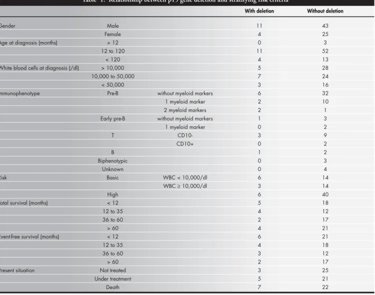
sub-Table 1. Relationship between p15 gene deletion and stratifying risk criteria
With deletion Without deletion
Gender Male 11 43
Female 4 25
Age at diagnosis (months) > 12 0 3
12 to 120 11 52
< 120 4 13
White blood cells at diagnosis (/dl) > 10,000 5 28
10,000 to 50,000 7 24
< 50,000 3 16
Immunophenotype Pre-B without myeloid markers 6 32
1 myeloid marker 2 10
2 myeloid markers 2 1
Early pre-B without myeloid markers 1 3
1 myeloid marker 0 2
T CD10- 3 9
CD10+ 0 2
B 1 2
Biphenotypic 0 3
Unknown 0 4
Risk Basic WBC < 10,000/dl 6 14
WBC ≥ 10,000/dl 3 14
High 6 40
Total survival (months) < 12 5 18
12 to 35 4 12
36 to 60 2 17
> 60 4 21
Event-free survival (months) < 12 6 21
12 to 35 4 18
36 to 60 3 12
> 60 2 17
Present situation Not treated 3 25
Under treatment 5 21
Death 7 22
WBC = white blood cells.
São Paulo Medical Journal — Revista Paulista de Medicina
205
mitted to amplification by the polymerase chain reaction. The p15 gene was present in 68 bone marrow samples and one cer-ebrospinal fluid sample. These samples were tested via single-strand conformational polymorphism and did not show any altera-tion in the electrophoretic migraaltera-tion pat-tern on polyacrylamide gels for any of the exons analyzed.
Fifteen samples (including one of cerebro-spinal fluid) originating from 15 patients that were not amplified after three replications were tested via the multiplex polymerase chain re-action using the beta-globin primers, which confirmed the deletions. All 15 cases were deletions of exon 2. Eleven deletions were de-tected in boys and four in girls.
Twenty-two of the 68 patients without deletions and seven of the 15 patients with deletions died during the study period. (33.8 versus 46.6%).
Table 1 lists the classic laboratory param-eters used for the definition of risk groups according to the occurrence of the exon 2 deletion. The occurrence of p15 gene dele-tion was also studied with respect to age and number of white cells at diagnosis, and with respect to immunophenotype, risk group, survival time (total and unfavorable event-free survival) and present situation, and no correlation was detected.
○ ○ ○ ○ ○ ○ ○ ○ ○ ○ ○ ○ ○ ○ ○ ○ ○ ○ ○ ○
DISCUSSION
Since the mid-1980s, the 9p21-22 region has been recognized as an important site of al-terations in patients with cancer, especially chil-dren. Through the identification of new tumor suppressor genes by analysis of non-random de-letions in this region,13 and through the analysis of the p15, p16 and p19 genes located in this region, the role of their inactivation in the gen-esis and evolution of lymphohematopoietic neoplasias, especially childhood acute lymphob-lastic leukemia, has been studied. Some stud-ies6,12-16 have emphasized the importance of p15 inactivation in T-cell acute lymphoblastic leukemia, whereas others17 have not found any difference between T-cell and B-cell precursor acute lymphoblastic leukemia. Another aspect is the interdependence of p15 and p16, since some studies have shown homologous deletion of both genes in most acute lymphoblastic leukemia cases,5,6,14,18,19 while in others p16 was the only target.6,20,21 Furthermore, while deletion seems to be one of the main mechanisms of in-activation for both genes, point mutations may play an important role in p16 inactivation, while epigenetic mechanisms such as hypermethylation are also considered for p15.6,20
These studies have opened up prospects for the definition of a diagnostic and/or prognos-tic gene marker. The p15 gene has been
identi-fied by some authors as a tumor suppressor gene that can be correlated with disease progno-sis.5,18,22 In the present study, deletion of exon 2 was identified in 18% of a sample of Brazil-ian children, mainly originating from the south-eastern region, where there is great ethnic di-versity in population groups. This result indi-cates that p15 inactivation by homologous de-letion of exon 2 might represent an important molecular marker for the population studied, as also observed by others.5,13 However, in con-trast to these other studies, we could not estab-lish an unequivocal association between inac-tivation and classic risk factors. This finding may be explained by the characteristics of the children studied, considering their ethnic di-versity and miscegenation, which differ from those observed in studies carried out in Swe-den and Japan.5,18 No study carried out in Bra-zil or Latin America to analyze tumor suppres-sor genes of the 9p21 region in children with acute lymphoblastic leukemia is available in the literature. Thus, since deletions are not the only mechanism for tumor suppressor genes inacti-vation, the present findings indicate the need for further multicenter studies investigating epigenetic and gene expression mechanisms in order to understand the role of these genes and their interactions in cell cycle progression and carcinogenesis in Brazilian children with acute lymphoblastic leukemia.
São Paulo Medical Journal — Revista Paulista de Medicina
206
Inativação do gene p15 em crianças com leucemia linfoblástica aguda
CONTEXTO: Os genes supressores tumorais atu-am no controle da progressão do ciclo celu-lar. Em neoplasias pediátricas, alguns destes genes podem ser considerados marcadores de diagnóstico ou de recaída, sendo, portanto, prováveis indicadores prognósticos. OBJETIVO: Estudar a inativação do gene p15
em crianças com leucemia linfóide aguda. TIPO DE ESTUDO: Estudo retrospectivo. LOCAL: Laboratório de Biologia Molecular,
De-partamento de Puericultura e Pediatria, Fa-culdade de Medicina de Ribeirão Preto, Uni-versidade de São Paulo.
PACIENTES: Foram estudadas 83 crianças e ado-lescentes com leucemia linfóide aguda, sen-do examinadas 83 amostras de medula óssea ao diagnóstico, quatro também na recaída e duas amostras de líquido cefalorraquidiano em dois casos de recaída isolada em sistema nervoso central.
VARIÁVEIS ESTUDADAS: Deleção homóloga
○ ○ ○ ○ ○ ○ ○ ○ ○ ○ ○ ○ ○ ○ ○ ○ ○ ○ ○ ○ ○ ○ ○ ○ ○ ○ ○ ○ ○ ○ ○ ○ ○ ○ ○ ○ ○ ○ ○ ○ ○ ○
RESUMO
Rosana Cipolotti, MD. Department of Pediatrics, Faculdade de Medicina de Ribeirão Preto, Universidade de São Paulo, Ribeirão Preto, São Paulo, Brazil.
José Alexandre Rodrigues Lemos, MD, PhD.
Faculdade de Medicina de Ribeirão Preto, Universidade de São Paulo, Ribeirão Preto, São Paulo, Brazil.
Ricardo Defavery, MD. Department of Pediatrics, Faculdade de Medicina de Ribeirão Preto, Universidade de São Paulo, Ribeirão Preto, São Paulo, Brazil.
Carlos Alberto Scrideli, MD. Department of Pediatrics, Faculdade de Medicina de Ribeirão Preto, Universidade de São Paulo, Ribeirão Preto, São Paulo, Brazil.
Amaury Lellis Dal Fabbro, MD. Department of Social Medicine, Faculdade de Medicina de Ribeirão Preto, Universidade de São Paulo, Ribeirão Preto, São Paulo, Brazil.
Luiz Gonzaga Tone, MD. Coordinator of the Pediatric Oncology Service, Department of Pediatrics, Faculdade de Medicina de Ribeirão Preto, Universidade de São Paulo, Ribeirão Preto, São Paulo, Brazil.
Sources of funding: None
Conflict of interest: None
Date of first submission: September 10, 2002
Last received: April 29, 2003
Accepted: May 30, 2003
Address for correspondence
Luiz Gonzaga Tone
Departamento de Puericultura e Pediatria Faculdade de Medicina de Ribeirão Preto Av. Bandeirantes, 3900
Ribeirão Preto/SP — Brasil — CEP 14049-900 Tel. (+55 16) 602-2772
E-mail: [email protected]
COPYRIGHT © 2003, Associação Paulista de Medicina
○ ○ ○ ○ ○ ○ ○ ○ ○ ○ ○ ○ ○ ○ ○ ○ ○ ○ ○ ○
Publishing information
do gene p15, por reação em cadeia de polimerase-multiplex e triagem para muta-ção de ponto por reamuta-ção em cadeia de polimerase-polimorfismo conformacional em fita simples.
RESULTADOS: Deleção do exon 2 do gene p15 foi observada em 15 crianças, incluindo um caso apenas na recaída isolada em sistema nervoso central. Nenhum caso de deleção do exon 1 foi verificado, bem como nenhum caso sugestivo de mutação de ponto. Não foi estabelecida associação entre inativação do gene p15 e fatores de riscos clássicos. CONCLUSÃO: De acordo com a literatura, a
inativação do gene p15 por deleção do exon 2 em leucemia linfóide aguda encontrada na população estudada pode ser considerada um marcador molecular de diagnóstico ou de re-caída. No entanto, não foi observada qual-quer correlação entre deleção do gene p15 e indicadores clínicos prognósticos. PALAVRAS-CHAVE: Leucemia linfóide. Genes
supressores de tumor. p15.
1. Schüz J, Kaatsch P, Kaletsch U, Meinert R, Michaelis J.
Asso-ciation of childhood cancer with factors related to pregnancy and birth. Int J Epidemiol 1999;28(4):631-9.
2. Stass SA, Mixson J. Oncogenes and tumor suppressor genes:
thera-peutic implications.Clin Cancer Res 1997;3(12 Pt 2):2687-95.
3. Hunter T, Pines J. Cyclins and cancer II: Cyclin D and CDK
inhibitors come of age. Cell 1994;79(4):573-82.
4. Hannon GJ, Beach D. p15INK4Bis a potential effector of
TGF-beta-induced cell cycle arrest. Nature 1994;371(6494):257-61.
5. Heyman M, Rasool O, Borgonovo Brandter L, et al.
Prognos-tic importance of p15INK4B and p16INK4 gene inactivation in childhood acute lymphocytic leukemia. J Clin Oncol 1996;14(5):1512-20.
6. Batova A, Diccianni MB, Yu JC, et al. Frequent and selective
methylation of p15and deletion of both p15and p16 in T-cell
acute lymphoblastic leukemia. Cancer Res 1997;57(5):832-6.
7. Maloney KW, McGavran L, Odom LF, Hunger SP. Different
patterns of homozygous p16INK4A and p15INK4B deletions in childhood acute lymphoblastic leukemias containing distinct E2A translocations. Leukemia 1998;12(9):1417-21.
8. Brandalise S, Odone V, Pereira W, et al. Treatment results of
three consecutive Brazilian cooperative childhood ALL protocols: GBTLI-80, GBTLI-82 and -85. ALL Brazilian Group. Leukemia 1993;7:(Suppl 2):S142-5.
○ ○ ○ ○ ○ ○ ○ ○ ○ ○ ○ ○ ○ ○ ○ ○ ○ ○ ○ ○ ○ ○ ○ ○ ○ ○ ○ ○ ○ ○ ○ ○ ○ ○ ○ ○ ○ ○ ○ ○ ○ ○ ○ ○ ○ ○ ○ ○ ○ ○ ○ ○ ○ ○ ○ ○ ○ ○ ○ ○ ○ ○ ○ ○
REFERENCES
9. Saiki RK, Gelfand DH, Stoffel S, et al. Primer-directed
enzymatic amplification of DNA with a thermostable DNA polymerase. Science 1988;239(4839):487-91.
10. Orita M, Iwahana H, Kanazawa H, Hayashi K, Sekiya T. De-tection of polymorphisms of human DNA by gel electrophore-sis as single-strand conformation polymorphisms. Proc Natl
Acad SciUSA 1989;86(8):2766-70.
11. Sambrook J, Fritsch EF, Maniatis J. Molecular Cloning: A
Labo-ratory Manual. 2nd ed. Philadelphia: Cold Spring Harbor
Labo-ratory Press; 1989.
12 - Rasool O, Heyman M, Brandter LB, et al. p15INK4B and p16INK4 gene inactivation in acute lymphocytic leukemia. Blood 1995;85(12):3431-6.
13. Hebert J, Cayuela JM, Berkeley J, Sigaux F. Candidate tumor suppressor genes MTS1 (p16INK4A) and MTS2 (p15INK4B) display frequent homozygous deletions in primary cells from T- but not from B-cell lineage acute lymphoblastic leukemias. Blood 1994;84(12):4038-44.
14. Otsuki T, Clark HM, Wellmann A, Jaffe ES, Raffeld M. In-volvement of CDKN2 (p16INK4A/MTS1) and p15INK4B/ MTS2 in human leukemias and lymphomas. Cancer Res 1995;55(7):1436-40.
15. Okuda T, Shurtleff SA, Valentine MB, et al. Frequent deletion of p16INK4a/MTS1 and p15INK4b/MTS2 in pediatric acute
lymphoblastic leukemia. Blood 1995;85(9):2321-30. 16. Takeuchi S, Bartram CR, Seriu T, et al. Analysis of a family of
cyclin-dependent kinase inhibitors: p15/MTS2/INK4B, p16/ MTS1/INK4A, and p18 genes in acute lymphoblastic leukemia of childhood. Blood 1995;86(2):755-60.
17. Iravani M, Dhat R, Price CM. Methylation of the multi tumor suppressor gene-2 (MTS2, CDKN1, p15INK4B) in childhood acute lymphoblastic leukemia. Oncogene 1997;15(21):2609-14. 18. Hangaishi A, Ogawa S, Imamura N, et al. Inactivation of mul-tiple tumor-suppressor genes involved in negative regulation of the cell cycle, MTS1/p16INK4A/CDKN2/, MTS2/p15INK4B, p53, and Rb genes in primary lymphoid malignancies. Blood 1996;87(12):4949-58.
19. Thullberg M, Bartkova J, Khan S, et al. Distinct versus redun-dant properties among members of the INK4 family of cyclin-dependent kinase inhibitors. FEBS Lett 2000;470(2):161-6. 20. Guo SX, Taki T, Ohnishi H, et al. Hypermethylation of p16
and p15 genes and RB protein expression in acute leukemia. Leuk Res 2000;24(1):39-46.
21. Bringold F, Serrano M. Tumor suppressors and oncogenes in cellular senescence. Exp Gerontol 2000;35(3):317-29. 22. Fink JR, LeBien TW. Novel expression of cyclin-dependent
ki-nase inhibitors in human B-cell precursors.Exp Hematol
2001;29(4):490-8.