○ ○ ○ ○ ○ ○ ○ ○ ○ ○ ○ ○ABSTRACT○ ○ ○ ○ ○ ○ ○ ○ ○ ○ ○ ○ ○ ○ ○ ○ ○ ○ ○ ○ ○ ○ ○ ○ ○ ○
INTRODUCTION
Early prediction of the risks to which a child is exposed at birth allows for better organiza-tion of perinatal care and optimizaorganiza-tion of avail-able resources, thus avoiding unnecessary con-trols and assuring maximum attention for those children who really need it.1With this objec-tive in mind, several indicators have been rec-ommended. Among these, the birth weight in relation to gestational age has frequently been used for classifying newborns according to the intrauterine growth experienced. The three cat-egories are normal intrauterine growth (AGA, or appropriate for gestational age), subnormal growth (SGA, or small for gestational age) or supranormal growth (LGA, or large for gesta-tional age).2Several criteria have been used for separating these three categories, the most com-mon being based on percentiles from weight for gestational age distribution in a reference population.3
Several studies have led to the conclusion that the newborn’s nutritional status is more important than birth weight alone for identi-fying perinatal risks.4,5 Perinatal risk assessment by weight percentile criteria has been shown to be insufficient, thus requiring the determina-tion of addidetermina-tional or alternative indices to im-prove this evaluation. The mid-arm circumfer-ence (MAC) measurement is less affected by subclinical edema than weight alone and is rela-tively easy to obtain. This has led several au-thors to employ it as an important tool for iden-tifying malnutrition and mortality risk.6-9 How-ever, most of the nutritional studies involving anthropometric parameters have used the weight/height ratio and its derivatives – body
mass index and ponderal index7,10 – in order to evaluate individual body proportionality.
With the aim of evaluating preschool chil-dren’s nutritional condition, Kanawati and McLaren11were the first authors to propose the mid-arm circumference/head circumfer-ence (MAC/HC) ratio for such an evaluation. This ratio is easily obtained by simple and non-expensive equipment, with minimal training requirements. Its use in the neonatal period was introduced in 1986, when Sasanow et al.12 established reference values for newborns of gestational age 25 to 42 weeks with appropri-ate growth for gestational age.
In Brazil, Dias13 and Alves et al.14 showed that the newborn’s mid-arm circum-ference was strongly related to birth weight, thus representing a good marker for low and very low birth weight. However, a large number of neonatologists are not aware of the potential usefulness of such measurements, with the result that these anthropometric pa-rameters are remarkably underused in Brazil. The objectives of the present study were the following:
• to establish mid-arm circumference val-ues and the mid-arm circumference/head circumference ratio among a population of term Brazilian newborns, according to gestational age and birth weight; • to investigate the occasional differences in
anthropometric variables found in the present study according to gender; • to evaluate the possibility of obtaining
cor-relation curves for the studied variables ac-cording to gestational age and birth weight.
○ ○ ○ ○ ○ ○ ○ ○ ○ ○ ○ ○ ○ ○ ○ ○ ○ ○ ○ ○ METHODS
Mattos Segre
mid-arm/head circumference
ratio in term newborns
Hospital Maternidade Leonor Mendes de Barros, São Paulo, Brazil, and
Instituto de Assistência Médica ao Servidor Público Estadual de São Paulo
(IAMSP), São Paulo, Brazil
Original Ar
ticle
CONTEXT: Mid-arm circumference of the newborn is strongly associated with birth weight and is a very good indicator of low and insufficient birth weight. However, there are few Brazilian studies on the relationship between mid-arm and head circum-ferences and, thus, this does not form part of the routine evaluation for newborns.
OBJECTIVES: To establish the mid-arm circumference and mid-arm/head circumference ratio in a popu-lation of term newborns.
TYPE OF STUDY: Cross-sectional study carried out between June 1997 and August 1999.
SETTING:Hospital Maternidade Leonor Mendes de Barros, São Paulo.
PARTICIPANTS: Term newborns (66 males and 65 fe-males) of appropriate growth for gestational age, whose mothers were healthy, were included in the study.
MAIN MEASUREMENTS: Arm circumference, arm circumference/head circumference ratio, birth weight and gestational age were measured within 48 hours of birth. Data were considered signifi-cant when p < 0.01.
RESULTS: The mean values for the mid-arm circumfer-ence were 10.76 cm (standard deviation, SD = 0.68) for females and 10.76 (SD = 0.81) for males. The mean value for the mid-arm/head circumfer-ence ratio was 0.31 (SD = 0.02) for both sexes. Mid-arm circumference values were significantly related to birth weight and gestational age, whereas mid-arm/head circumference ratio was related only to birth weight.
CONCLUSIONS: Mid-arm circumference and mid-arm/head circumference ratio values were estab-lished for the studied population. It was possible to obtain curves for both mid-arm circumference and mid-arm/head circumference ratio in relation to birth weight. However, for mid-arm circumfer-ence, it was only possible to obtain curves in rela-tion to gestarela-tional age. The use of the regression curves did not seem powerful enough to predict the mid-arm circumference and mid-arm/head cir-cumference ratio in this population of term newborns. There were no gender differences for either of the measurements studied.
newborns whose mothers agreed to partici-pate in the study were included.
Newborns whose mothers presented com-plications during pregnancy, such as previous or pregnancy-related arterial hypertension, in-fection, previous or pregnancy-related diabe-tes, or had a history of illegal drug abuse or smoking habits, if more than 10 cigarettes per day, were excluded. Newborns with major mal-formations, hydropic appearance or presenting signs of intrauterine growth restriction such as an Apgar score of less than 7 in the fifth minute of life, hypoglycemia, hypocalcemia or poly-cythemia and those whose mothers denied au-thorization were also excluded.
Thus, the total sample included in the study comprised 131 newborns.
Gestational age determination
In order to determine the gestational age, a daily service neonatologist or a duly trained pediatric resident examined the children 6 to 12 hours after birth. The “term” concept was applied to those newborns whose gestational ages ranged from 37 weeks to less than 42 completed weeks: from 259 to 293 gestational days, as calculated by Naegele’s rule.15 The gestational age was also estimated via clinical-neurological examination of the newborn (us-ing Capurro’s method)15 and was expressed as “completed weeks”.16
Intrauterine growth adequacy determina-tion
For this purpose, the weight-gestational age criterion was applied, considering the 10th percentile as the lower limit for newborns to be appropriate for gestational age, at the bor-der with small for gestational age, and the 90th percentile as the upper limit for newborns to be appropriate for gestational age, at the border with large for gestational age. The newborns were classified according to the weight-gestational age curve routinely used in the hospital where the study was car-ried out.17
Anthropometric measurements: Birth weight
The weight was obtained with the naked infant in dorsal decubitus, soon after birth, still in the delivery room, using an electronic balance with a maximum capacity of 15 kg and a minimum of 125 g, and 5 g subdivi-sions, previously calibrated by the Brazilian National Institute of Weights and Measures (Inmetro). The measurements were taken by an attending nurse or the neonatologist at-tending the delivery room.
Circumferences
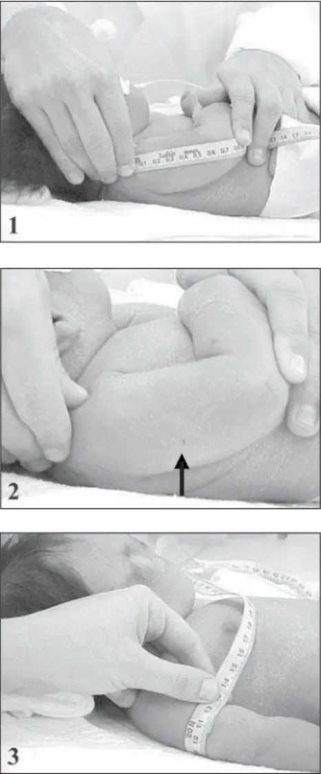
All measurements were taken by the main author or the neonatology resident, according to the technique previously described,12 so that this could not represent an impediment in com-paring the final results. The arm and head cir-cumferences were measured within the first 48 hours of life, using a fiberglass non-extendable measuring tape, with a width of 1.0 cm and subdivisions of 0.1 cm. The mid-arm circum-ference was obtained from the left arm, at the mid point between the acromion and ole-cranon, with the newborn in dorsal decubitus with the arm lying laterally to the trunk. The midpoint was located by measuring the distance between the acromion and olecranon extremi-ties, with the elbow flexed at an angle of 90°. A small mark was made at the identified point (Figure 1). A total of three consecutive meas-urements were taken for each newborn, and the mean value (rounded to the nearest 0.1 cm) was considered for analysis.
The head circumference was measured with the newborn in dorsal decubitus. The measuring tape was placed along the occipi-tal-frontal circumference, just over the eye-brows and the occiput, in order to obtain the largest measurement. The maximum value of three consecutive measurements was consid-ered, rounded to the nearest 0.1 cm.18
Statistical analysis
The data processing was done using the Sta-tistical Package for the Social Sciences (SPSS) version 9.0 software. The probability level p < 0.01 was considered to be significant. Statistical analyses were performed to estimate the arith-metic mean and standard deviation, followed by the Kolmogorov-Smirnov test to determine the normal distribution of the variables studied: arm circumference and arm circumference/head cir-cumference ratio. The Student t test was used to compare genders, and a correlation matrix was built in order to test associations with gestational age and birth weight among the studied vari-ables. Linear regression was applied considering birth weight as an independent variable. Multi-ple regression analyses were used, in which ges-tational age (GA), gesges-tational age squared (GA2) and gestational age cubed (GA3) were consid-ered as independent variables.
The present study was duly approved by the Research Ethics Committee of Hospital Maternidade Leonor Mendes de Barros.
○ ○ ○ ○ ○ ○ ○ ○ ○ ○ ○ ○ ○ ○ ○ ○ ○ ○ ○ ○ RESULTS
The study comprised 131 newborns: 66 males and 65 females. The mean birth weight
Figure 1. Mid-arm circumference: obtaining the measure-ment. 1. length of the arm; 2. mid-arm point (arrow); 3. measurement of circumference at the mid-arm point.
A cross-sectional study was performed among term live birth newborns, from June 1997 to August 1999, at Hospital Materni-dade Leonor Mendes de Barros, São Paulo, Brazil, a public maternity hospital within the healthcare system that serves a low-income population and is used as a reference center for high-risk pregnancies.
Inclusion and exclusion criteria
of the sample was 3,177 g, ranging from 2,330 g to 3,910 g. The average gestational age was 39 complete weeks, ranging from a minimum of 37 weeks to a maximum of 41 weeks.
Table 1 shows the mean and standard de-viations for male and female newborns. The normality test (Kolmogorov-Smirnov) showed that both variables studied followed the nor-mal distribution. The Student t test, used in order to identify possible differences between sexes, showed no significant differences for any of the evaluated parameters, and therefore the sample was considered as a whole.
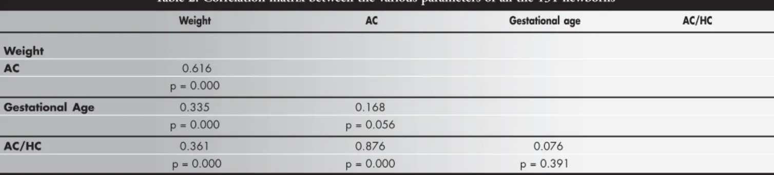
The correlation matrix for the parameters of the overall group is shown in Table 2. Birth weight was positively correlated with both arm circumference and arm/head circumference ratio (p < 0.0001 for both), whereas the cor-relation coefficients between gestational age and arm circumference or arm/head circum-ference ratio were low and not significant. The quadratic regression for arm circumference proved to be significant (r2= 0.056; f = 3.77; p = 0.026), thereby showing that the best ad-justment for the curve could be obtained by using the squared gestational age (Figure 4). For the mid-arm circumference/head circum-ference ratio, the regression analysis did not prove to be significant (r2= 0.0028; f = 1.86; p = 0.159). It was possible to obtain linear equations and their graphs using quadratic regression (Figure 4), including the individual confidence interval calculations for the mid-arm circumference and mid-mid-arm circumfer-ence/head circumference ratio versus birth
Table 1. Patient characteristics — mean values and standard deviation by gender
Characteristics Gender Calculated “t” significance (p)
Female Male
Age (hours) 28.52 ± 11.68 29.18 ± 11.08 0.34 0.737 (NS) Birth weight (g) 3154.54 ± 296.82 3200.20 ± 311.98 0.86 0.393 (NS) Gestational age (weeks) 39.15 ± 1.11 39.26 ± 1.06 0.55 0.845 (NS) Arm circumference (cm) 10.76 ± 0.68 10.76 ± 0.81 0.04 0.965 (NS) AC/HC 0.31 ± 0.02 0.31 ± 0.02 1.41 0.160 (NS)
NS = Not significant; AC/HC = arm circumference/head circumference.
Table 2. Correlation matrix between the various parameters of all the 131 newborns*
Weight AC Gestational age AC/HC
Weight
AC 0.616 p = 0.000
Gestational Age 0.335 0.168 p = 0.000 p = 0.056
AC/HC 0.361 0.876 0.076
p = 0.000 p = 0.000 p = 0.391
* Person multiple correlation; correlation is significant at the 0.01 level, two-tailed; AC = arm circumference; AC/HC: arm circumference/head circumference.
Figure 2. Regression line corresponding to the mid-arm circumference (AC) of newborns with gestational ages between 37 and 41 completed weeks, in relation to birth weight.
r = 0.616; p < 0.001; 95% confidence interval for the mean value weight (Figures 2 and 3), and for the mid-arm circumference versus gestational age. Al-though significant, the regression coefficient (r2) for mid-arm circumference over gesta-tional age was low and, consequently, the
ac-curacy of estimating arm circumference by gestational age was not recommended.
Figure 3. Regression line corresponding to the arm circumference/head circumference ratio (AC/HC) of newborns with gesta-tional ages between 37 and 41 completed weeks, in relation to birth weight.
r = 0.361; p = 0.0001; 95% confidence interval for the mean value
then calculate the mean and standard deviation for the adjusted variable, thereby obtaining an arm circumference value independent of gesta-tional age. Thus, the range from the average minus two standard deviations to the average plus two standard deviations, which corresponds to 95% of the distribution, could be used as the parameter for the normal range. The corrected value was obtained as follows:
observed arm circumference + b x (mean
ges-tational age – gesges-tational age) + c x (mean
ges-tational age2 – gestational age2),
where: b = regression constant for gestational age and c = regression constant for gestational age2. This new distribution presented a mean value of 10.82 cm and standard deviation of 0.73. Thus, 9.36 cm to 12.28 cm could be con-sidered as the normal range, which included 95% of the studied population.
○ ○ ○ ○ ○ ○ ○ ○ ○ ○ ○ ○ ○ ○ ○ ○ ○ ○ ○ ○ DISCUSSION
As a general rule, anthropometric meas-urements present systematic differences be-tween genders. Therefore, whenever the in-tention is to study such measurements, it is essential to report the gender of the studied population and study any possible differences between sexes.18 However, mid-arm circum-ference seems to show lesser variation related to gender, and the differences were not sig-nificant in a study among infants aged 3 months to 4-years by Kanawati et al.19 Among studies of the neonatal period, our data also match the findings of the majority of authors, who did not find any difference between gen-ders in relation to the mid-arm circumference and mid-arm circumference/head circumfer-ence ratio values.6,17,20 However, in a study among newborns of gestational ages ranging from 34 to 42 weeks, Jiménez Garcia et al.21 found different mid-arm circumference val-ues according to gender, with male measure-ments greater than female ones. Our study did not corroborate these findings.
Mid-arm circumference increases as preg-nancy progresses and this is mainly due to fat ac-cumulation in the subcutaneous deposits of the upper extremity.22 In the present study, mid-arm circumference showed a linear increase in relation to birth weight, agreeing with the literature on this subject,12,20but the correlation with gestational age was low. It was thus hypothesized that the use of the mean value plus or minus two standard deviations, would better predict the normal val-ues in this population.
Significant variation exists in mid-arm cir-cumference values among different popula-tions. These differences in measurements may
Figure 4. Quadratic regression line corresponding to the arm circumference (AC) of newborns with gestational ages between 37 and 41 completed weeks, in relation to gestational age.
be due to several factors, including each popu-lation’s genetic characteristics and nutritional status, as well as possible differences in meas-urement procedures.23 The mid-arm circum-ference values found in the present study showed lesser dispersion of means and stronger correlation coefficient when considered in re-lation to birth weight, rather than in rere-lation to gestational age. Our data differ from those described by most of the authors who have already studied this association.12,20 However, our findings were similar to those described by Sánchez et al.24in a Chilean population comprised mainly of term newborns. In the Chilean study, in addition to identifying greater scattering of mid-arm circumference values when associated with gestational age, rather than with birth weight, the authors showed that there was progressive increase in those measurements as gestational age pro-gressed and also found slightly lower values for the mid-arm circumference among the 42-week gestational age newborns. Similar find-ings were also demonstrated for birth weight. This fact matches some authors’ descriptions of the reduction in intrauterine growth rate that takes place by the end of the gestational period. Normal newborns tend to present minor variations in their subcutaneous fat content at term,22,25 thus resulting in lesser mid-arm circumference variation and ac-counting for the low correlation coefficients found in this study.
Malnutrition is characterized by a remark-able lack of body proportions and the mid-arm circumference/head circumference ratio identifies this characteristic, since both arm and head circumferences are affected by mal-nutrition in different ways. Kanawati and McLaren11 demonstrated that, among nor-mal infants and preschool children ranging from 3 months to 4 years old, the mid-arm circumference/head circumference ratio re-mains practically constant, with a mean value of 0.31. In the neonatal period, however, sev-eral authors have demonstrated that this ra-tio varies directly with gestara-tional age.12,20,26 In the present study, the mid-arm circum-ference/head circumference ratio also showed direct correlation with birth weight, but the correlation was not significant regarding ges-tational age.
Considering that, from a nutritional point of view, mid-arm circumference provides in-formation similar to weight,26 it can be ex-pected that the growth in this measurement will present a pattern similar to what is seen
for the weight. Therefore, instead of showing indefinitely increasing values as the gestational age progresses, the mid-arm circumference and consequently the mid-arm circumference/ head circumference ratio would tend to present a reduction in their growth rate when reaching term, in keeping with weight behavior. In the present study, although only term newborns were analyzed, this behavior is represented by the quadratic regression graph shown in Figure 4, which shows a slowdown after 38 weeks of gestational age and a drop after 40 weeks. Such behavior was also described by Golebiowska et al.26 and Balcazar et al.,27 in their studies consisting of pre-term and term newborns, and by Ramos28 in his paper on ponderal index.
Yau & Chang25studied a Chinese new-born population ranging from 27 to 42 weeks of gestational age in order to obtain refer-ence indices for body proportions. They found that, except for the head circumfer-ence/length ratio, all other indices, includ-ing the mid-arm circumference/head circum-ference ratio, showed a significant correla-tion with gestacorrela-tional age, when the popula-tion was considered as a whole. However, when analyzing these correlation coefficients separately for pre-term and term newborns, the latter group showed no significant varia-tion in the ponderal index and a weaker cor-relation coefficient of the mid-arm circum-ference/head circumference ratio, in com-parison with the pre-term group. These au-thors found that there was a reduction in the mid-arm circumference/head circumference ratio values after the gestational age of 40 weeks, similar to our findings.
The correlation coefficient found for term newborns in the present study was positively associated with birth weight and, although statistically significant, was a much weaker association than those described in the stud-ies previously mentioned. The reason for these findings may be the characteristics of the population studied, which comprised only term newborns. It is possible that the new-born population evaluated by our group showed similar behavior to the newborns stud-ied in Poland,26 with mid-arm circumference/ head circumference ratio showing an associa-tion with both weight and gestaassocia-tional age until reaching term and after that, only with birth weight. The explanation for this may lie in the mid-arm circumference behavior found in term newborns that was mentioned earlier: these values would be determinant in
calcu-lating the mid-arm circumference/head cir-cumference ratio. Such findings may imply the need for differentiated curves and criteria for evaluating pre-term and term newborns via these anthropometric parameters.
Reports including only term newborns do not describe the construction of regres-sion curves for the parameters of the present study, and only determine mean values for the mid-arm circumference and/or mid-arm circumference/head circumference ratio.29 Gueri et al.30 even mentioned the impossi-bility of obtaining a linear equation for their cases. In the present study, although we were able to obtain regression equations for the data by using only term infants, the correla-tion coefficients found here were weaker than those described in the literature. This may suggest that, for term newborns, the use of fixed parameters such as the mean value plus or minus two standard deviations would bet-ter fit the behavior of these anthropometric measurements.
○ ○ ○ ○ ○ ○ ○ ○ ○ ○ ○ ○ ○ ○ ○ ○ ○ ○ ○ ○ CONCLUSIONS
The present findings showed the direct association of mid-arm circumference with both birth weight and gestational age, in ac-cordance with descriptions presented in the literature. However, this correlation was stronger for birth weight than for gestational age. The mid-arm circumference/head circum-ference ratio was associated only with birth weight and not with gestational age.
No significant associations were noted between gender and either mid-arm circum-ference or mid-arm circumcircum-ference/head cir-cumference ratio.
It was possible to obtain regression curves for the mid-arm circumference, and for the mid-arm circumference/head circumference ratio in relation to birth weight alone. The correlation coefficients in this term newborn population were weaker than those reported in literature for populations including both pre-term and term newborns, thereby result-ing in a low degree of predictability for the studied variables. Thus, our findings suggest that the use of curves obtained by linear re-gressions may not be a reliable way to predict the mid-arm circumference and mid-arm/ head circumference ratio in term newborns.
1. Georgieff MK, Sasanow SR, Chockalingam UM, Pereira GR. A comparison of the mid-arm circumference/head circumfer-ence ratio and ponderal index for the evaluation of newborn infants after abnormal intrauterine growth. Acta Paediatr Scand. 1988;77(2):214-9.
2. Lubchenco LO, Hansman C, Boyd E. Intrauterine growth in length and head circumference as estimated from live births at gestational ages from 26 to 42 weeks. Pediatrics. 1966;37(3):403-8. 3. Battaglia FC, Lubchenco LO. A practical classification of
new-born infants by weight gestational age. J Pediatr. 1967; 71(2):159-63.
4. Patterson RM, Pouliot MR. Neonatal morphometrics and peri-natal outcome: who is growth retarded? Am J Obstet Gynecol. 1987;157(3):691-3.
5. Patterson RM, Prihoda TJ, Gibbs CE, Wood RC. Analysis of birth weight percentile as a predictor of perinatal outcome. Obstet Gynecol. 1986;68(4):459-63.
6. Sharma JN, Sharma BS, Gupta ML, Saxena S, Sharma U. Mid-arm circumference at birth as a predictor of low birth weight babies and early neonatal mortality. Indian Pediatr. 1986;23(11):915-9.
7. Fay RA, Dey PL, Saadie CM, Buhl JA, Gebski VJ. Ponderal index: a better definition of the “at risk” group with intrauter-ine growth problems than birth-weight for gestational age in term infants. Aust N Z J Obstet Gynaecol. 1991;31(1):17-9. 8. Leichtig A, Ibarra A, Gupta M, Klein R. Indicadores de riesgo
de morir durante el primer año de vida en areas rurales de Gua-temala. [Indicators of the risk of death during the first year of life in rural areas of Guatemala]. Arch Latinoam Nutr. 1980;30(4):677-81.
9. De Vaquera MV, Townsend JW, Arroyo JJ, Lechtig A. The rela-tionship between arm circumference at birth and early mortal-ity. J Trop Pediatr. 1983;29(3):167-74.
10. Wales JK, Carney S, Gibson AT. The measurement of neonates. Horm Res. 1997;48(Suppl 1):2-10.
11. Kanawati AA, McLaren DS. Assessment of marginal
malnutri-○ ○ ○ ○ ○ ○ ○ ○ ○ ○ ○ ○ ○ ○ ○ ○ ○ ○ ○ ○ ○ ○ ○ ○ ○ ○ ○ ○ ○ ○ ○ ○ ○ ○ ○ ○ ○ ○ ○ ○ ○ ○ ○ ○ ○ ○ ○ ○ ○ ○ ○ ○ ○ ○ ○ ○ ○ ○ ○ ○ ○ ○ ○ ○ REFERENCES
tion. Nature. 1970;228(271):573-5.
12. Sasanow SR, Georgieff MK, Pereira GR. Mid-arm circumfer-ence and mid-arm/head circumfercircumfer-ence ratios: standard curves for anthropometric assessment of neonatal nutritional status. J Pediatr. 1986;109(2):311-5.
13. Dias MLCM. Perímetros do braço da coxa e da panturrilha do recém nascido como indicadores do baixo peso e do peso insatisfatório ao nascer [thesis]. Recife: Universidade Federal de Pernambuco; 1986.
14. Alves JG, Lima GM, Acevedo GN, Cabral VB, Moggi RS, Nunes R. Avaliação do perímetro braquial em recém-nascidos como método de verificação de baixo peso ao nascer. [Determination of the arm circumference in newborn infants as a method of verifying low birth weight]. Bol Oficina Sanit Panam. 1991;111(3):215-7.
15. Capurro H, Konichezky S, Fonseca D, Caldeyro-Barcia R. A simplified method for diagnosis of gestational age in the new-born infant. J Pediatr. 1978;93(1):120-2.
16. World Health Organization (WHO). Cid 10. Organização Mundial da Saúde trad Centro Colaborador da OMS para Classificação de doenças em português. São Paulo: EDUSP; 1994.
17. Roselli CAM, Segre CAM. Avaliação da idade gestacional. Classificação do RN. In: Segre CAM, editor. Perinatologia: fundamentos e prática. São Paulo: Sarvier; 2002. p.374-84. 18. World Health Organization (WHO). Physical Status. The use
and interpretation of anthropometry. Report of a WHO Ex-pert Committee. Technical Report Series 854. Available from URL: www.who.int/bookorders/anglais/detart1.jsp?sesslan=1& codlan=1&codcol=10&codcch=854. Accessed in 2004 (Jan 24). 19. Kanawati AA, McLaren DS, Abu-Jawdeh I. Failure to thrive in Lebanon. I. Experience with some simple somatic measurements. Acta Paediatr Scand. 1971;60(3):309-16.
20. Sharma JN, Saxena S, Sharma U. Standard curves for mid arm circumference and mid-arm/head circumference ratio in newborns. Indian J Pediatr. 1990;57(3):389-93.
21. Jiménez Garcia R, Gallestey J, Garcia JRM, Rubi MCM, Rivero MA. Perfiles somatometricos de las circunferencias y los pliegues de grasa en el recién nacido. Rev Latinoam Perinatol. 1992;12:9-14.
22. Ziegler EE, O’Donnell AM, Nelson SE, Fomon SJ. Body com-position of the reference fetus. Growth. 1976;40(4):329-41. 23. Pereira-da-Silva L, Veiga Gomes J, Clington A, Videira-Amaral
JM, Bustamante SA. Upper arm measurements of healthy neonates comparing ultrasonography and anthropometric meth-ods. Early Hum Dev. 1999;54(2):117-28.
24. Sánchez D, Ignacio CSMF, Tapia IJL, Juez GG. Relación entre perímetro braquial y algunos indicadores de crecimiento intrauterino. [Mid-arm circumference and intrauterine growth]. Rev Chil Pediatr. 1988;59(5):295-8.
25. Yau KI, Chang MH. Weight to length ratio - a good parameter for determining nutritional status in preterm and full-term newborns. Acta Paediatr. 1993;82(5):427-9.
26. Golebiowska M, Ligenza I, Kobierska I, et al. Zastosowanie wskaznika ramienno-glowowego dla oceny wieku ciazowego i stanu odzywienia noworodka. [Use of mid-arm and head cir-cumference to estimate gestational age and nutritional status of the newborns]. Ginekol Pol. 1992; 63(5): 221-6. 27. Balcazar H, Keefer L, Chard T. Use of anthropometric
indica-tors and maternal risk facindica-tors to evaluate intrauterine growth retardation in infants weighing more than 2500 grams at birth. Early Hum Dev. 1994;36(3):147-55.
28. Ramos JLA. Avaliação do crescimento intra-uterino por medidas antropométricas do recém-nascido. [Thesis]. São Paulo (SP): Faculdade de Medicina da Universidade de São Paulo; 1983. 29. Meadows NJ, Till J, Leaf A, Hughes E, Jani B, Larcher V.
Screen-ing for intrauterine growth retardation usScreen-ing ratio of mid-arm circumference to occipitofrontal circumference. Br Med J. 1986;292(6527):1039-40.
O perímetro braquial e a relação perímetro braquial/perímetro cefálico em recém-nas-cidos de termo
CONTEXTO: O perímetro braquial do recém-nascido está fortemente relacionado ao peso de nascimento e constitui bom indicador de baixo peso e de peso insatisfatório ao nascer. No entanto, há carência de estudos nacionais que forneçam informações acerca da relação perímetro braquial/perímetro cefálico no pe-ríodo neonatal, de modo que essa medida não faz parte da rotina de avaliação do recém-nas-cido.
OBJETIVOS: Estudar a medida do perímetro braquial e a relação perímetro braquial/perí-metro cefálico, em uma população de recém-nascidos de termo.
TIPO DE ESTUDO: Estudo de corte transver-sal, realizado entre junho de 1997 e agosto de 1999.
LOCAL: Hospital Maternidade Leonor Mendes de Barros. São Paulo e Instituto de Assistên-cia Médica ao Servidor Estadual de São Pau-lo (IAMSP — SP)
PARTICIPANTES: Foram incluídos no estudo 131 recém-nascidos: 66 do sexo masculino e 65 do feminino.
PROCEDIMENTOS: Foram incluídos recém-nascidos de mães sadias, no termo, de gesta-ção única, adequados para a idade gestacional, sem malformações graves ou alterações clíni-cas sugestivas de restrição de crescimento intra-uterino. Os recém-nascidos foram me-didos nas primeiras 48 horas de vida e os da-dos, analisados mediante um nível de significância de 1%.
VARIÁVEIS ESTUDADAS: Perímetro braquial, relação perímetro braquial/perímetro cefálico, peso ao nascer, idade gestacional, sexo.
RESULTADOS: Foram incluídos no estudo 131 recém-nascidos: 66 do sexo masculino e 65 do feminino. O perímetro braquial apresentou
va-lor médio de 10,76 ± 0,68 no sexo feminino e 10,76 ± 0,81 no sexo masculino. A relação pe-rímetro braquial/pepe-rímetro cefálico apresentou valor médio de 0,31 ± 0,02 para ambos os se-xos. Não se observou diferença significativa em nenhuma das medidas em relação ao sexo. Os valores do perímetro braquial apresentaram cor-relação positiva com o peso de nascimento e com a idade gestacional ao passo que a relação perímetro braquial/perímetro cefálico relacio-nou-se apenas com o peso ao nascer. Foram obtidas curvas de regressão para o perímetro braquial em relação ao peso e à idade gestacional, e para a relação perímetro braquial/perímetro cefálico em relação ao peso de nascimento, ape-nas. A associação entre o perímetro braquial e a idade gestacional foi melhor representada atra-vés de regressão quadrática.
CONCLUSÕES: Foram estabelecidos valores de perímetro braquial e da relação perímetro braquial/perímetro cefálico na população es-tudada. Foi possível obter curvas de regres-são para o perímetro braquial em relação tanto ao peso de nascimento quanto à idade gestacional. No entanto, para a relação perí-metro braquial/períperí-metro cefálico, foi possí-vel obter curva de regressão linear apenas em relação ao peso de nascimento. Não se de-monstrou diferença entre os sexos para ne-nhuma das medidas estudadas. Os baixos coeficientes de correlação encontrados, bem como o comportamento das medidas em re-lação à idade gestacional, sugerem que a uti-lização de curvas de regressão linear não pa-rece um bom método para predizer os valo-res do perímetro braquial ou da relação entre o perímetro braquial e o perímetro cefálico, em recém-nascidos de termo.
PALAVRAS-CHAVES: Perímetro braquial. Me-dição do perímetro braquial. Cefalometria. Antropometria. Retardo do crescimento fetal. Recém-nascido.
○ ○ ○ ○ ○ ○ ○ ○ ○ ○ ○ ○ ○ ○ ○ ○ ○ ○ ○ ○ ○ ○ ○ ○ ○ ○ ○ ○ ○ ○ ○ ○ ○ ○ ○ ○ ○ ○ ○ ○ ○ ○ RESUMO ○ ○ ○ ○ ○ ○ ○ ○ ○ ○ ○ ○ ○ ○ ○ ○ ○ ○ ○ ○
PUBLISHING INFORMATION
Bettina Barbosa Duque Figueira, MD. MSc in peri-natology from Instituto de Assistência Médica ao Servidor Público Estadual de São Paulo (IAMSP), Neonatologist of Hospital Maternidade Leonor Mendes de Barros, São Paulo, Brazil.
Conceição Aparecida de Mattos Segre, MD. PhD in neonatal pediatrics from Universidade Federal de São Paulo — Escola Paulista de Medicina. Coordinator of the perinatology specialization course, Institute for Teach-ing and Research of Hospital Israelita Albert Einstein, São Paulo, Brazil.
Sources of funding: None
Conflict of interest: None
Date of first submission: September 16, 2002
Last received: May 12, 2003
Accepted:October 23, 2003
Address for correspondence:
Bettina Barbosa Duque Figueira Av. Santa Inês, 945 — Apto 41 São Paulo/SP — Brasil — CEP 02415-001 Tel./Fax (+55 11) 6204-2794 E-mail: [email protected]