○ ○ ○ ○ ○ ○ ○ ○ ○ ○ ○ ○ABSTRACT○ ○ ○ ○ ○ ○
C
ase Repor
t
○ ○ ○ ○ ○ ○ ○ ○ ○ ○ ○ ○ ○ ○ ○ ○ ○ ○ ○ ○
INTRODUCTION
A lingual thyroid gland is an ectopic thy-roid tissue located on the midline of the base of the tongue.1-4 When not located in the sec-ond, third and fourth tracheal rings along the midline of the ventral portion of the neck, the thyroid gland is characterized as ectopic.2-4
Although the pathogenesis of lingual thyroid is unclear, some authors have pos-tulated that maternal antithyroid immunoglobulins may impair gland de-scent.2,3 Ectopic thyroid glands may be found within four general groups in the upper aerodigestive tract. These categories are based upon the natural descent of the thyroid from its embryological starting point at the base of the tongue to its final resting position, anterior to the trachea: lin-gual (most frequent), sublinlin-gual, thyroglos-sal and intralaryngotracheal.2,3Lingual thy-roid is found in approximately 1 in 100,000 people and there is a marked female sex pre-dominance ratio of 4:1 to 7:1.2,3 The loca-tion of the ectopic thyroid can be identified by thyroid scan. Other rare locations described in the literature are the oropharynx, esophagus, pericardium, mediastinum and diaphragm.2,3 Only rarely there is a report of carcinoma originating in a lingual thyroid gland.2
○ ○ ○ ○ ○ ○ ○ ○ ○ ○ ○ ○ ○ ○ ○ ○ ○ ○ ○ ○
CASE REPORT
A 41-year-old woman was referred with a history of dysphonia and mild dysphagia to solid foods. She also reported that she had been using thyroid hormone replacement for
• Alfio José Tincani
• Antonio Santos Martins
• André Del Negro
• Priscila Pereira Costa Araújo
• Gilson Barretto
Lingual thyroid causing
dysphonia: evaluation and
management. Case report
Faculdade de Ciências Médicas da Universidade Estadual de Campinas
(Unicamp), Campinas, São Paulo, Brazil
CONTEXT: Lingual thyroid gland is a rare clinical entity that is caused by the failure of the thy-roid gland to descend to a normal cervical location during embryogenesis. The occur-rence of an ectopic thyroid gland located at the base of the tongue may cause problems for the patient, with symptoms of dysphagia, dysphonia, upper airway obstruction or even hemorrhage at any time from infancy through adulthood.
CASE REPORT: We report on a case of lingual thyroid gland in a 41-year-old female patient. The embryology and diagnosis of ectopic thy-roid are discussed and its management is out-lined. Features of the diagnostic and thera-peutic evaluation are described with attention to the clinical findings, laboratory tests, thy-roid scan and computed tomography imaging studies employed in the confirmation of diag-nosis and planning of appropriate treatment. The history of the condition is reviewed and a treatment strategy is outlined. Surgical exci-sion of the gland is reserved for cases of gland enlargement that result in compromised air-ways (dysphagia or dysphonia) or recurrent hemorrhage.
KEY WORDS:. Thyroid. Dysphonia. Head and neck tumors. Malformations. X-Ray Computed Tomography.
eight years, as prescribed by a doctor, due to thyroid dysfunction. She was unaware of her thyroid status.
Upon physical examination, it was no-ticed that she had a 6 x 5 cm smooth, rub-bery and reddish mass on the midline at the base of the tongue, covered by normal mu-cosae, just posterior to the circumvallate pa-pillae. During neck examination, the thyroid gland was not palpable.
Thyroid function tests demonstrated normal T3, T4 and thyroid-stimulating hor-mone levels. Other laboratory tests were within normal limits. Additional testing in-cluded technetium (Tc99m) thyroid scan, which revealed isotope uptake at the base of the tongue and no uptake in the normal thy-roid location (Figure 1).
A computed tomography imaging scan revealed an oval-shaped mass at the base of the tongue causing sub-occlusion in the oropharynx and absence of the normal thy-roid gland in its usual location (Figure 2). No other imaging tests, like magnetic reso-nance imaging (MRI) or ultrasound, were performed.
The patient was diagnosed as having lin-gual thyroid and was submitted to surgical re-section of the gland under general anesthesia. Intubation was performed, with fiberoptic en-doscopy and insertion of a nasotracheal tube.
The ectopic thyroid was resected by means of an oral approach (Figure 3), us-ing a harmonic scalpel to diminish the bleeding. No temporary tracheostomy was needed. Her postoperative evolution was uneventful, and voice and diet restoration were immediate.
São Paulo Medical Journal — Revista Paulista de Medicina
68
○ ○ ○ ○ ○ ○ ○ ○ ○ ○ ○ ○ ○ ○ ○ ○ ○ ○ ○ ○
DISCUSSION
Lingual thyroid gland is a rare clinical entity caused by the failure of the gland to descend from its anlage, early in the course of embryogenesis.1-3
The symptoms may be varied, most of them related to oropharyngeal obstruction, and may include dysphagia (mild or severe), dyspnea and, as reported by us, dysphonia.4 Stridor is most common in neonates.2,3 A rarely described symptom is bleeding.3 De-pending on the patient’s age, the symptoms may be drastic: infants and young children whose lingual thyroid is detected via routine screening may suffer from failure to thrive and mental retardation, or even severe respi-ratory distress, resulting in a medical emer-gency.3 Other cases may present with onset of slowly progressing dysphagia and
symp-toms of oropharyngeal obstruction before or during puberty, or even during pregnancy. This occurs as a response to the increased demand for thyroid hormone in these hypermetabolic states.
The evaluation of such patients includes thorough head and neck examination with special attention to the base of the tongue. Endoscopic examination of the upper airway is opportune, in order to determine gland size and whether ulceration or hemorrhage is present, and to view the larynx and hypophar-ynx. Palpation of the neck is absolutely essen-tial, in order to check the presence or absence of the thyroid gland in its normal position. Thyroid function tests must also be per-formed, but these often demonstrate normal gland functions.1-4
Technetium scanning confirms the pres-ence of ectopic thyroid tissue at the base of
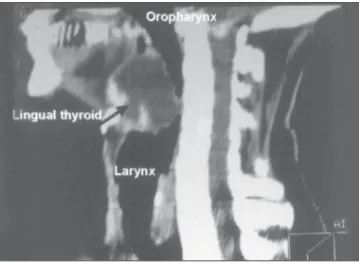
the tongue. In our case report, the computed tomography (CT) scan demonstrated the size of the gland at the base of the tongue, its in-filtration of the muscle and the absence of nor-mal thyroid tissue in the neck (Figure 3). In this situation and in our opinion, an ultra-sound examination is unnecessary.
Although controversial, in small oligosym-ptomatic lingual thyroid glands, clinical treat-ment may be attempted, using suppressive therapy with exogenous thyroid hormone.1,2
The surgical management of lingual thy-roid depends on the severity of the symptoms present. Preoperative tracheostomy and nasotracheal intubation are both effective. In our case, patient intubation was done by means of fiberoptic endoscopy with nasotracheal tube access, thus not requiring any temporary tracheostomy (Figure 4).
Suprahyoid access, or combined
cervi-Figure 1. Thyroid scan with technetium (Tc99m) revealing isotope uptake at the base of the tongue and no uptake in the normal thyroid location (uptake in 2 hours: 5.8%; uptake in 6 hours: 6.3%).
Figure 3. Computed tomography of the larynx, demostrating thyroid absence.
Figure 2. Computed tomography imaging scan demonstrating an oval-shaped mass at the base of the tongue causing sub-occlusion in the oropharynx.
Figure 4. Lingual thyroid in the base of the tongue ready for resection via oral approach (A). The surgical specimen after resection (B).
A B
São Paulo Medical Journal — Revista Paulista de Medicina
69
Tireóide lingual causando disfonia – avaliação e conduta. Apresentação de caso
CONTEXTO: Tireóide lingual é uma entidade rara, devida a falha na migração da glândula em sua fase embriogênica. A presença da glân-dula tireóide na base da língua pode acarretar sérios problemas aos pacientes, como disfagia e disfonia, obstrução de vias aéreas superiores e até hemorragia, em qualquer fase da vida.
RELATO DE CASO: Apresentamos um caso de tireóide lingual em paciente de 41 anos de idade, discutindo-se a embriogênese, diagnós-tico e conduta adequada para tratamento.
○ ○ ○ ○ ○ ○ ○ ○ ○ ○ ○ ○ ○ ○ ○ ○ ○ ○ ○ ○ ○ ○ ○ ○ ○ ○ ○ ○ ○ ○ ○ ○ ○ ○ ○ ○ ○ ○ ○ ○ ○ ○
RESUMO
Alfio José Tincani, MD, PhD. Professor, Discipline of Head and Neck Surgery, Faculdade de Ciências Médicas da Universidade Estadual de Campinas (Unicamp), Campinas, São Paulo, Brazil.
Antonio Santos Martins, MD, PhD. Head of Disci-pline of Head and Neck Surgery, Faculdade de Ciências Médicas da Universidade Estadual de Campinas (Unicamp), Campinas, São Paulo, Brazil.
André Del Negro. Resident of Discipline of Head and Neck Surgery, Faculdade de Ciências Médicas da Universidade Estadual de Campinas (Unicamp), Campinas, São Paulo, Brazil.
Priscila Pereira Costa Araújo. Resident of Discipline of Head and Neck Surgery, Faculdade de Ciências Médicas da Universidade Estadual de Campinas (Unicamp), Campinas, São Paulo, Brazil.
Gilson Barreto, MD. Head and Neck Surgeon, Hospi-tal Centro Médico de Campinas, Campinas, São Paulo, Brazil.
Sources of funding: Not declared
Conflict of interest: Not declared
Date of first submission: February 24, 2003
Last received: July 22, 2003
Accepted:October 5, 2003
Address for correspondence: Alfio José Tincani
Rua Geraldo Trefiglio 140, casa 6A — Jardim Aruan Campinas/SP — Brasil — CEP 13083-793 Tel. (19) 3287 5228 / 9113-0007 E-mail: [email protected]
COPYRIGHT © 2004, Associação Paulista de Medicina ○ ○ ○Publishing information○ ○ ○ ○ ○ ○ ○ ○ ○ ○ ○ ○ ○ ○ ○ ○ ○
Elementos para o diagnóstico e avaliação te-rapêutica são descritos com especial atenção aos achados clínicos, testes laboratoriais, além de metodologia de imagem como medicina nuclear e tomografia computadorizada, rea-lizados para confirmação diagnóstica e pla-nejamento da melhor conduta operatória. A excisão cirúrgica da tireóide ectópica é reser-vada para casos de aumento glandular, que podem resultar em disfunção das vias aéreas superiores (disfonia ou disfagia), além de he-morragia recorrente.
PALAVRAS-CHAVE: Tireóide. Disfonia. Tomografia computadorizada por raio x.
1. Buckland RW, Pedley J. Lingual thyroid – a threat to the air-way. Anaesthesia 2000;55(11):1103-5.
2. Massine RE, Durning SJ, Koroscil TM. Lingual thyroid carci-noma: a case report and review of the literature. Thyroid
○ ○ ○ ○ ○ ○ ○ ○ ○ ○ ○ ○ ○ ○ ○ ○ ○ ○ ○ ○ ○ ○ ○ ○ ○ ○ ○ ○ ○ ○ ○ ○ ○ ○ ○ ○ ○ ○ ○ ○ ○ ○ ○ ○ ○ ○ ○ ○ ○ ○ ○ ○ ○ ○ ○ ○ ○ ○ ○ ○ ○ ○ ○ ○
REFERENCES
2001;11(12):1191-6.
3. Williams JD, Sclafani AP, Slupchinskij O, Douge C. Evalua-tion and management of the lingual thyroid gland. Ann Otol Rhinol Laryngol 1996;105(4):312-6.
4. Zitsman JL, Lala VR, Rao PM. Combined cervical and intraoral approach to lingual thyroid: a case report. Head Neck 1998;20(1):79-82.
cal and intraoral access,1,2,4 is described as excellent for a surgical approach. We achieved good exposure by transoral route without intra or postoperative complica-tions, and this was very important for im-mediate patient recovery, with total excision of the ectopic thyroid.
○ ○ ○ ○ ○ ○ ○ ○ ○ ○ ○ ○ ○ ○ ○ ○ ○ ○ ○ ○
CONCLUSION
Lingual thyroid is a rare entity that may cause serious problems for the patient. When diagnosed in adults, thyroid function tests and radionuclide scanning are essen-tial. Computed tomography scans are
nec-essary for planning surgical intervention and the approach.
Although other types of surgical access have been described,1-4 the transoral ap-proach, in our opinion, provides good ex-posure and is less traumatic for the patient, with better postoperative recovery.