www.reumatologia.com.br
REVISTA BRASILEIRA DE
REUMATOLOGIA
☆ Study conducted at Universidade de São Paulo, São Paulo, SP, Brazil.
* Corresponding author.
E-mail: i [email protected] (F.M. Mello).
0482-5004/$ - see front matter. © 2013 Elsevier Editora Ltda. All rights reserved.
Original article
Gout in the spine
☆Eduardo Massato Hasegawa, Filipe Martins de Mello*, Cláudia Goldenstein-Schainberg,
Ricardo Fuller
Discipline of Rheumatology, Universidade de São Paulo, São Paulo, SP, Brazil
a r t i c l e i n f o
Article history:
Received 29 March 2012 Accepted 24 May 2012
Keywords:
Gout Spine Tophus Radiculopathy
a b s t r a c t
Axial gout can affect all segments of the spine. It is manifested as back pain, as pain associ-ated with neurological symptoms, and as neurological impairment without pain in 17.9%, 75.8% and 4.2% of cases, respectively. These manifestations were the i rst presentation of gout in many patients. Although X-rays as well as computed tomography and especially magnetic resonance scans can be very suggestive, histopathological, cytological and crystal analyses are the diagnostic gold standard. In most cases involving neurological manifesta-tions, the patient underwent surgery, leading to satisfactory results. There are, however, some reports of full recovery following the usual clinical treatment for gout, suggesting that such treatment may be the initial option for those subjects with a history of gout and radiological i ndings of axial involvement.
© 2013 Elsevier Editora Ltda. All rights reserved.
Gota axial
Palavras-chave:
Gota
Coluna vertebral Tofo
Radiculopatia
r e s u m o
A gota axial pode afetar todos os segmentos da coluna vertebral. Ela se manifesta como dor nas costas, dor associada com sintomas neurológicos, e como comprometimento neu-rológico sem dor em 17,9%, 75,8% e 4,2% dos casos, respectivamente. Essas manifestações foram a primeira apresentação da gota em muitos pacientes. Embora radiograi as, bem como tomograi a computadorizada e especialmente ressonância magnética, possam ser muito sugestivos, análises histopatológicas, citológicas e pesquisa de cristais são o padrão ouro de diagnóstico. Na maioria dos casos que envolveram manifestações neurológicas, o paciente foi submetido à cirurgia, levando a resultados satisfatórios. Há, no entanto, alguns relatos de recuperação total após o tratamento clínico habitual para gota, o que sugere que esse tratamento pode ser a opção inicial para os indivíduos com histórico de gota e sinais radiológicos de envolvimento axial.
Introduction
Spinal gout was i rst described by Kersley et al.1 in 1950, and
in 1953 Koskoff et al.2 reported the i rst case of myelopathy
due to gout. Since then, several cases of spinal gout have been reported, with manifestations ranging from an asymptomatic clinical picture to serious complications, such as paraplegia and quadriparesis (see Table 1 with case reports cited). This study reviews the literature related to clinical manifestations, diagnosis and treatment of spinal involvement due to gout.
Methods
A bibliographical search for the terms spine, gout, tophus, and
myelopathy was carried out on PubMed and Medline, and a
selection of articles describing spinal gout, including case re-ports, letters to the editor, radiological i ndings, systematic reviews and observational trials was made. Cross-referencing cases among these reports, that were absent in initial search, were also included in the current review. The i nal number of articles was 94, and a total of 113 subjects were included in these reports. Our search also found two larger studies regard-ing axial gout and radiological image, one with a retrospective design and the other one being a prospective study. No previ-ous reviews regarding this subject included these two studies, and neither had such a comprehensive search for case reports.
Results
Clinical manifestations
The mean age of the 113 patients was 60.3 ± 14.4 years ranging from 17 to 85 years,3,4 and 70.8% were male. A previous history
of gout was observed in 62 patients (65.9% of the reports that mentioned this information), and 31 of them were tophaceous. There was no reference to a history of hyperuricemia and/or gout in 19 (16.8%) patients.
Serum uric acid was measured at the diagnosis in 69 pa-tients and 48 (69.6%) presented with high levels. Renal insuf-i cinsuf-iency was reported insuf-in 25 cases (22.1%) and a previnsuf-ious hinsuf-is- his-tory of renal transplant in another 7 (6.2%).4-6 Alcohol use and
diuretic regimen was reported only in 7 (6.2%) and 12 (10.6%) cases, respectively.
Spinal involvement was the initial manifestation of gout in 28 patients (24.8%). All spinal segments were affected: the lum-bar spine in 66 of the 113 patients (58.4%); the cervical spine in 28 (24.8%); and the thoracic spine in 24 (21.2%). The involve-ment of S1 was observed in 15 cases (13.3%), associated with lumbar spine lesions in 13 (86.7%) of them. Two patients (1.8%) had both cervical and thoracic lesions,7,8 and another four
(3.5%) had simultaneous thoracic and lumbar involvement.5,9-11
Gout may affect any spinal structure, such as intervertebral discs, facet joints, laminae, vertebral bodies, pedicles, ligamen-tum l avum, i lum terminale and the soft tissues adjacent to the spinal column.12
Neurological symptoms were observed in 88 patients (77.9%), and there was an association with cervical, thoracic or
lumbar pain in 80 (90.9%) of these cases. Pain with no neuro-logic symptom was reported in 23 patients (20.4%), 3 (2.7%) in the cervical spine, 1 (0.9%) in the thoracic spine, 20 (17.7%) in the lumbar spine (one case with both cervical and lumbar pain, and one report with both lumbar and thoracic involve-ment) and 1 (0.9%) with pain over the sacrum. Two (1.8%) pa-tients were asymptomatic and were only diagnosed on au-topsy (Table 2).13,14
Radiculopathy (motor dysfunction or dysesthesia along the course of a specii c nerve caused by compression of its root) was the most frequent neurological symptom, occurring in 39 patients (34.5%), followed by claudication in 23 (20.4%), crural paraparesis in 14 (12.4%), quadriparesis in 8 (7.1%), and paraplegia in 5 (4.4%). Atlanto-axial subluxation with the presence of tophus was observed in two cases of cervical pain leading to quadriparesis,15,16 and in one case of multiple
cra-nial nerves palsy.17 Neurologic symptoms without pain were
related in 8 patients (7.1%).
Bladder and/or bowel dysfunction was observed in 10 sub-jects, all of them presenting with other neurological mani-festations, and 5 with back pain. Thirty-four (38.6%) of the 88 cases with neurological impairment exhibited acute onset of symptoms (four weeks or less prior to diagnosis).
Fever higher than 38 °C was reported in 15 patients (10.6%), all of them with elevated erythrocyte sedimentation rate (ESR) and C reactive protein; therefore, exclusion of an infec-tious process was mandatory. Additionally, further 12 cases without fever had elevated ESR.
Imaging studies
As observed by King et al.,18 X-rays may be normal or reveal
soft tissue edema, signs compatible with osteoarthritis (new bone formation and/or reduction of intervertebral space), clearly-dei ned subchondral bone cysts, erosions with scle-rotic borders, erosion of the odontoid process, atlanto-axial subluxation and pathologic fracture.18,19 Among the i ndings
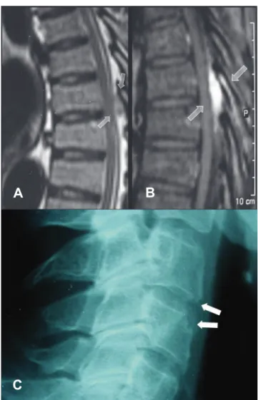
listed above, the most common were those suggestive of os-teoarthritis, seen in 26 (65%) of the 40 reports in which a spi-nal radiography was described. Fig. 1 shows a thoracic radiog-raphy of a patient followed at our clinic due to longstanding gout and thoracic (T7) spinal involvement.
Although not frequently mentioned in the literature, com-puted tomography may show erosions located in facet joints20
and damage to soft tissue with the presence of low-density nodule or mass in some cases.21
In magnetic resonance imaging (MRI), tophaceous gout is usually characterized by a homogeneous image with a signal ranging from intermediate to low on T1 (with the same signal intensity as muscle), and on T2, the image appears homoge-neous and may show low or high intensity.22-25 When contrast
(gadolinium) is used, peripheral heterogeneous or homoge-neous contrast enhancement may occur, revealing reactive vascularization.22,26 Fig. 2 shows the MRI of the lumbar spine
of the same patient described above.
Table 1 – Spinal gout case reports and series reviewed but not cited in the text.
No. Authors No. of cases reported
Year Reference
1 Adenwalla HN, Usman MH, Bagir M et al. 1 2007 South Med J. 2007 Apr;100(4):413-4 2 Arnold MH, Brooks PM, Savvas P et al. 1 1988 Aust N Z J Med. 1988 Dec;18(7):865-7 3 Barrett K, Miller ML, Wilson JT 1 2001 Neurosurgery. 2001 May;48(5):1170-2 4 Beier CP, Hartmann A, Woertgen C et al. 1 2005 J Neurosurg Spine. 2005 Dec;3(6):485-7
5 Bonaldi VM, Duong H, Starr MR et al. 1 1996 AJNR Am J Neuroradiol. 1996 Nov-Dec;17(10):1949-52 6 Burnham J, Fraker K, Steinbach H 1 1977 AJR Am J Roentgenol. 1977 Dec;129(6):1116-9 7 Cabot J, Mosel L, Kong A et al. 1 2005 Skeletal Radiol. 2005 Dec;34(12):803-6 8 Chan AT, Leung JL, Sy AN 1 2009 Hong Kong Med J. 2009 Apr;15(2):143-5
9 Chang IC 4 2005 Clin Orthop Relat Res. 2005 Apr(433):106-10
10 Clerc D, Marfeuille M, Labous E et al. 1 1998 Clin Exp Rheumatol. 1998 Sep-Oct;16(5):621
11 Das De S 1 1988 J Bone Joint Surg Br. 1988 Aug;70(4):671
12 Dhote R, Roux FX, Bachmeyer C et al. 1 1997 Clin Exp Rheumatol. 1997 Jul-Aug;15(4):421-3 13 Diaz A, Porhiel V, Sabatier P 1 2003 Neurochirurgie. 2003 Dec;49(6):600-4 14 Draganescu M, Leventhal LJ 1 2004 J Clin Rheumatol. 2004 Apr;10(2):74-9 15 Duprez TP, Malghem J, Vande Berg BC et al. 1 1996 AJNR Am J Neuroradiol. 1996 Jan;17(1):151-3 16 El Sandid M, Ta H 1 2004 Ann Intern Med. 2004 Apr 20;140(8):W32 17 Ferreira A, Silva Junior BA, Braga FM et al. 1 1989 Arq Neuropsiquiatr. 1989 Dec;47(4):479-83 18 Fontenot A, Harris P, Macasa A et al. 1 2008 J Clin Rheumatol. 2008 Jun;14(3):188-9 19 Gines R, Bates DJ. 1 1998 Am J Emerg Med. 1998 Mar;16(2):216 20 Hasegawa EM, Goldenstein-Schainberg C, Fuller R 1 2007 Rev Bras Reumatol. 2007 Jul-Aug;47(4):300-2 21 Jacobs SR, Edeiken J, Rubin B et al. 1 1985 Arch Phys Med Rehabil. 1985 Mar;66(3):188-90 22 Justiniano M, Colmegna I, Cuchacovich R 1 2007 J Rheumatol. 2007 May;34(5):1157-8
23 Kao MC, Huang SC, Chiu CT et al. 1 2000 J Formos Med Assoc. 2000 Jul;99(7):572-5 24 Kelly J, Lim C, Kamel M et al. 1 2005 J Neurosurg Spine. 2005 Feb;2(2):215-7 25 Kern A, Schunk K, Thelen M 1 1999 Rofo. 1999 May;170(5):515-7
26 Ko KH, Huang GS, Chang WC 1 2009 Arthritis Rheum. 2009 Jan;60(1):198 27 Ko KH, Huang GS, Chang WC 1 2010 J Clin Rheumatol. 2010 Jun;16(4):200
28 Ko PJ, Huang TJ, Liao YS et al. 1 1996 Changgeng Yi Xue Za Zhi. 1996 Sep;19(3):272-6 29 Lam HY, Cheung KY, Law SW et al. 4 2007 J Orthop Surg (Hong Kong). 2007 Apr;15(1):94-101 30 Leaney BJ, Calvert JM 1 1983 J Neurosurg. 1983 Apr;58(4):580-2
31 Lievre JA, Leroux-Robert J, Bacri J 1 1961 Presse Med. 1961 Jul 8;69:1525-6 32 Litvak J, Briney W 1 1973 J Neurosurg. 1973 Nov;39(5):656-8 33 Miller JD, Percy JS 1 1984 J Rheumatol. 1984 Dec;11(6):862-5 34 Niva M, Tallroth K, Konttinen YT 1 2006 Clin Exp Rheumatol. 2006 Jan-Feb;24(1):112 35 Nygaard HB, Shenoi S, Shukla S 1 2009 Neurology. 2009 Aug 4;73(5):404
36 Paquette S, Lach B, Guiot B 1 2000 Neurosurgery. 2000 Apr;46(4):986-8
37 Peeters P, Sennesael J 1 1998 Nephrol Dial Transplant. 1998 Dec;13(12):3245-7 38 Pi ster AK, Schlarb CA, O'Neal JF 1 1998 AJR Am J Roentgenol. 1998 Nov;171(5):1430-1 39 Reynolds AF, Jr., Wyler AR, Norris HT 1 1976 Arch Neurol. 1976 Nov;33(11):795
40 Riddell CM, Elliott M, Cairns AP 1 2008 J Rheumatol. 2008 Oct;35(10):2076-7 41 Sabharwal S, Gibson T 1 1988 Br J Rheumatol. 1988 Oct;27(5):413-4 42 Saketkoo LA, Robertson HJ, Dyer HR et al. 1 2009 J Med Sci. 2009 Aug;338(2):140-6 43 Samuels J, Keenan RT, Yu R et al. 1 2010 Bull NYU Hosp Jt Dis. 2010;68(2):147-8 44 Sequeira W, Bouffard A, Salgia K et al. 1 1981 Arthritis Rheum. 1981 Nov;24(11):1428-30 45 Souza AW, Fontenele S, Carrete H et al. 1 2002 Clin Exp Rheumatol. 2002 Mar-Apr;20(2):228-30 46 St George E, Hillier CE, Hati eld R 1 2001 Rheumatology (Oxford). 2001 Jun;40(6):711-2 47 Staub-Schmidt T, Chaouat A, Rey et al. 1 1995 Arthritis Rheum. 1995 Jan;38(1):139-41 48 Suk KS, Kim KT, Lee SH et al. 1 2007 Spine J. 2007 Jan-Feb;7(1):94-9
49 Tkach S 1 1970 Clin Orthop Relat Res. 1970;71:81-6
50 Vaccaro AR, An HS, Cotler JM et al. 1 1993 Orthopedics. 1993 Nov;16(11):1273-6 51 van de Laar MA, van Soesbergen RM, Matricali B 1 1987 Arthritis Rheum. 1987 Feb;30(2):237-8 52 Varga J, Giampaolo C, Goldenberg DL 1 1985 Arthritis Rheum. 1985 Nov;28(11):1312-5 53 Vervaeck M, De Keyser J, Pauwels P et al. 1 1991 Clin Neurol Neurosurg. 1991;93(3):233-6 54 Vinstein AL, Cockerill EM 1 1972 Radiology. 1972 May;103(2):311-2 55 Wald SL, McLennan JE, Carroll RM et al. 1 1979 J Neurosurg. 1979 Feb;50(2):236-9 56 Wang LC, Hung YC, Lee EJ et al. 1 2001 J Formos Med Assoc. 2001 Mar;100(3):205-8 57 Yasuhara K, Tomita Y, Takayama A et al. 1 1994 J Spinal Disord. 1994 Feb;7(1):82-5
The most common i ndings in imaging study are listed in Table 3.
Diagnosis
In 103 of the 113 cases (91.2%), a diagnosis was achieved through cytological or histopathological studies. A histologi-cal study of the tissue removed during the surgihistologi-cal excision of the lesion or decompressive laminectomy was performed in 87 patients (77%). Guided puncture was done in 16 patients (14.2%), biopsy being unnecessary; and an open biopsy was performed in one (0.9%).27 A pasty, chalky-white mass was
usually observed during the surgery.
In seven patients (6.2%),8,17,23,24,28-30 histological or
cytologi-cal tests were not performed, and diagnosis was presumed based on clinical and imaging i ndings or on arthrocentesis of
A
B
C
Fig. 1 – (A and B) MRI of thoracic spine on lateral view. Note the presence of a low signal lesion in T1 indicated by the arrows (A) before and (B) after contrast administration, with a homogeneous enhancement. (from: Hasegawa EM, Goldenstein-Schainberg C, Fuller R. Rev Bras Reumatol. 2007;47(4):300-2). (C) Lateral radiography of cervical spine in a patient with chronic tophaceous gout. The arrows show erosions in the anterior aspects of the vertebral bodies.
other joints. In another four cases (3.5%) diagnosis was made during autopsy,1,2,13,14 but none of them died because of spinal
involvement by gout.
In 17 patients who reported pain without neurological manifestation, guided puncture or surgical intervention was performed based on the presence of abnormalities in CT and/ or MRI (mass lesion), and eventually fever and elevation in ESR at onset.
The histological description includes classic gout aspects, such as the presence of histiocytes and multinucleated giant cells and i broblasts surrounding eosinophilic debris or amor-phous material, and may contain needle-shaped crystals with negative birefringence under polarized light. Occasion-ally, only the negative image of the crystals is observed in the neutrophils, as they are dissolved during i xation in aqueous medium.31-33
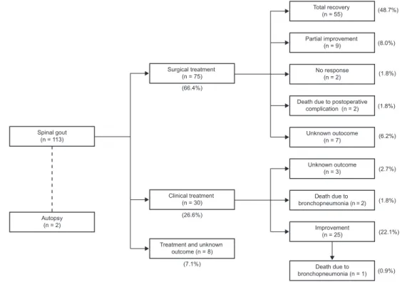
Treatment
Among the 88 patients with neurological symptoms, 74 (84.1%) underwent surgery. The most common procedure was decom-pressive laminectomy. Full recovery of neurological manifes-tations was observed in 55 of 74 patients (74.3%); partial re-covery followed surgery in nine, and two patients31,34 reported
no recovery after surgery, although one of them34 improved
after subsequent clinical treatment with non-steroidal anti-inl ammatory (NSAIDs) and hypouricemic drugs. In another six cases there was no follow-up description and thus no data about response to surgery could be retrieved. Only one pa-tient without neurological symptoms was managed surgically but there was no mention to outcomes. Two patients died in the post operatory status due to bronchopneumonia,15,28 one
of them had not shown any improvement, while the other was actually getting better from the neurological symptoms before the infectious complication.
Twenty-i ve patients (22.1%) received clinical treatment alone (NSAIDs, colchicines and oral, intravenous or epidural corticosteroid) with recovery from neurological dei cits and/ or pain. In three cases managed conservatively the outcome was not reported.6,23,35 Three patients who did not undergo
surgery died from bronchopneumonia,1,9,17 one of them had
been improving from the symptoms related to gout, whilst the other two had not. In eight cases there was no mention to management and outcome. Treatment outcomes are sum-marized in Fig. 2.
Discussion
Spinal involvement in gout arthritis is increasingly recog-nized as an unusual manifestation; however its prevalence is clearly underestimated because only those patients with neurological dei cits and/or fever, and those who do not im-prove with clinical treatment are investigated with imaging and subsequent histopathological studies to coni rm the di-agnosis.
Fig. 2 – Treatment and outcome summary.
Total recovery (n = 55)
Surgical treatment (n = 75)
Spinal gout (n = 113)
Autopsy (n = 2)
Clinical treatment (n = 30)
Treatment and unknown outcome (n = 8)
(66.4%)
(26.6%)
(7.1%)
(0.9%) (22.1%) (1.8%) (2.7%) (6.2%) (1.8%) (1.8%) (8.0%) (48.7%)
Death due to bronchopneumonia (n = 1)
Partial improvement (n = 9)
No response (n = 2)
Death due to postoperative complication (n = 2)
Unknown outocome (n = 7)
Unknown outcome (n = 3)
Death due to bronchopneumonia (n = 2)
Improvement (n = 25)
Table 2 – Clinical manifestations.
Symptoms n %
Neurologic dei cit with back pain 80
Radiculopathy 39 34.5
Claudication 23 20.4
Paraparesis 14 12.4
Tetraparesis 8 7.1
Paraplegia 5 4.4
Pain without neurologic dei cit 23 20.4
Cervical 3 2.7
Thoracic 1 0.9
Lumbar 20 17.7
Sacral 1 0.9
Neurologic dei cit only 8 7.1
Unknown* 2 1.8
* Diagnosis made by autopsy
Table 3 – Imaging fi ndings in spinal gout.
Method Findings
X – Ray Osteoarthritic changes: osteophytes, subchondral cysts and bony sclerosis
CT Osteoarthritic changes Low-density mass Facet joint erosion
MRI T1 – homogeneous, low to intermediate signal T2 – homogeneous, low to high signal
Contrast (Gadolinium): homogeneous, peripheral enhancement
Normal bone marrow signal of the adjacent vertebrae
CT, computed tomography; MRI, magnetic resonance imaging.
alterations in gout might be much more common than previ-ously thought. Konatalapalli et al.36 retrospectively reviewed
64 CT images of the spine from patients with gout, and found out that 14% of them presented features of spinal gout. The same research group performed a prospective study in which 48 subjects with a diagnosis of gout had a spinal CT scan.37
Thirty-i ve percent of the patients had CT scan evidence of spinal gout erosions or tophi. Since these two studies were not case reports and therefore did not mention any individual clinical info on each patient, they were not included in our analysis.
Axial gout should be considered in the differential diag-nosis of patients with previous diagdiag-nosis of gout or a history of hyperuricemia who present symptoms suggesting spinal cord involvement and lumbar or cervical pain. Although usu-ally not mentioned in many case reports, risk factors for the
development of acute gout such a as renal failure, drugs (di-uretics, low-dose aspirin), diet, alcohol intake, and infection should also be considered for the presumptive diagnosis of spinal gout.
The mechanism associated with axial gout is not yet clear. However, it is assumed that the same factors leading to the peripheral picture such as pH, temperature, trauma and joint degeneration are involved in crystal deposits.33,38 Finally, the
presence of spinal osteoarthritis perhaps also facilitates fur-ther crystal deposition.
In patients with no history of gout or hyperuricemia, the diagnosis may be presumed from CT scan, MRI study, clinical and laboratorial i ndings, previous medical history, and as-sociated risk factors. Plain radiography is a relatively limited diagnostic resource.
especially in patients without history of gout and/or those with signs and symptoms of red l ags for back pain. The sam-ple should be preserved in alcohol to prevent urate crystals dissolution.33
Patients without neurological involvement may initially be treated with NSAIDs, as indicated in acute gout attacks,39,40
and subsequently with hypouricemic drugs. In the presence of neurological symptoms, clinical treatment may also be tried, as it leads to improvement in some patients. The favor-able response to clinical treatment alone may suggest that the inl ammatory process, rather than a compression due to tophi itself, should be the main mechanism for the develop-ment of symptoms.
In a previous review of spinal gout made by Houet al.,22
af-ter an initial clinical, laboratorial and imaging evaluation, bi-opsy is suggested as dei nite diagnosis procedure. If there are progressive neurologic dei cits, surgery may be preferred. If biopsy coni rms the diagnosis of gout, conservative treatment may be tried, and when symptoms persist or recur after initial improvement, surgery must be performed. We recommend guided puncture rather than open biopsy in those patients without severe or progressive neurological involvement, and in those with pain as sole manifestation that did not improve with clinical treatment.
In conclusion, gout should be included in the differential diagnosis of acute spinal pain episodes, associated or not with neurological manifestations in patients with a history of gout and hyperuricemia. In the cases without or with mild to moderate neurological manifestations, we recommend guided puncture as initial diagnostic procedure, and conser-vative treatment with NSAIDs and/or corticosteroids. Sur-gery must be reserved for those patients with no improve-ment or those with progressive neurologic dei cits, despite clinical treatment.
R E F E R E N C E S
1. Kersley GD, Mandel L, Jeffrey MR. Gout; an unusual case with
softening and subluxation of the i rst cervical vertebra and splenomegaly. Ann Rheum Dis. 1950;9(4):282-304.
2. Koskoff YD, Morris LE, Lubic LG. Paraplegia as a complication of gout. J Am Med Assoc. 1953;152(1):37-8.
3. Lagier R, Mac Gee W. Spondylodiscal erosions due to gout: anatomico-radiological study of a case. Ann Rheum Dis. 1983;42(3):350-3.
4. Wald SL, McLennan JE, Carroll RM, Segal H. Extradural spinal involvement by gout. Case report. J Neurosurg. 1979;50(2):236-9.
5. Hausch R, Wilkerson M, Singh E, Reyes C, Harrington T. Tophaceous gout of the thoracic spine presenting as back pain and fever. J Clin Rheumatol. 1999;5(6):335-41.
6. Thornton FJ, Torreggiani WC, Brennan P. Tophaceous gout of the lumbar spine in a renal transplant patient: a case report and literature review. Eur J Radiol. 2000;36(3):123-5.
7. Magid SK, Gray GE, Anand A. Spinal cord compression by tophi in a patient with chronic polyarthritis: case report and literature review. Arthritis Rheum. 1981;24(11):1431-4. 8. Coulier B, Tancredi MH. Articular tophaceous gout of the
cervical spine: CT diagnosis. JBR-BTR. 2010;93(6):325. 9. Marinho F, Zeitoun-Eiss D, Renoux J, Brasseur JL, Genestie
C, Grenier P. Tophaceous gout of the spine: Case report and
review of the literature. J Neuroradiol. 2011 [Epub ahead of print].
10. Tsai CH, Chen YJ, Hsu HC, Chen HT. Bacteremia coexisting with tophaceous gout of the spine mimicking spondylodiscitis: a case report. Spine (Phila Pa 1976). 2009;34(2):E106-9.
11. Nygaard HB, Shenoi S, Shukla S. Lower back pain caused by tophaceous gout of the spine. Neurology. 2009;73(5):404. 12. Hasturk AE, Basmaci M, Canbay S, Vural C, Erten F. Spinal
gout tophus: a very rare cause of radiculopathy. Eur Spine J. 2011 [Epub ahead of print].
13. Levin MH, Lichtenstein L, Scott HW. Pathologic changes in gout; survey of eleven necropsied cases. Am J Pathol. 1956;32(5):871-95.
14. Hall MC, Selin G. Spinal involvement in gout: case report with autopsy. J Bone Joint Surg Am. 1960;42:341-3.
15. Wazir NN, Moorthy V, Amalourde A, Lim HH. Tophaceous gout causing atlanto-axial subluxation mimicking rheumatoid arthritis: a case report. J Orthop Surg (Hong Kong). 2005;13(2):203-6.
16. Thavarajah D, Hussain R, Martin JL. Cervical arthropathy caused by gout: stabilisation without decompression. Eur Spine J. 2011;20(Suppl 2):S231-4.
17. Tran A, Prentice D, Chan M. Tophaceous gout of the odontoid process causing glossopharyngeal, vagus, and hypoglossal nerve palsies. Int J Rheum Dis. 2011;14(1):105-8.
18. King JC, Nicholas C. Gouty arthropathy of the lumbar spine: a case report and review of the literature. Spine (Phila Pa 1976). 1997;22(19):2309-12.
19. Bret P, Ricci AC, Saint-Pierre G, Mottolese C, Guyotat J. [Thoracic spinal cord compression by a gouty tophus. Case report. Review of the literature]. Neurochirurgie. 1999;45(5):402-6.
20. Fenton P, Young S, Prutis K. Gout of the spine. Two case reports and a review of the literature. J Bone Joint Surg Am. 1995;77(5):767-71.
21. Feydy A, Liote F, Carlier R, Chevrot A, Drape JL. Cervical spine and crystal-associated diseases: imaging i ndings. Eur Radiol. 2006;16(2):459-68.
22. Hou LC, Hsu AR, Veeravagu A, Boakye M. Spinal gout in a renal transplant patient: a case report and literature review. Surg Neurol. 2007;67(1):65-73.
23. Hsu CY, Shih TT, Huang KM, Chen PQ, Sheu JJ, Li YW. Tophaceous gout of the spine: MR imaging features. Clin Radiol. 2002;57(10):919-25.
24. Miller LJ, Pruett SW, Losada R, Fruauff A, Sagerman P. Clinical image. Tophaceous gout of the lumbar spine: MR i ndings. J Comput Assist Tomogr. 1996;20(6):1004-5.
25. Popovich T, Carpenter JS, Rai AT, Carson LV, Williams HJ, Marano GD. Spinal cord compression by tophaceous gout with l uorodeoxyglucose-positron-emission tomographic/MR fusion imaging. AJNR Am J Neuroradiol. 2006;27(6):1201-3. 26. Yen PS, Lin JF, Chen SY, Lin SZ. Tophaceous gout of the lumbar
spine mimicking infectious spondylodiscitis and epidural abscess: MR imaging i ndings. J Clin Neurosci. 2005;12(1):44-6. 27. Nakajima A, Kato Y, Yamanaka H, Ito T, Kamatani N. Spinal
tophaceous gout mimicking a spinal tumor. J Rheumatol. 2004;31(7):1459-60.
28. Dharmadhikari R, Dildey P, Hide IG. A rare cause of spinal cord compression: imaging appearances of gout of the cervical spine. Skeletal Radiol. 2006;35(12):942-5. 29. Buenzli D, So A. Inl ammatory sciatica due to spinal
tophaceous gout. BMJ Case Rep. 2009. doi:10.1136/ bcr.07.2008.0492.
30. Schorn C, Behr C, Schwarting A. [Fever and back pain--a case report of spinal gout]. Dtsch Med Wochenschr. 2010;135(4):125-8.
32. Kuo YJ, Chiang CJ, Tsuang YH. Gouty arthropathy of the cervical spine in a young adult. J Chin Med Assoc. 2007;70(4):180-2.
33. Becker MA. Clinical manifestations and diagnosis of gout. UpToDate (v.19.1); 2011 [updated February 13, 2011; cited 2010 January 11]; Available from: www.uptodateonline.com. 34. Mekelburg K, Rahimi AR. Gouty arthritis of the spine:
clinical presentation and effective treatments. Geriatrics. 2000;55(4):71-4.
35. Alarcon GS, Reveille JD. Gouty arthritis of the axial skeleton including the sacroiliac joints. Arch Intern Med. 1987;147(11):2018-9.
36. Konatalapalli RM, Demarco PJ, Jelinek JS, Murphey M, Gibson M, Jennings B, et al. Gout in the axial skeleton. J Rheumatol. 2009;36(3):609-13.
37. Konatalapalli RM, Lumezanu E, Jelinek JS, Murphey M, Carter E, Weinstein A. A Prospective Studyof Correlates of Axial Gout. ACR/ARHP 2010 Scientii c Meeting; Atlanta, USA2010. 38. Choi HK, Mount DB, Reginato AM. Pathogenesis of gout. Ann
Intern Med. 2005;143(7):499-516.
39. Cannella AC, Mikuls TR. Understanding treatments for gout. Am J Manag Care. 2005;11(15 Suppl):S451-8; quiz S65-8. 40. Terkeltaub RA. Clinical practice. Gout. N Engl J Med.