1093 Arq Neuropsiquiatr 2009;67(4):1093-1096
Letter
AlicAtA DiSEASE
Neuroinfestation by
Angiostrongylus cantonensis
in Recife, Pernambuco, Brazil
Ana Rosa Melo Correa Lima
1, Solange Dornelas Mesquita
2, Silvana Sobreira Santos
1,
Eduardo Raniere Pessoa de Aquino
1, Luana da Rocha Samico Rosa
3, Fábio Souza Duarte
3,
Alessandra Oliveira Teixeira
1, Zenize Rocha da Silva Costa
4, Maria Lúcia Brito Ferreira
5DOENÇA DE AlicAtA: NEUROiNFEStAÇÃO POR Angiostrongylus cantonensis EM REciFE, PERNAMBUcO, BRASil.
Neurology Service of Hospital da Restauração, Recife PE, Brazil: 1Neurologist; 2Neurologist and Liquorologist; 3Neurology Resident; 4Nurse Coordinator of Meningitis Program of Health Department of Pernambuco State, Recife PE, Brazil; 5Chairwoman Neurologist.
Received 3 July 2009, received in inal form 4 August 2009. Accepted 24 September 2009.
Dra. Maria Lúcia Brito Ferreira – Rua Neto de Mendonça 230 / 802 - 52050-100 Recife PE - Brasil. E-mail: lucabrito@uol.com.br Angiostrongylus cantonensis, is a nematode in the
Secernentea class, Strongylidae order, Metastrongylidæ superfamily and Angiostrongylidæ family1, and is the most common cause of human eosinophilic meningi-tis worldwide. This parasite has rats and other mammals as definitive hosts and snails, freshwater shrimp, fish, frogs and monitor lizards as intermediate hosts1. Mam-mals are infected by ingestion of intermediate hosts and raw/undercooked snails or vegetables, contain-ing third-stage larvae2. Once infested, the larvae pen-etrate the vasculature of the intestinal tract and pro-mote an inflammatory reaction with eosinophilia and lymphocytosis. This produces rupture of the blood-brain barrier, changes to nervous tissue and damage to the Purkinje cells in the cerebellum, thereby promot-ing eosinophilic menpromot-ingoencephalitis or Alicata disease3. A. cantonensis has been found in Southeast Asia and the South Paciic, where it is endemic2, as well as in Af-rica, India, Caribbean, Australia, North AmeAf-rica, Jamaica, Haiti, Cuba, Puerto Rico3 and Brazil4.
cASE
A female from Olinda, Pernambuco, Brazil, who was born on April 27, 1982, was seen in the neurological emergency room of Hospital da Restauração on May 24, 2008. The person
accom-panying the patient reported that she had presented a rash as-sociated with joint pain, followed by progressive dificulty in walking for 30 days, which was associated with sleepiness over the last 15 days.
In the patient’s past history, there were references to mental retardation and lack of ability to understanding simple orders. She presented independent gait and had frequently run away from home into the surrounding area. There was mention of in-voluntary movements, predominantly of the upper limbs, which intensiied after the change of health status that motivated the current search for medical assistance. In November 2007, the pa-tient presented with generalized tonic-clonic seizures and was medicated with carbamazepine, 200 mg/twice a day.
On clinical examination, the patient was febrile and had ery-thema and heat in her right knee. On neurological examination, the patient was comatose, with Glasgow index 8, tonic deviation of the head and gazing to the right, associated with intermittent masticatory movements, suggestive of seizures. There were also choreoathetotic movements of the upper limbs. The ocular fun-di were normal. There was no resistance to neck movement. She presented laccid paraplegia, which was associated with Ash-worth grade 2 hypertonia in the upper limbs that was more ev-ident on the right side. Relex examination showed absence of knee relex, normal adductor and Achilles relex, indifferent cu-taneous-plantar relex and brisk upper-limb relex.
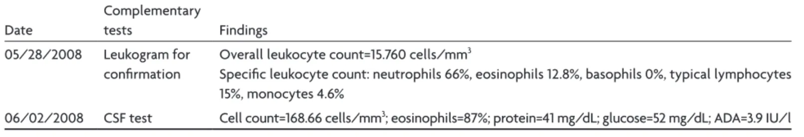
Table 1. Results from additional tests for diagnosis.
Date
Complementary
tests Findings 05/28/2008 Leukogram for
conirmation
Overall leukocyte count=15.760 cells/mm3
Speciic leukocyte count: neutrophils 66%, eosinophils 12.8%, basophils 0%, typical lymphocytes 15%, monocytes 4.6%
Arq Neuropsiquiatr 2009;67(4)
1094
Alicata disease at Recife, Brazil: Angiostrongylus cantonensis
Lima et al.
The overall leukocyte count on admission was 13,380 cells/ mm³, consisting of: 48.9% neutrophils, 20% eosinophils, 0% ba-sophils, 22.4% typical lymphocytes and 8.4% monocytes.
The diagnostic hypotheses, based on the history, neurologi-cal examination and leukocyte count were meningo-encephalo-radiculoneuritis of infectious or inlammatory etiology, or acute disseminated encephalomyelitis (ADEM). The patient was admit-ted to the ward for more detailed investigation by means of ad-ditional tests (Table 1).
The spinal luid eosinophils associated with marked eosino-philia provided the basis for diagnosing neuroinfestation, for which new additional tests were requested (Table 2).
The patient’s symptoms worsened and she was transferred to the intensive care unit, where she received assisted ventilation.
In parallel, through the Ministry of Health, the Epidemiolog-ical Surveillance Division of the Environmental Health Depart-ment of Pernambuco was mobilized to carry out an active search in the vicinity of the patient’s home. The surveillance technicians located and collected adult snails of the species Achantina fu-lica. The snails were sent to Fiocruz, Rio de Janeiro, Brazil, for infestations to be investigated. A sample of the patient’s cere-brospinal luid was sent to the Molecular Parasitology Laborato-ry and Parasitic Biology LaboratoLaborato-ry of the Institute of Biomedi-cal Research and School of Biosciences, Pontifícia Universidade Católica of Rio Grande do Sul, for further investigation.
The results and the inding of snails suggested the diagnos-tic hypothesis of parasidiagnos-tic eosinophilic meningo-encephalo-ra-diculoneuritis. Treatment with methylprednisolone 1 g/day for 5 days, and ivermectin 6 mg/ 2 tablets/day in a single dose was
instituted. Carbamazepine was replaced by valproic acid 500 mg, 2 tablets/day, because of the possibility of drug interaction.
The results from the additional tests for clinical follow-up are shown in Table 3.
On August 4, 2008, the diagnosis of neuroinfestation by An-giostrongyluscantonensis was made because of positive indings in CSF shown by the real-time polymerase chain reaction.
The patient presented an improvement in clinical conscious-ness level, and her knee relex reappeared. However, the laccid paraplegia remained, with accentuation of the previous chore-oathetotic movements and ankylosis in the right knee, with a limitation of 90 degrees of lexion.
To investigate the possibility of nerve root involvement, be-cause of the presence of arelexia and laccid paraplegia, the pa-tient underwent upper and lower-limb electromyography. This was suggestive of subacute symmetrical sensory-motor axonal polyneuropathy, associated with active reinnervation and radic-ular involvement, as shown by lumbosacral paravertebral mus-cle fasciculation and ibrillation.
The patient then presented clinical worsening and died on October 5, 2008.
DiScUSSiON
The clinical manifestations of angiostrongyloidiasis are similar to the symptoms and signs of meningitis or radic-ulomyeloencephalitis. For example, there may be head-ache, vomiting, paresthesia, weakness and, occasionally, visual disturbances and paralysis of the ocular extrinsic muscles2,5. As noted in this case, eosinophilic pleocytosis
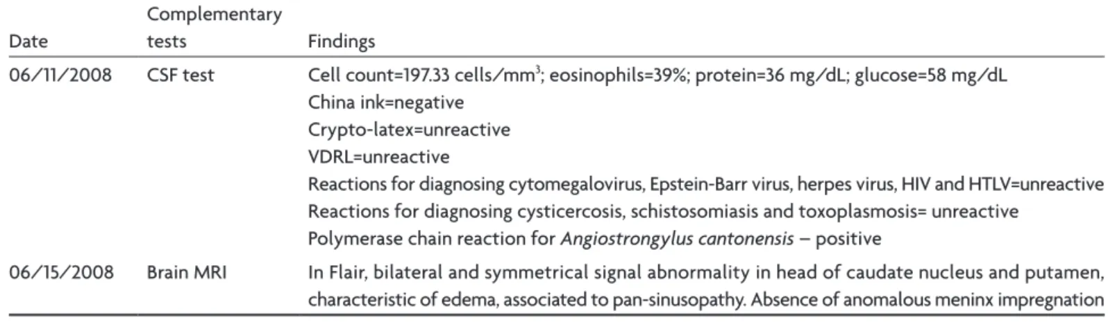
Table 2. Results of the second battery of complementary tests for diagnosis.
Date
Complementary
tests Findings
06/11/2008 CSF test Cell count=197.33 cells/mm3; eosinophils=39%; protein=36 mg/dL; glucose=58 mg/dL
China ink=negative Crypto-latex=unreactive VDRL=unreactive
Reactions for diagnosing cytomegalovirus, Epstein-Barr virus, herpes virus, HIV and HTLV=unreactive Reactions for diagnosing cysticercosis, schistosomiasis and toxoplasmosis= unreactive
Polymerase chain reaction for Angiostrongylus cantonensis – positive
06/15/2008 Brain MRI In Flair, bilateral and symmetrical signal abnormality in head of caudate nucleus and putamen, characteristic of edema, associated to pan-sinusopathy. Absence of anomalous meninx impregnation
Table 3. Results of the third battery of additional tests for clinical follow-up.
Date
Complementary
tests Findings
07/02/2008 CSF test Cell count=10.66 cells/mm3; eosinophils=0%; protein=39 mg/dL; glucose=63 mg/dL
08/08/2008 CSF test Cell count=24 cells/mm3; eosinophils=36%; protein=33 mg/dL; glucose=59 mg/dL; ADA=1.7 IU/l
Arq Neuropsiquiatr 2009;67(4)
1095
Alicata disease at Recife, Brazil: Angiostrongylus cantonensis
Lima et al.
is associated with this because of parasite migration to the spinal cord and cranial nerves5. This occurs under the action of interleukin 5, which stimulates the maturation of eosinophil function and acts as a potent and selective eosinophil chemotaxis factor6.
Spinal luid eosinophils associated with a history of exposure to larval infestation should be the basis of the presumptive diagnosis of eosinophilic meningoencepha-litis. This is deined as the presence of 10 or more eosino-phils/mm3 in CSF or spinal luid eosinophils represent-ing at least 10% of the total spinal luid leukocytes. The causes of eosinophilic meningoencephalitis may be par-asitic, fungal, bacterial, viral, rickettsial, malignant, drug-related or hypereosinophilic. This range of causes justi-ied the additional tests required in this case. The results from the tests ruled out the non-infectious and infec-tious hypotheses, and left the identiication of the par-asite agent.
In this case, magnetic resonance images only rein-forced the clinical evidence of meningoencephalitis. This corroborated the indings of Jin et al.6 that MRI is not spe-ciic and that clinical and epidemiological characteristics should be considered more important. However, we must emphasize that MRI changes may relect edema caused by the immune process, triggered by the presence of parasite larvae in the brain. This process includes: (a) increased syn-thesis of eotaxin (especially eotaxin-2), a chemokine that increases blood eosinophil chemotaxis to the brain, (b) in-trathecal synthesis of IgE that attaches to the wall of L3-stage larvae and facilitates the binding of eosinophils that secrete enzymes to destroy the parasite7, (c) eosinophilic enzyme action on nervous tissue, stimulating edema8.
Because there was mention in the patient’s history of leaks around the home, we sought to investigate the pres-ence of Achantina fulica in the vicinity, since this mollusc is the parasite’s deinitive host. The positive inding from the polymerase chain reaction for A. cantonensis in CSF and the isolation of parasites in the snails (Figure) helped to establish a deinitive diagnosis.
It should be emphasized that notifying the Environ-mental Surveillance Division regarding this neuroinfesta-tion was important as a preventive public health measure for the community where the patient lived, given the se-verity of this infestation and the possibility that the par-asite might spread.
The hypotheses of infectious or inlammatory menin-go-encephalo-radiculoneuritis, or acute disseminated en-cephalomyelitis (ADEM), led us to administer a methyl-prednisolone pulse, in an attempt to reduce the inlamma-tory process and improve the patient’s neurological symp-toms. An oral ivermectin regimen was instituted, because Brazil is a country with high prevalence of strongyloidia-sis. The latter is caused by an intestinal parasite that can be exacerbated by the use of steroids, through two mech-anisms: activation of the parasite’s ecdysteroid receptors, thereby increasing its virulence; or reduction of the im-mune response mediated by T cells, thereby facilitating the spread of the parasite to the central nervous system9. Ivermectin is the drug of choice for treating strongyloid-iasis because it has fewer adverse effects than shown by albendazole or thiabendazole, and higher rates of larval clearance10. This aspect of treatment deserves emphasis, because it helped the patient, since the ivermectin is also indicated for treating infestation by A. cantonensis.
The methylprednisolone pulse and ivermectin admin-istration may have been responsible for the absence of spinal luid eosinophils in subsequent CSF tests, as a leet-ing effect and transient improvement, because high lev-els of spinal luid eosinophils appeared again in other CSF tests, at levels compatible with previous levels.
Arq Neuropsiquiatr 2009;67(4)
1096
Alicata disease at Recife, Brazil: Angiostrongylus cantonensis
Lima et al.
ingitis. Eosinophilic encephalitis affects less than 10% of cases and is usually fatal11, which may explain this death.
In summary, this report of meningo-encephalo-radicu-loneuritis due to A. cantonensis in northeastern Brazil was the inding of the etiological agent in infested snails. The main objective of this report was to alert general physi-cians and neurologists to the need to pay special atten-tion to spinal luid eosinophil counts or marked blood eo-sinophilia. This is especially important in the presence of neurological symptoms and signs associated with the epi-demiological aspects of the patient‘s environment, which may facilitate the identiication of the etiological agent.
REFERENcES
1. Hüttemann M, Schmahl G, Mehlhorn H. Light and electron microscop-ic studies on two nematodes, Angiostrongylus cantonensis and Trichuris muris, differing in their mode of nutrition. Parasitol Res 2007;103(Suppl 2):S225-S232.
2. Baheti NN, Sreedharan M, Krishnamoorthy T, Nair MD, Radhakris-hanan K. Eosinophilic meningitis and an ocular worm in a patient from Kerala, South India. J Neurol Neurosurg Psychiatry 2008;79:271.
3. Kliks MM, Palumbo NE. Eosinophilic meningitis beyond the Paciic
Basin: the global dispersal of a peridomestic zoonosis caused by
An-giostrongylus cantonensis, the nematode lungworm of rats. Soc Sci Med 1992;34:199-212.
4. Caldeira RL, Mendonça CLGF, Goveia CO, et al. First record of mol-luscs naturally infected with Angiostrongylus cantonensis (Chen, 1935) (Nematoda: Metastrongylidae) in Brazil. Mem Inst Oswaldo Cruz Rio de Janeiro 2007;102:887-889.
5. Intapan PM, Kittimongkolma S, Niwattayakul K, Sawanyawisuth K,
Maleewong W. Cerebrospinal luid cytokine responses in human eo
-sinophilic meningitis associated with angiostrongyliasis. J Neurolol Sci 2008;267:17-21.
6. Jin EH, Ma Q, Ma DQ, He W, Ji AP, Yin CH. Magnetic resonance imag-ing of eosinophilic menimag-ingoencephalitis caused by Angiostrongylus can-tonensis following eating freshwater snails. Chin Med J 2008;121:67-72.
7. Padilla-Docal B, Dorta-Contreras AJ, Bu-Coiiu-Fanego R, et al. Intrathecal
synthesis of IgE in children with eosinophilic meningoencephalitis caused by Angiostrongylus cantonensis. Cerebrospinal Fluid Res 2008;5:18-22. 8. Dorta-Contreras AJ, Núñez-Fernández FA, Pérez-Martin O, et al. Pecu-liaridades de la meningoencefalitis por Angiostrongylus cantonensis en América. Rev Neurol 2007;45:755-763.
9. Mora CS, Segami MI, Hidalgo JA. Strongyloides stercoralis hyperinfec-tion in systemic lupus erythematosus and the antiphospholipid syn-drome. Semin Arthritis Rheum 2006;36:135-143.
10. Pacanowski J, Santos M, Roux A, et al. Subcutaneous ivermectin as a safe salvage therapy in Strongyloides stercoralis hyperinfection syn-drome: a case report. Am J Trop Med Hyg 2005;73:122-124.