164
&KDQJLQJ3UR¿OHRI3URVWDWLF$EVFHVV
6XUHVK.%KDJDW1LWLQ6.HNUH*DQHVK*RSDODNULVKQDQ9%DODML0DU\60DWKHZV
Department of Urology (SKB, NSK, GG) and Department Clinical Microbiology (VB, MSM), Christian Medical College, Vellore, India
$%675$&7
Purpose:To compare the clinical presentation of prostatic abscess and treatment outcome in two different time frames with regards to etiologies, co-morbid factors and the impact of multidrug resistant organism.
Materials and Methods: We retrospectively assessed the charts of 48 patients with the diagnosis of prostatic abscess from 1991 to 2005. The period was divided arbitrarily into two different time frames; phase I (1991-1997) and phase II (1998-2005). Factors analyzed included presenting features, predisposing factors, imaging, bacteriological and antibiotic VXVFHSWLELOLW\SUR¿OHWUHDWPHQWDQGLWVRXWFRPH
Results:The mean patient age in phase I (n = 18) and phase II (n = 30) were 59.22 ± 11.02 yrs and 49.14 ± 15.67 respec-± 11.02 yrs and 49.14 ± 15.67 respec- 11.02 yrs and 49.14 ± 15.67 respec-± 15.67 respec- 15.67 respec-tively (p = 0.013). Diabetes mellitus was most common predisposing factor in both phases. Eleven patients in phase II had QRFRPRUELGIDFWRURIZKLFKQLQHZHUHLQWKH\RXQJHUDJHJURXS\HDUV2IWKHVHHOHYHQSDWLHQWV¿YHSUHVHQWHG with pyrexia of unknown origin and had no lower urinary tract symptoms LUTS Two patients with HIV had tuberculous prostatic abscess along with cryptococcal abscess in one in phase II. Two patients had melioidotic prostatic abscess in SKDVH,,7KHRUJDQLVPVFXOWXUHGZHUHSUHGRPLQDQWO\VXVFHSWLEOHWR¿UVWOLQHDQWLELRWLFVLQSKDVH,ZKHUHDVVHFRQGRU third line in phase II.
Conclusion:The incidence of prostatic abscess is increasing in younger patients without co-morbid factors. The bacterio-ORJLFDOSUR¿OHUHPDLQHGJHQHUDOO\XQFKDQJHGEXWUHFHQWO\PXOWLGUXJUHVLVWDQWRUJDQLVPVKDYHHPHUJHG$ZRUU\LQJWUHQG of HIV infection with tuberculous prostatic abscess and other rare organism is also emerging.
Key words: prostate; infection; abscess; antibiotics; predisposing factors
Int Braz J Urol. 2008; 34: 164-70
,1752'8&7,21
7KH LQFLGHQFH RI SURVWDWLF DEVFHVV 3$
has declined markedly with the widespread use of antibiotics and the decreasing incidence of urethral
JRQRFRFFDOLQIHFWLRQV3UHGLVSRVLQJIDFWRUVIRU3$
include indwelling catheter, instrumentation of lower urinary tract, bladder outlet obstruction, acute and chronic bacterial prostatitis, chronic renal failure,
hemodialysis, diabetes mellitus, cirrhosis and more
UHFHQWO\WKHDFTXLUHGLPPXQRGH¿FLHQF\V\QGURPH 7KHFOLQLFDOGLDJQRVLVRI3$KDVKLVWRULFDOO\ EHHQ UHJDUGHG DV GLI¿FXOW EHFDXVH RI WKH ODFN RI SDWKRJQRPRQLFV\PSWRPVRUVSHFL¿FFOLQLFDOVLJQV
With the advent of transrectal ultrasound (TRUS) (3) and computed tomography (CT), the diagnosis of prostatic abscess has been greatly facilitated (4,5).
7KHSDWKRORJLFVSHFWUXPRI3$UDQJHVIURP
treat-165
ment alone to large multilocular abscesses requiring
GUDLQDJH$OWKRXJKUDUHSURVWDWLFDEVFHVVFDQUHVXOW
in severe complications, including rupture into the periprostatic space, urethra, rectum
(rectoure-WKUDO ¿VWXOD SHULYHVLFDO VSDFH SHULQHXP DV ZHOO
as into the peritoneum and bladder due to either delayed diagnosis or inadequate drainage (6-8).
The spectrum of organisms responsible for the causation of prostatic abscess has changed. In the past, Neisseria gonorrhoeae and Staphylococ-cus aureus were common (6), nowadays the most
FRPPRQ RUJDQLVPV UHVSRQVLEOH IRU 3$ KDYH EHHQ
gram-negative bacteria, especially Escherichia coli (1,8,9). Recently, we encountered several cases of
3$FDXVHGE\.OHEVLHOODSQHXPRQLDH(QWHUHURFRFFL
spp, Mycobacteria spp and Burkholderia pseudomallei suggesting the possibility of a shift in the pattern of causation of the disease that prompted us to review the clinical and laboratory data therapeutic details on prostatic abscess over a fourteen-year period.
0$7(5,$/6$1'0(7+2'6
$UHWURVSHFWLYHVWXG\ZDVFDUULHGRXWRQ
patients with prostatic abscess diagnosed between June 1991 and June 2005. In order to determine changes in disease pattern over time, the 14-year study period was arbitrarily divided into two 7-year periods, phase I (1991 - 1997) and phase II (1998 - 2005). Institutional review board approval is not required for a retrospective study in our country. The factors analyzed were age, presenting features, digital rectal examinations, diagnostic imaging, associated
co-mor-ELGLW\EDFWHULRORJLFDOSUR¿OHDQWLELRWLFVXVFHSWLELOLW\
pattern, treatment modalities and its outcome during each phase. Urine samples were collected as clean catch midstream voided sample and catheter specimen by sterile technique. Pus from prostatic abscess was collected in a sterile culture bottle during transurethral resection of the prostate or ultrasound guided
aspira-WLRQE\DVHSWLFWHFKQLTXH,GHQWL¿FDWLRQRIFDXVDWLYH
organisms was performed by standard microbiologic
PHWKRGV $QWLPLFURELDO VXVFHSWLELOLW\ WHVWLQJ
was carried out using disk diffusion method (11). The interpretation was based on the recommendations of
Clinical Laboratory Standards Institute (CLSI) (12).
(FROL$PHULFDQ7\SH&XOWXUH&ROOHFWLRQ$7&& 3 DHUXJLQRVD$7&& DQG 6 DXUHXV $7&&ZHUHXVHGDVTXDOLW\FRQWUROV
Statistical analysis was performed using SPSS
9HUVLRQVRIWZDUH$JHZDVFRPSDUHGXVLQJ
Mann-Whitney U Test between the two phases. Other variables like lower urinary tract symptoms (LUTS), acute urinary retention, pain localization, fever, chills, sepsis, diabetes and digital rectal examination (DRE)
¿QGLQJVZHUHFRPSDUHGEHWZHHQWKHWZRSKDVHVXV
-LQJ&KL6TXDUH7HVW$OOSYDOXHVOHVVWKDQZHUH FRQVLGHUHGVLJQL¿FDQW7KHGDWDDUHH[SUHVVHGDVPHDQ
and ± SD or median and range.± SD or median and range. SD or median and range.
5(68/76
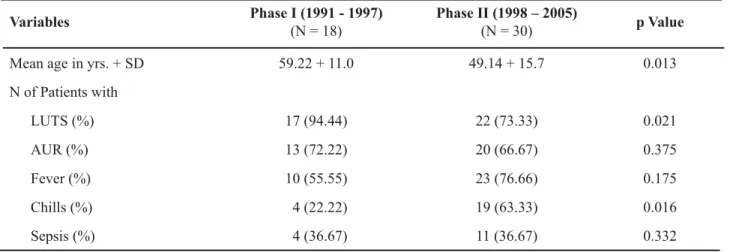
The baseline data and clinical presentations in both the phases are shown in Table-1. There was a
UHFHQWVWDWLVWLFDOO\VLJQL¿FDQWVKLIWWR\RXQJHUDJHDW
presentation (p = 0.013). The clinical presentations in both phases were similar except LUTS and chills (Table-1). DRE measured size, tenderness and
indu-UDWLRQDQGUHYHDOHGVLPLODU¿QGLQJVLQERWKSKDVHV $OWKRXJKWKHGLDEHWHVPHOOLWXVZDVWKHPRVW
common factor in both phases, it was seen less fre-quently in phase II (53.33%) than in phase I (77.77%) (Table-2). There were four patients with no co-mor-bidity in phase I. There were 11 patients in phase II with no co-morbid factor, of which nine were in the younger age group (22 - 44 years). Of these 11 patients
¿YHSUHVHQWHGZLWKS\UH[LDRIXQNQRZQRULJLQ382
and the cause was prostatic abscess with no LUTS. There were two patients with HIV infection, two with perinephric abscess and one each with chronic liver disease and end stage renal disease in Phase II.
Urine culture was available in 13 of 18 patients in phase I and 28 of 30 patients in phase II (Table-3), it was positive in 9 and 23 respectively. The pus culture was performed in eight patients in phase I and 16 patients in phase II that was positive in two and 14, respectively (Table-3). The urine culture and pus culture were similar in only six cases. The
organ-LVPVFXOWXUHGZHUHSUHGRPLQDQWO\VXVFHSWLEOHWR¿UVW
166
and quinolones) in phase I whereas organisms were predominantly susceptible to second line (amikacin, ceftazidime) or third line antibiotics (imipenem or meropenem), in phase II. In phase I, of nine isolates four were E. coli and three of them were sensitive
WRFRPPRQO\XVHG¿UVWOLQHGUXJVDPSLFLOOLQFLS
-URÀR[DFLQFRWULPR[D]ROHDQGJHQWDPLFLQDQGRQH
was resistant. In phase II , of nine E. coli isolates only
WZRZHUHVHQVLWLYHWR¿UVWOLQHDQWLELRWLFVDPSLFLO -lin, cefuroxime, co-trimoxazole, gentamicin) and rest 7 were resistant and were susceptible to second line (amikacin, ceftazidime) or third line antibiotics (imipenem and meropenem).
Of four Pseudomonas spp two were sus-ceptible to gentamicin and amikacin, and rests were susceptible only to ceftazidime, imipenem and
PHURSHQHP.OHEVLHOODVSSZDVVXVFHSWLEOHRQO\WR
ceftazidime, imipenem and meropenem. Burkholderia pseudomallei was susceptible only to ceftazidime, imipenem and meropenem However, the susceptibil-ity of the gram positive organisms remained the same in both the phases.
,QSKDVH,GLDJQRVLVZDVFRQ¿UPHGE\DE -dominal ultrasound in nine patients and transrectal ultrasound (TRUS) in three whereas in phase II the common mode of diagnosis was TRUS in 17,
trans-Table 1 – Clinical presentation.
9DULDEOHV 3KDVH,
(N = 18)
3KDVH,,±
(N = 30) S9DOXH
Mean age in yrs. + SD 59.22 + 11.0 49.14 + 15.7 0.013
N of Patients with
LUTS (%) 17 (94.44) 22 (73.33) 0.021
$85 13 (72.22) 20 (66.67) 0.375
Fever (%) 10 (55.55) 23 (76.66) 0.175
Chills (%) 04 (22.22) 19 (63.33) 0.016
Sepsis (%) 04 (36.67) 11 (36.67) 0.332
AUR = acute urinary tension; LUTS = lower urinary tract symptoms.
Table 2 – Predisposing factors.
9DULDEOHV 3KDVH,
(N = 18)
3KDVH,,
(N = 30)
Diabetes mellitus 14 16
HIV 00 02
Chronic liver disease 00 01
End-stage renal disease 00 01
Perinephric abscess 00 02
abdominal ultrasound in six (Figure-1) and CT scan in one patient with perinephric abscess and other two with ruptured prostatic abscess making it possible to
H[DFWO\GH¿QHWKHH[WUDSURVWDWLFH[WHQWRISXVLQWKH
ischiorectal fossa and perirectal tissue (Figure-2). The value of DRE in diagnosing prostatic abscess remained the same in both phases demon-strated by the fact that six patients in phase I and four in phase II were diagnosed based solely on DRE.
$VWUHDWPHQWLQDGGLWLRQWRDSSURSULDWHDQWL -biotics, in phase I, 10 patients underwent transurethral resection of the prostate (TURP) along with trans-urethral drainage of pus, as they were older and with symptoms of prostatic enlargement. Three patients had TUR drainage, four had spontaneous rupture and one patient underwent transperineal aspiration. In phase II, TUR drainage was the most common mode of treatment, which was performed in 14 patients as
Table 3 –%DFWHULRORJLFDOSUR¿OHRIXULQHDQGSXVFXOWXUHV
&XOWXUH 3KDVH,
(N = 18)
3KDVH,,
(N = 30)
Urine 13 28
Escherichia coli 04 09
Staphylococcus aureus 03 02
Pseudomonas aeruginosa 00 04
.OHEVLHOODVSS 00 03
Serratia spp 01 00
Citrobacter spp 00 01
Burkholderia pseudomallei 00 02
Fungus 01 02
No Growth 04 05
Culture not available 05 02
Pus 08 16
Escherichia coli 01 06
Staphylococcus aureus 01 03
Pseudomonas aeruginosa 00 01
Ps. aeruginosa + Enterococci 00 01
.OHEVLHOODVSS 00 02
Cryptococcus spp 00 01
No growth 06 02
patients were of a younger age group, only four el-derly patients with concomitant prostatic enlargement had TURP. Three had TRUS guided aspiration, one with distal penile urethral stricture had transperineal aspiration with statistical process control and four had spontaneous rupture. Two patients with microab-scesses and one with melioidosis were treated exclu-sively with antibiotics. One patient who underwent transperineal aspiration in phase II developed septic shock requiring ventilatory and vasopressure support in intensive care unit. None of the patients in phase I or phase II had septicemia due to formal TURP and TUR drainage.
In phase I, four patients had spontaneous rupture due to delayed diagnosis. One developed
perineal abscess and one pararectal abscess requiring open drainage, and in two patients abscesses had rup-tured into the prostatic urethra. In phase II, there were four patients with spontaneous rupture due to delayed diagnosis. One developed horse shoe perineal abscess that required open drainage and temporary sigmoid colostomy. One had pararectal abscess that was man-aged by incision and drainage. One had rectourethral
¿VWXODWKDWZDVWUHDWHGZLWKDQWLELRWLFDQGVXSUDSXELF
drainage for three months. In one abscess ruptured into the prostatic urethra. In phase II, there were two patients of HIV infection with tuberculous prostatic abscess, along with tuberculous pyocele and Cryptococcus neoformans isolated on pus culture in one.
$OOSDWLHQWVUHFRYHUHGZHOOLQERWKWKHSKDVHV
except one death in phase II who had melioidosis. Three young patients in phase II following TUR drain-age of prostatic abscess developed retrograde ejacula-tion. Mean duration of hospital stay were similar in both the phases, 11.37 days (range 6 - 23 days) and 9.33 days (range 2 - 28 days ) as was the duration of antibiotic therapy 28 days (14 - 42 days) and 30 days (9 - 90 days) in phase I and phase II respectively.
&200(176
Prostatic abscess is an infrequent condition in the modern antibiotic era with an incidence of 0.5% to 2.5% of all prostatic disease (8). Prostatic abscess can occur in patients of any age but is mainly found in men in their 5th and 6thGHFDGHRIOLIH$VVHHQLQ
our series, prostatic abscess is occurring in a younger age group.
Predisposing factors for development of prostatic abscess are diabetes mellitus, bladder outlet obstruction, indwelling catheter, chronic renal failure, patients on hemodialysis, chronic liver disease and more recently HIV infection (14). In our series, dia-betes was the most common predisposing factor, with HIV causing tuberculous abscesses, in two patients. In phase II 53% of patients were diabetic. They were younger and keeping with the WHO report (15) of diabetes occurring in younger individuals in the Indian subcontinent. Three patients (21.42%) in phase I and 7 patients(43.75%) in phase II were diagnosed to be
GLDEHWLFIRUWKH¿UVWWLPHZKHQWKH\SUHVHQWHGZLWK
Figure 1 – Transrectal ultrasound showing prostatic abscess.
Figure 2 – CT scan showing prostatic abscess ruptured into left
169
prostatic abscess. This could be a major new form of presentation in keeping with the increased incidence of diabetes.
Prostatic abscess should be considered as a possible etiology when evaluating for PUO in
\RXQJHUPHQDV¿YHRISDWLHQWVZLWKRXWSUHGLVSRV -ing factor presented with PUO.
The clinical diagnosis of prostatic abscess is
VRPHWLPHVGLI¿FXOWEHFDXVHRIQRQVSHFL¿FV\PSWRPV
(8). This condition usually presents as an irritative voiding symptoms, perineal pain, and fever and occasionally as acute urinary retention (1). In our series, 17 patients (94.44%) in phase I and 22 patients (73.33%) in phase II presented with irritative LUTS. This may be due to the fact that patients were of the older age group in phase I than in phase II. The patients with prostatic abscess presented more com-monly with fever and chills in phase II than in phase I. The number of patients with sepsis was higher in phase II (36.67%) than in phase I (22.2%). This is most probably due to infection caused by multi drug resistant bacteria related to the misuse of antibiotics in the community. In our series in phase I, 75% of E.
FROLZHUHVHQVLWLYHWRFRPPRQO\XVHG¿UVWOLQHGUXJV DPSLFLOOLQFLSURÀR[DFLQFRWULPR[D]ROHDQGJHQWD -micin) and in phase II, more than 75% of E. coli were
UHVLVWDQWWR¿UVWOLQHDQWLELRWLFVDQGZHUHVXVFHSWLEOH
to second line (amikacin, ceftazidime) or third line antibiotics (imipenem and meropenem).
The microbiology of prostatic abscess has undergone a complete metamorphosis in the antibiotic era. More recently, various reports have shown that the common organisms causing prostatic abscess are E. coli and other enteric gram negative bacilli (1,8,9). More recently we have reported two cases of prostatic abscess due to Burkholderia pseudomallei (16).
However, the prevalence of immunocompro-mised individuals has increased in the modern era (phase II), and the potential for uncommon fastidious pathogens, particularly mycobacterial, fungal and anaerobic pathogens, melioidosis, in addition to typi-cal gram-negative bacilli, will make the diagnosis of prostatic abscess more complicated (14,16,17).
Surprisingly urine culture and pus culture isolates were similar in only six cases (all in phase II). Of these, 4 were gram negative bacilli, this includes E.
FROLQ .OHEVLHOODVSSQ DQG3VHXGRPRQDV
spp (n = 1) and 2 were gram positive cocci (S. aureus).
2IVL[SDWLHQWV¿YHZHUHGLDEHWLFDQGIRXUKDGVHSVLV
at presentation. It is important to send material for culture (pus, urine, and/or prostatic chips) in order to identify the etiologic agent, especially in immuno-compromised patients because they usually present with uncommon microorganisms (18). Urine culture may be negative unless the abscess ruptures into urethra or bladder. Thus it is important to emphasize that pus culture and sensitivity should be performed routinely for management of prostatic abscess.
$OWKRXJK WKH EDFWHULRORJLFDO SURILOH ZDV
similar in both phases, it is important to note that the
DQWLELRWLF VXVFHSWLELOLW\ SUR¿OH KDG FKDQJHG ZLWK RUJDQLVPVUHVLVWDQWWR¿UVWOLQHGUXJVDQGVHQVLWLYH
only to higher antibiotics.
In our series, trans-abdominal USG was the most common modality of diagnosis in phase I, but in phase II, TRUS became the major diagnostic tool and was performed in 56.67% of patients and has now become a standard protocol as the transrectal probe was acquired later part of 1st phase.
&21&/86,216
Prostatic abscess should be considered in the differential diagnosis of young men who pres-ent with pyrexia of unknown origin. It could be the primary presentation in a recently diagnosed diabetic. The incidence of prostatic abscess is increasing in younger males. This is probably related to the higher incidence of diabetes in younger males in this
UHJLRQ&OLQLFDO¿QGLQJVFRXOGEHVXEWOHHVSHFLDOO\
in younger men who may not present with LUTS. While the bacteriology remains largely unchanged, the emergence of multi drug resistant organisms points to the rampant misuse of antibiotics. The emergence of HIV brings the added concern that some of the abscesses could be the result of tuberculous infection.
&21)/,&72),17(5(67
None declared.
5()(5(1&(6
$QJZDIR )) UG 6RVVR$0 0XQD:) (G]RD7 -XLPR$*3URVWDWLFDEVFHVVHVLQVXE6DKDUDQ$IULFD a hospital-based experience from Cameroon. Eur Urol. 1996; 30: 28-33.
2. Leport C, Rousseau F, Perronne C, Salmon D, Joerg $9LOGH-/%DFWHULDOSURVWDWLWLVLQSDWLHQWVLQIHFWHG ZLWKWKHKXPDQLPPXQRGH¿FLHQF\YLUXV-8URO 141: 334-6.
3. Cytron S, Weinberger M, Pitlik SD, Servadio C: Value of transrectal ultrasonography for diagnosis and treat-ment of prostatic abscess. Urology. 1988; 32: 454-8. 'DYLGVRQ.&*DUORZ:%%UHZHU-&RPSXWHUL]HG
tomography of prostatic and periurethral abscesses: 2 case reports. J Urol. 1986; 135: 1257-8.
9DFFDUR-$%HOYLOOH:'.LHVOLQJ9--U'DYLV5 Prostatic abscess: computerized tomography scanning as an aid to diagnosis and treatment. J Urol. 1986; 136: 1318-9.
6. Sargent JC, Irwin R: Prostatic abscess: a clinical study RIFDVHV$P-6XUJ
7. Mitchell RJ, Blake JR: Spontaneous perforation of prostatic abscess with peritonitis. J Urol. 1972; 107: 622-3.
*UDQDGRV($5LOH\*6DOYDGRU-9LQFHQWH-3URV-tatic abscess: diagnosis and treatment. J Urol. 1992; 148: 80-2.
-DFREVHQ-'.YLVW(3URVWDWLFDEVFHVV$UHYLHZRI literature and a presentation of 5 cases. Scand J Urol Nephrol. 1993; 27: 281-4.
0\HU¶VDQG.RVKL¶V0DQXDORI'LDJQRVWLF3URFHGXUH in Medical Microbiology and Immunology/Serology. Faculty, Department of Clinical Microbiology, Chris-WLDQ0HGLFDO&ROOHJH9HOORUH,QGLD3RQGLFKHUU\$OO India Press, 2001.
%DXHU$:.LUE\:06KHUULV-&7XUFN0$QWLELRWLF susceptibility testing by a standardized single disk PHWKRG$P-&OLQ3DWKRO
12. Clinical Laboratory Standards Institute (formerly, National committee for Clinical Laboratory Standards. 3HUIRUPDQFH6WDQGDUGVIRU$QWLPLFURELDOV'LVF6XV -FHSWLELOLW\7HVWVWKHG$SSURYHG6WDQGDUGV1&&/6 'RFXPHQW0$:D\QH3$1DWLRQDO&RPPLWWHH for Clinical Laboratory Standards, 2000.
13. Pai MG, Bhat HS: Prostatic abscess. J Urol. 1972; 108: 599-600.
7UDX]]L6-.D\&-.DXIPDQ'*/RZH)&0DQDJH-ment of prostatic abscess in patients with human immu-QRGH¿FLHQF\V\QGURPH8URORJ\ 15. Mohan V, Deepa M, Deepa R, Shanthirani CS, Farooq
6*DQHVDQ$HWDO6HFXODUWUHQGVLQWKHSUHYDOHQFH of diabetes and impaired glucose tolerance in urban South India--the Chennai Urban Rural Epidemiology Study (CURES-17). Diabetologia. 2006; 49: 1175-8. 9LVZDURRS%6%DODML90DWKDL(.HNUH160HOLRL-dosis presenting as genitourinary infection in two men with diabetes. J Postgrad Med. 2007; 53: 108-10. 17. Ludwig M, Schroeder-Printzen I, Schiefer HG, Weidner
W: Diagnosis and therapeutic management of 18 patients with prostatic abscess. Urology. 1999; 53: 340-5. 2OLYHLUD3$QGUDGH-$3RUWR+&3HUHLUD)LOKR-(
9LQKDHV$) 'LDJQRVLV DQG WUHDWPHQW RI SURVWDWLF abscess. Int Braz J Urol. 2003; 29: 30-4.
Accepted after revision: March 6, 2008
&RUUHVSRQGHQFHDGGUHVV
Dr. Ganesh Gopalakrishnan Department of Urology Christian Medical College Vellore, S. India
Fax: + 914 162 232-103