SUMMARY
Objective: To assess the prevalence of uterine anatomical abnormalities found by oice diagnostic hysteroscopy in a population of patients experiencing more than two consecu-tive miscarriages and compare the prevalence of uterine abnormalities between patients with two miscarriages and those with three or more consecutive miscarriages. Methods: A cross-sectional study of 66 patients with two or more consecutive miscarriages diag-nosis was conducted. Patients were divided into two groups: Group A (up to two miscar-riages, 23 patients), and Group B (3 miscarmiscar-riages, 43 patients). hey underwent an out-patient diagnostic hysteroscopy study, with either congenital or acquired abnormalities of the uterine cavity being identiied. Results: Uterine changes were found in 22 (33.3%) patients, with 9 cases of congenital changes [arcuate uterus (4 cases), septate uterus (2 cases), and bicornuate uterus (1 case)], and 13 patients with acquired changes [intrauter-ine adhesions (7 cases), endometrial polyp (4 cases), and uter[intrauter-ine leiomyoma (2 cases)]. No signiicant diferences were found between the groups as regarding both acquired and congenital uterine changes. A positive correlation was found between anatomical changes on hysteroscopy and number of miscarriages (r = 0.31; p = 0.02). Conclusion: Patients with more than two miscarriages have a high prevalence of uterine cavity abnor-malities diagnosed by hysteroscopy; however there are no diferences in prevalence or distribution of these lesions related to the number of recurrent miscarriages.
Keywords: Abortion, habitual; hysteroscopy; uterine diseases; congenital abnormalities.
Study conducted at the Department of Gynecology and Obstetrics, Clinical Hospital, Porto Alegre, Porto Alegre, RS, Brazil
Submitted on: 02/13/2011
Approved on: 05/03/2011
Correspondence to:
Carlos Augusto Bastos de Souza R. Ramiro Barcelos, 2350/s 1125 Porto Alegre, RS, Brazil CEP: 90035-003 [email protected]
Conlict of interest: None.
©2011 Elsevier Editora Ltda. All rights reserved.
Ofice hysteroscopy study in consecutive miscarriage patients
CARLOS AUGUSTO BASTOSDE SOUZA1, CARLA SCHMITZ2, VANESSA KREBS GENRO3, ANA MARTINS4, CAMILA SCHEFFEL4, MARIA LUCIA OPPERMANN5, JOÃO SABINO CUNHA FILHO6
1Post-Doctorate in Endometriosis and Minimally Invasive Gynecology; Attending Gynecologist, Clinical Hospital, Porto Alegre, Porto Alegre, RS, Brazil 2M.Sc. Student in Human Reproduction; Attending Gynecologist, Pompeia Hospital, Caxias do Sul, RS, Brazil
3Ph.D. in Human Reproduction; Attending Gynecologist, Insemine Clinic, Porto Alegre, RS, Brazil
4Medicine Undergraduate Student, Universidade Federal do Rio Grande do Sul (UFRGS), Porto Alegre, RS, Brazil 5Ph.D. in Epidemiology; Professor, UFRGS, Porto Alegre, RS, Brazil
INTRODUCTION
Recurring miscarriages are considered when pregnancy is spontaneously interrupted in three consecutive episodes either previously to 20 weeks of gestational age or before the fetus reaches 500 g in weight1,2. More recently, there
has been a tendency to include into this diagnosis those patients with two early spontaneous pregnancy losses, mainly if they occur later than the age of 35 years3. his
new approach prevents delays in recognizing the disease in a more critical age group; however, it can contribute to a higher number of studies and invasive procedures ordered in this population, with no beneits necessarily resulting
from the case management3-5. Repeated miscarriages can
occur due to a set of factors, such as: genetic, endocrine, and immune diseases, coagulation system disorders or
anatomical factors3. Immune changes were more
preva-lently found in patients with repeated miscarriages, and the frequency of indings was similar when patients with two miscarriages were compared with those with three or more miscarriages4.
Prevalence of congenital or acquired anatomical changes in patients with repeated miscarriages is high, ranging from 6.3% to 67%6,7, depending on the type of the
study and the study population. Usually, anatomical as-sessment in these patients is performed through hystero-salpingography, ultrasonography, hysteroscopy, and lapa-roscopy, with further studies possibly being used, such as tridimensional ultrasonography, hysterosonography, and magnetic resonance8-10. Congenital uterine anomalies are
correctly diagnosed by ultrasonography, especially when it is combined with a tridimensional resource; on the other hand, diagnostic hysteroscopy allows the diagnosis of ac-quired anomalies, in addition to congenital anomalies8,11.
Recently, a reduction in hysteroscopy cost associated with reduced optical diameters has allowed hysteroscopy to be performed in an outpatient basis, with no anesthetics use, minimal discomfort and optimal acceptance by pa-tients10,12-15. his study was conducted in order to assess the
prevalence of uterine anatomical abnormalities diagnosed by hysteroscopy in a population of patients with more than two consecutive miscarriages. We further looked for a pos-sible diference in prevalence of uterine changes in patients with two miscarriages, compared with those with three or more miscarriages.
METHODS
A cross-sectional study was conducted from January 2007 to December 2010 and 74 patients of the Department of Gynecology with consecutive miscarriages were assessed. Only patients with consecutive losses were included, and they were classiied according to the number of losses. Miscarriage was considered as a spontaneous gestational loss occurred up to 20 weeks or with a fetal weight lower than 500 g1,7. According to the number of miscarriages,
patients were divided into two groups: Group A (two mis-carriages, n = 23) and Group B (three or more miscarriag-es, n = 43) for purposes of comparison1,4. Patients whose
gestational age at the time of the loss was unknown (n = 4), patients with a current pregnancy diagnosed (n = 1), prior uterine surgery other than curettage or C-section (n = 1), patients who refused to participate in the study or patients who did not tolerate the assessment without anesthesia (n = 2) were excluded16. Demographics of the sample, such
as age, menarche age, cycle characteristics, obstetric histo-ry (parousity, gestational age when prior losses occurred), smoking and alcohol consumption were collected at the time the test was ordered. Table 1 shows the distribution of the sample demographics.
DIAGNOSTICPROCEDURE
he patients underwent a diagnostic hysteroscopy at the follicular phase of the menstrual cycle (days 3-15) and all procedures were performed by skilled gynecologists (CAS, JSCF). he examiner did not know the test indication when it was performed. A k index was calculated among the ex-aminers and no signiicant diference was found (p = 0.83). In summary, the procedure was performed with 2.6 mm optics with an angle of view of 30º (Karl Storz Endoscopy, Germany). Normal saline was used as a distending medium with a pressure of 20 mmHg to 50 mmHg. Hysteroscopy was performed in an outpatient basis, with neither use of anesthesia nor antibiotic prophylaxis, with cervical grasping by using a Pozzi tenaculum being avoided17,18. In case the
patient did not tolerate the procedure, it would be discon-tinued and rescheduled using procedural sedation and an-esthesia, and that patient would be excluded from the study.
CLASSIFICATIONOFFINDINGS
Changes found by hysteroscopy were subdivided into congenital or acquired abnormalities. Congenital changes were classiied as arcuate uterus, didelphic uterus, bicor-nuate uterus, unicorbicor-nuate uterus, and septate uterus. he acquired changes found received the following diagnoses: uterine polyp, leiomyoma, intrauterine adhesions, endo-metritis, and hyperplasia8,17.
STATISTICALANALYSIS
Table 1 – Distribution of demographics in the sample (median, interquartile range)
Two miscarriages (n = 23)
Three or more miscarriages (n = 43)
Total
n = 66 p
Age (years) 35 (19.7-35.7) 32.7 (29-35) 34 (31-39) 0.64ª
Menarche age (years) 12.5 (12-16) 11.1 (11-13) 12 (11-13) 0.16ª
Race 0.49b
White 17 (74.0) 36 (83.7) 53 (80.3)
Afrodescendant 3 (13.0) 5 (11.6) 8 (12.1)
Mixed 3 (13.0) 2 (4.7) 5 (7.6)
Regular cycles 18 (78.3) 38 (88.4) 56 (84.8) 0.27b
Pregnancies 2 (2.0-3.0) 3 (3.0-4.0) 3 (3-4) 0.0001a
Deliveries 0.5 (0.5-1.0) 0.5 (0.5-1.0) 0.5 (0.5-1) 0.86 a
C-sections 0.1 (0.1-0.5) 0.2 (0.1-0.5) 0.1 (0.1-0.5) 0.17 a
Miscarriage 2 (2.0-2.0) 3 (3.0-4.0) 3.0 (2.0-3.2) 0.0001 a
GA at the miscarriage (weeks) 11 (9.0-12.0) 11 (9.5-13) 11 (9.0-12.0) 0.7a
Weight (kg) 55 (53.5-64.7) 60 (56.5-64.7) 60 (55.0-67.5) 0.43ª
Height (m) 1.57 (1.56-1.68) 1.61 (1.57-1.65) 1.58 (1.57-1.65) 0.41ª
BMI (kg/m2) 22.6 (20.1-24.2) 23.04 (22.3-23.9) 23.1 (22.3-26.4) 0.83ª
Smoking 18 (78.3) 38 (88.4) 12 (18.2) 0.27b
Alcohol consumption 5 (21.7) 7 (16.3) 2 (3.0) 0.7 b
a Mann-Whitney; b Chi-squared.
GA, gestational age.
RESULTS
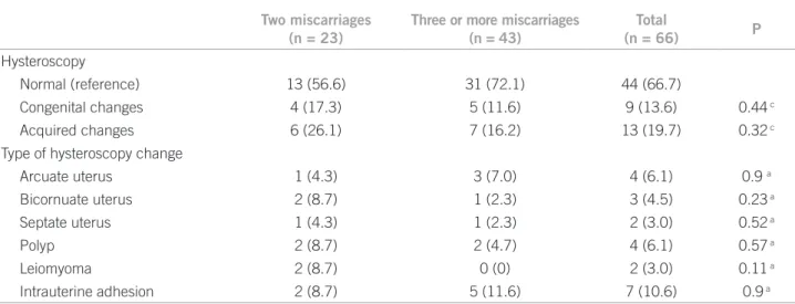
Twenty-two (33.3%) patients in the sample were found to have uterine cavity changes, with 9 of them being congeni-tal and 13 acquired anomalies. By evaluating the congenicongeni-tal changes in the uterine cavity, the following diagnoses were found: arcuate uterus (n = 4), bicornuate uterus (n = 3) and septate uterus (n = 2). By considering the acquired anoma-lies, the most frequent diagnoses were: intrauterine adhe-sion (n = 7), polyp (n = 4), leiomyoma (n = 2) (Table 2).
When groups with two miscarriage episodes were compared with groups with three or more miscarriages,
the sample characteristics found had a similar distribu-tion between the groups. Regarding hysteroscopy indings, both congenital and acquired changes had no signiicant diferences between the groups (Table 2). Hysteroscopy anomalies in patients in Group A were no diferent from anomalies in Group B (10 vs. 12, respectively, p = 0.2, chi-squared). When the number of miscarriages was correlated with hysteroscopy indings, a correlation coeicient r = 0.31 (p = 0.02 – Spearman’s) was found, the correlation of the number of miscarriages with the number of patients having intrauterine adhesions was r = 0.11 (p = 0.39 – Spearman’s).
Table 2 – Distribution of hysteroscopy indings between the groups (median, interquartile range)
Two miscarriages
(n = 23)
Three or more miscarriages (n = 43)
Total
(n = 66) P
Hysteroscopy
Normal (reference) 13 (56.6) 31 (72.1) 44 (66.7)
Congenital changes 4 (17.3) 5 (11.6) 9 (13.6) 0.44 c
Acquired changes 6 (26.1) 7 (16.2) 13 (19.7) 0.32 c
Type of hysteroscopy change
Arcuate uterus 1 (4.3) 3 (7.0) 4 (6.1) 0.9 a
Bicornuate uterus 2 (8.7) 1 (2.3) 3 (4.5) 0.23 a
Septate uterus 1 (4.3) 1 (2.3) 2 (3.0) 0.52 a
Polyp 2 (8.7) 2 (4.7) 4 (6.1) 0.57 a
Leiomyoma 2 (8.7) 0 (0) 2 (3.0) 0.11 a
Intrauterine adhesion 2 (8.7) 5 (11.6) 7 (10.6) 0.9 a
DISCUSSION
In our study, we demonstrated consecutive miscarriages are associated with uterine cavity anomalies, as about one-third of the sample had congenital or acquired chan-ges on hysteroscopy. We further demonstrated chanchan-ges are equally distributed in patients with two miscarriages compared with those with three or more consecutive miscarriages.
Studies have sought to analyze if the traditional dei-nition for repeated miscarriage, considering three con-secutive episodes, should be reviewed; however, indings are still incipient1-5. Our study is in accordance with
pre-vious studies demonstrating that although there is a high incidence of anatomical changes in the population of pa-tients with repeated miscarriages1,3,19, there is no
difer-ence in inciddifer-ence of indings regarding patients with two miscarriages compared with those with three or more events1,2. Jaslow et al.4, by evaluating a large series of
re-peated miscarriage cases demonstrated immune changes were similarly distributed, regardless the number of mis-carriages. his set of indings indicates the assessment of patients with repeated miscarriages can be reviewed, by trying to identify the patients earlier and in a more par-ticular way.
Over the last years, hysteroscopy has been shown as an excellent diagnostic and therapeutic tool in gynecol-ogy2,15,20. We have found a high prevalence of acquired
anatomical abnormalities, particularly intrauterine ad-hesions. his fact is likely associated with these patients having usually undergone uterine emptying procedures. Uterine curettage is known to produce intrauterine
ad-hesions20,21. Although the intrauterine manual vacuum
aspiration procedure is increasingly prevalent here in Porto Alegre, a large number of patients still undergo standard uterine curettage procedures20. In our study, a
correlation between hysteroscopy anomalies and number of miscarriages was present (r = 0.31); thus, we can as-sume there is an association between anatomical changes and increased miscarriage incidence. Unfortunately, the correlation is not sustained in cases of intrauterine adhe-sions (r = 0.11).
Our study has several points to be highlighted. We could show a homogeneous series of repeated miscar-riage cases. he data collect was appropriate, controlling the methodology employed to carry out the tests and interexaminer variability. As our practice is a reference center in endoscopy, with studies being performed for various indications, the examiners were unaware of the test indication as it was performed; however, the patient’s obstetric and surgical history was informed, preventing the examiner’s total blinding.
Despite we were careful about methodology, our study has limitations. Our incidence of repeated miscar-riage cases, as well as the hysteroscopy abnormal indings,
is supposedly higher than that found in the general pop-ulation. Moreover, endoscopy availability likely allowed uterine anomalies which otherwise could go undetected or be diagnosed later to be diagnosed earlier. Another noticeable point in our study was a higher number of pa-tients with more than three miscarriages over the group
with only two miscarriages16. In our sample, we do not
have the patients’ hysterosalpingography data. his is an easily available, non-invasive, and low-cost study show-ing a correlation with indshow-ings in other tests, such as ul-trasonography and hysteroscopy. However, hysterosal-pingography has a high false-positive and false-negative rates as a disadvantage, in addition to being a more pain-ful test for most patients22,23.
CONCLUSION
hus, repeated miscarriage cases have an increased preva-lence of acquired and congenital uterine anomalies diag-nosed by outpatient diagnostic hysteroscopy. It is shown as an applicable and easily performed test for that population. Changes in the uterine cavity have already been present from two miscarriages; thus, starting earlier the anatomi-cal investigation in repeated miscarriages can be suitable as managing these cases. Prospective studies with a higher number of patients are still required so that changes in management of repeated miscarriages can be deined.
REFERENCES
1. Weiss A, Shalev E, Romano S. Hysteroscopy may be justiied ater two miscarriages. Hum Reprod 2005;20(9):2628-31.
2. Dendrinos S, Grigoriou O, Sakkas EG, Makrakis E, Creatsas G. Hys-teroscopy in the evaluation of habitual abortions. Eur J Contracept Reprod Health Care 2008;13(2):198-200.
3. Li TC, Makris M, Tomsu M, Tuckerman E, Laird S. Recurrent mis-carriage: aetiology, management and prognosis. Hum Reprod Up-date 2002;8(5):463-81.
4. Jaslow CR, Carney JL, Kutteh WH. Diagnostic factors identiied in 1020 women with two versus three or more recurrent pregnancy losses. Fertil Steril 2010;93(4):1234-43.
5. Stephenson MD. Management of recurrent early pregnancy loss. J Reprod Med 2006;51(4):303-10.
6. Stephenson MD. Frequency of factors associated with habitual abor-tion in 197 couples. Fertil Steril 1996;66(1):24-9.
7. Tulppala M, Palosuo T, Ramsay T, Miettinen A, Salonen R, Ylikork-ala O. A prospective study of 63 couples with a history of recurrent spontaneous abortion: contributing factors and outcome of subse-quent pregnancies. Hum Reprod 1993;8(5):764-70.
8. Raga F, Bauset C, Remohi J, Bonilla-Musoles F, Simon C, Pellicer A. Reproductive impact of congenital Mullerian anomalies. Hum Re-prod 1997;12(10):2277-81.
9. Propst AM, Hill JA. 3rd. Anatomic factors associated with recurrent pregnancy loss. Semin Reprod Med 2000;18(4):341-50.
10. El-Mazny A, Abou-Salem N, El-Sherbiny W, Saber W. Outpatient hysteroscopy: a routine investigation before assisted reproductive techniques? Fertil Steril 2011;95(1)272-6.
11. Salim R, Regan L, Woelfer B, Backos M, Jurkovic D. A comparative study of the morphology of congenital uterine anomalies in women with and without a history of recurrent irst trimester miscarriage. Hum Reprod 2003;18(1):162-6.
13. Koskas M, Mergui JL, Yazbeck C, Uzan S, Nizard J. Oice hysteros-copy for infertility: a series of 557 consecutive cases. Obstet Gynecol Int 2010;2010:168096.
14. Lasmar RB, Dias R, Barrozo PR, Oliveira MA, Coutinho Eda S, Rosa DB. Prevalence of hysteroscopic indings and histologic di-agnoses in patients with abnormal uterine bleeding. Fertil Steril 2008;89(6):1803-7.
15. Yela DA, Ravacci SH, Monteiro IM, Pereira KC, Gabiatti JR. [Com-parative study of transvaginal sonography and outpatient hysteros-copy for detection of pathologic endometrial lesions in postmeno-pausal women]. Rev Assoc Med Bras 2009;55(5):553-6.
16. von Elm E, Altman DG, Egger M, Pocock SJ, Gotzsche PC, Vanden-broucke JP. he Strengthening the Reporting of Observational Stud-ies in Epidemiology (STROBE) statement: guidelines for reporting observational studies. J Clin Epidemiol 2008;61(4):344-9.
17. Fatemi HM, Kasius JC, Timmermans A, van Disseldorp J, Fauser BC, Devroey P et al. Prevalence of unsuspected uterine cavity abnormali-ties diagnosed by oice hysteroscopy prior to in vitro fertilization. Hum Reprod 2010;25(8):1959-65.
18. Kasius JC, Broekmans FJ, Fauser BC, Devroey P, Fatemi HM. Anti-biotic prophylaxis for hysteroscopy evaluation of the uterine cavity. Fertil Steril 2011;95(2)792-4 .
19. Portuondo JA, Camara MM, Echanojauregui AD, Calonge J. Mulle-rian abnormalities in fertile women and recurrent aborters. J Reprod Med 1986;31(7):616-9.
20. Traina EMR, Moron AF, Albuquerque Neto LC, Matheus ED. Acu-rácia diagnóstica da histerossalpingograia e da ultra-sonograia para avaliaçao de doenças da cavidade uterina em pacientes com aborta-mento recorrente. RBGO 2004;26(7):7.
21. Salzani A, Yela DA, Gabiatti JR, Bedone AJ, Monteiro IM. Prevalence of uterine synechia ater abortion evacuation curettage. São Paulo Med J. 2007;125(5):261-4.
22. Siristatidis C, Chrelias C, Salamalekis G, Kassanos D. Oice hyster-oscopy: current trends and potential applications: a critical review. Arch Gynecol Obstet 2010;282(4):383-8.