immunohistochemical features
in situ
and invasive
components of ductal carcinoma of breast
Comparação do grau nuclear e perfil imunoistoquímico nos
componentes in situ e invasivo de carcinoma mamário
Carlos e. BaCCHi FiloMena Marino CarvalHo4
Study carried out at the Pathology Department, Faculdade de Medicina, Univerisdade de São Paulo – USP – São Paulo (SP), Brazil. 1Instituto do Câncer do Estado de São Paulo – ICESP – São Paulo (SP), Brazil; Department of Pathology, Faculdade de Medicina,
Universidade de São Paulo – USP – São Paulo (SP), Brazil.
2Faculdade de Medicina, Universidade de São Paulo – USP – São Paulo (SP), Brazil. 3Consultancy in Pathology – Botucatu (SP), Brazil.
4Department of Pathology, Faculdade de Medicina, Universidade de São Paulo – USP – São Paulo (SP), Brazil. Correspondence
Filomena Marino Carvalho Faculdade de Medicina da Universidade de São Paulo, Departamento de Patologia Avenida Doutor Arnaldo, 455, sala 1149 Zip code: 02146-903 São Paulo (SP), Brazil
Received 01/11/2013
Accepted with modiications 02/01/2013
Abstract
PURPOSE:To compare the prognostic and predictive features between in situ and invasive components of ductal breast carcinomas. METHODS: We selected 146 consecutive breast samples with ductal carcinoma in situ (DCIS) associated with adjacent invasive breast carcinoma (IBC). We evaluated nuclear grade and immunohistochemical expression of estrogen receptor (ER), progesterone receptor (PR), human epidermal growth factor receptor 2 (HER2), cytokeratin 5/6 (CK5/6), and epidermal growth factor receptor (EGFR) in both components, in situ and invasive, and the Ki-67 percentage of cells in the invasive part. The DCIS and IBC were classiied in molecular surrogate types determined by the immunohistochemical proile as luminal (RE/PR-positive/ HER2-negative), triple-positive (RE/RP/HER2-positive), HER2-enriched (ER/PR-negative/HER2-positive), and triple-negative (RE/RP/HER2-negative). Discrimination between luminal A and luminal B was not performed due to statistical purposes. Correlations between the categories in the two groups were made using the Spearman correlation method. RESULTS: There was a signiicant correlation between nuclear grade (p<0.0001), expression of RE/RP (p<0.0001), overexpression of HER2 (p<0.0001), expression of EGFR (p<0.0001), and molecular proile (p<0.0001) between components in situ and IBC. CK 5/6 showed different distribution in DCIS and IBC, presenting a signiicant association with the triple-negative phenotype in IBC, but a negative association among DCIS. CONCLUSIONS: Our results suggest that classical prognostic and predictive features of IBC are already determined in the preinvasive stage of the disease. However the role of CK5/6 in invasive carcinoma may be different from the precursor lesions.
Resumo
OBJETIVO: Comparar características prognósticas e preditivas entre os componentes in situ e invasivo de carcinomas ductais da mama. MÉTODOS: Selecionamos 146 amostras mamárias consecutivas com carcinoma ductal in situ (CDIS) associado com carcinoma invasivo (CI) adjacente. Avaliamos grau nuclear e a expressão imunoistoquímica de receptor de estrogênio (RE), receptor de progesterona (RP), receptor do fator de crescimento epidérmico humano 2 (HER2), citoqueratina 5/6 (CK5/6) e o receptor do fator de crescimento epidérmico (EGFR) em ambos componentes, in situ e invasor, e a porcentagem de células marcadas pelo Ki-67 no componente invasivo. CDIS e CI foram classiicados nos tipos moleculares, determinados pelo peril imunoistoquímico, como luminal (RE/RP-positivo/HER2-negativo), triplo-positivo (RE/RP/HER2-triplo-positivo), HER2-puro (RE/RP-negativo/HER2-triplo-positivo) e triplo-negativo (RE/RP/HER2-negativo). A discriminação entre luminal A e Luminal B não foi feita por motivos estatísticos. Correlações entre as categorias dos dois grupos foram feitas pelo método de correlação de Spearman. RESULTADOS: Houve signiicante associação entre grau nuclear (p<0,0001), expressão de RE/RP) (p<0,0001), superexpressão de HER2 (p<0,0001), expressão de EGFR (p<0,0001) e peril molecular (p<0,0001) entre os componentes in situ e invasivo. CK5/6 mostrou distribuição distinta em CDIS e CI, apresentando signiicante associação com o fenótipo triplo-negativo em CI, mas uma associação negativa ente os CDIS. CONCLUSÕES: Nossos resultados sugerem que as características prognósticas e preditivas clássicas dos CI estão já determinadas no estágio pré-invasivo da doença. Entretanto, o papel da CK5/6 no carcinoma invasivo pode ser diferente daquele das lesões precursoras.
Artigo Original
Keywords
Breast neoplasms/chemistry Carcinoma, ductal, breast/chemistry Immunoenzyme techniques Receptors, estrogen/analysis Receptor, erbB-2/analysis keratin-5/analysis keratin-6/analysis Ki-67 antigen/analysis Tumor makers, biological
Palavras-chave
Introduction
Ductal carcinomas in situ (DCIS) are immediate pre-cursors of most breast cancer, but they are heterogeneous regarding morphology and invasiveness risk1. The
preva-lence of DCIS has been rising in the last decades, probably due to better screening programs and now accounts for approximately 20‒25% of all breast cancer diagnoses2.
The formerly accepted linear multi-step process of breast carcinogenesis, from hyperplasia, atypical hyperplasia, and carcinoma in situ, to invasive and metastatic carcinoma, changed to a more complex process involving a series of stochastic genetic events that lead to distinct and diver-gent pathways towards invasive carcinoma3-7. Although
the progression of DCIS to invasive breast carcinoma (IBC) is believed to be an important aspect feature of tumor aggressiveness, identiication of biomarkers and molecular proiles of IBC and DCIS is yet far to be fully elucidated6,8,9. Previous studies indicate that DCIS may
be classiied in a similar manner to invasive breast can-cer10-13. The understanding of the transition between
the preinvasive and invasive stages in breast carcinomas is the key to more eficient strategies for early diagnosis and treatment, as well as it expands the knowledge about the complex mechanisms of carcinogenesis. In this study our aim was to compare the prognostic and predictive pathological features between the in situ and invasive components of ductal breast carcinoma.
Methods
This study was approved by the Department of Pathology Scientiic Committee of the Faculdade de Medicina da Universidade de São Paulo and by the Ethical Committee for Research Projects of the Hospital das Clínicas da Faculdade de Medicina da Universidade de São Paulo (CAPPesq, process 2011/14741-7). As the study was retrospective, informed patient consent was waived and any form of patient identiication was abolished.
We selected breast samples from patients with con-irmed diagnosis of IBC after an initial sample represented by DCIS only. Cases were obtained from the iles of the Division of Surgical Pathology of Faculdade de Medicina da Universidade de São Paulo in the period from 2000 to 2009. All tissues had been ixed in 10% buffered formalde-hyde and embedded in parafin. The slides were rigorously reviewed and classiied by the same pathologist with exper-tise in breast pathology (FNA). For cases with discordant interpretation in relation to original report, a consensus was determined by simultaneous examination under a dual-head microscope (FNA and FMC). We included only carcinomas of non-special type according criteria of the histological classiication of tumors of World Health Organization,
201214. Carcinomas of special types and cases with
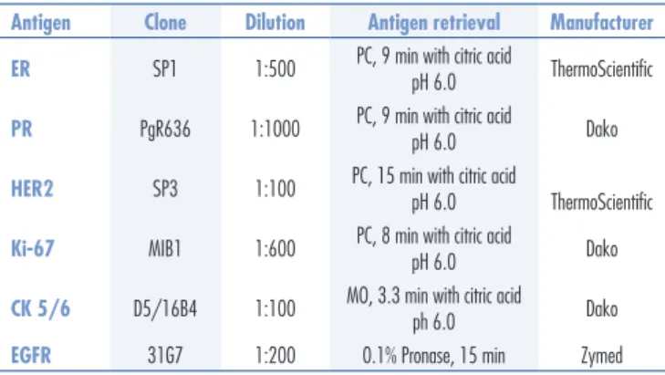
insuf-icient material to immunohistochemistry evaluation, signs of tissue autolysis and from pregnant patients were excluded from the study. We obtained 146 breast samples that met the criteria of inclusion. The age of the patients ranged from 29 to 87 years (median=59 years). Nuclear grades 1 (G1) and 2 (G2) were grouped as low-grade category, while nuclear grade 3 (G3) was deined as high-grade category (Figure 1). Immunohistochemistry was performed on 3 µm-thick histological sections containing in situ and in-vasive neoplasia. The source and dilutions of the antibodies and epitope retrieval methods used are listed in Table 1. Novolink® was used as the dectection system (Leica,
Bannockburn, IL, USA). Nuclear positivity was consid-ered speciic for estrogen receptor (ER) (Figure 1B and 1E), progesterone receptor (PR) and Ki-67; membranous positivity for human epidermal growth factor receptor 2 (HER2) (Figure 2) and epidermal growth factor receptor (EGFR), and cytoplasmic, for cytokeratin 5/6 (CK5/6) (Figure 1). Lesions with at least 10% of cells stained were considered positive for ER and PR. For CK5/6 expression we considered any positivity above 1% of epithelial cells. For HER2 and EGFR we only considered samples positive if they scored 3+ according to American Society of Clinical Oncology (ASCO) and College of American Pathologists (CAP) recommendations15.
An approximation of breast cancer molecular subtypes was used according to the following immunohistochemi-cal surrogate criteria modiied from St. Gallen consen-sus16: Luminal A (ER/PR-positive and Ki-67<14%),
Luminal B (ER/PR-positive, HER2-negative and Ki-67≥14%), triple-positive (ER/PR/HER2 positive), HER2-enriched (ER/PR-negative and HER2-positive), and triple-negative (ER/PR/HER2-negative) (TN). For DCIS we considered luminal A as being the lesions with at least 50% of ER and/or PR positive neoplastic cells, and HER2 negative; Luminal B as being the lesions with less than
Table 1. Source, dilutions of the antibodies and epitope retrieval methods used
ER: estrogen receptor; PR: progesterone receptor; HER2: human epidermal growth factor receptor 2; CK: cytokeratin 5/6; EGFR: epidermal growth factor receptor; PC: pressure cooker; MO: microwave oven.
Antigen Clone Dilution Antigen retrieval Manufacturer ER SP1 1:500 PC, 9 min with citric acid
pH 6.0 ThermoScientiic
PR PgR636 1:1000 PC, 9 min with citric acid
pH 6.0 Dako
HER2 SP3 1:100 PC, 15 min with citric acid
pH 6.0 ThermoScientiic
Ki-67 MIB1 1:600 PC, 8 min with citric acid
pH 6.0 Dako
CK 5/6 D5/16B4 1:100 MO, 3.3 min with citric acid ph 6.0 Dako
50% of ER and/or RP positive cells, and HER2 negative; Triple-positive as being ER and/or PR positive associated with HER2 positivity; HER2-enriched as being ER and/ or PR negative and HER2 positive, and TN as being negative for ER, PR and HER2. The subgroups luminal A and B were grouped as luminal category, as only three cases expressed less than 50% of hormonal receptors.
Associations between CK5/6 with the TN phenotype were determined by chi-square test and Odds Ratio with 95% conidence interval was calculated for the DCIS and IBC. Correlations between the categories of DCIS and IBC were made using the Spearman correlation method. Statistical analyses were performed using MedCalc for Windows (ver-sion 11.5.0.0; MedCalc Software, Mariakerke, Belgium), and a p-value less than 0.05 was considered signiicant.
Results
The distribution of the variables among in situ and invasive components of our cases of breast carcinoma is summarized in Table 2. Three cases of IBC G3 were as-sociated with DCIS G2. All DCIS cases revealing G3 were associated with G3 in the invasive component as well. Two ER-negative DCIS cases showed invasive component ER-positive. Only one case of DCIS HER2-positive was associated with IBC HER2-negative. This was a 46 years-old patient with a luminal B tumor presenting with ER-negative, PR-positive and 30% cell proliferation index by Ki-67 immunostaining. Fifteen DCIS cases expressed CK5/6, but only 5/15 expressed this cytokeratin in the invasive component. On the other hand, of the 14 IBC with expression of CK5/6, 7 expressed it also in the DCIS.
The expression of CK5/6 was associated with the triple-negative phenotype in IBC (OR=7.8; 95%CI 2.4–25.3; p=0.0006). Oppositely, the expression of CK5/6 was negatively associated with TN phenotype among DCIS (OR=0.2; 95%CI 2.4–25.3; p=0.02). Only 2 cases out of 16 DCIS-EGFR-positive were negative for this marker in the IBC. Among the 100 cases of DCIS without expression of EGFR, only 5 had an invasive component positive for this growth factor. All TN DCIS were associated with TN IBC. Only one TN breast cancer had a luminal DCIS component char-acterized by G3 and presence of ER/PR in 10% of the tumor cells. The basal-like proile deined by ER/PR/ HER2 negative and CK5/6 and/or EGFR positive was identiied in 10 cases of DCIS, all of them with the same proile in the invasive component. There were 2 cases of IBC TN basal-like with an unknown status of the DCIS component, and 3 cases with a TN non-basal DCIS. Six cases of TN DCIS with non-basal-like phenotype were associated with 3 basal-like and 3 non-basal-like phenotypes in the invasive components.
Figure 1. Upper row shows a low grade DCIS (A), ER-positive (B) and CK 5/6-positve (C). The second row presents a high grade DCIS (D), ER-positive (E) and CK5/6-positive (F).
A
D
B
E
C
F
Figure 2. HER2 score 3+ in DCIS (A) and invasive carcinoma (B), both components nuclear grade 3.
B A
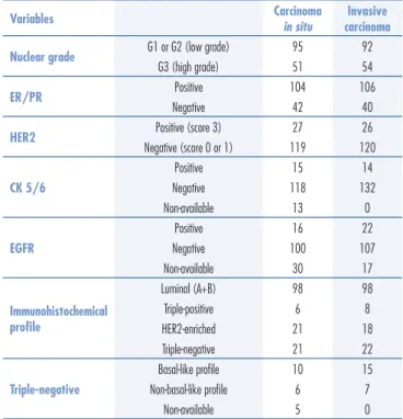
Table 2. Comparison of pathological and immunohistochemical features in situ and invasive components of 146 ductal carcinomas
Variables Carcinoma in situ carcinomaInvasive Nuclear grade G1 or G2 (low grade) 95 92
G3 (high grade) 51 54
ER/PR Positive 104 106
Negative 42 40
HER2 Positive (score 3) 27 26
Negative (score 0 or 1) 119 120
CK 5/6
Positive 15 14
Negative 118 132
Non-available 13 0
EGFR
Positive 16 22
Negative 100 107
Non-available 30 17
Immunohistochemical proile
Luminal (A+B) 98 98 Triple-positive 6 8 HER2-enriched 21 18 Triple-negative 21 22
Triple-negative
Basal-like proile 10 15 Non-basal-like proile 6 7
Non-available 5 0
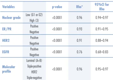
There was a signiicant correlation between nuclear grade (p<0.0001), expression of ER/PR (p<0.0001), overexpression of HER2 (p<0.0001) and molecular proile (p<0.0001) between components in situ and invasive. The value of the correlation coeficient ranged from 0.918 (HER2) to 0.967 (molecular proile) (Table 3).
Discussion
In this study we observed similar distribution of the nuclear grade and immunohistochemical variables in DCIS and the correspondent invasive component. This observation suggests that these classical prognostic and predictive factors may be predetermined in the preinvasive stage of the disease.
The management of breast cancer is largely based on some clinical and pathological parameters, including age of patient, size of tumor, nodal status, histologic and nuclear grade, and immunohistochemical evalua-tion of ER, PR, HER2, and Ki-67, either as individual information, or as panel of markers to approximate the molecular subtyping classiication16. DCIS are genuine
precursors of breast cancer, but the mechanisms involved in this transition are mostly unknown. DCIS is a very heterogeneous disease with variable risk of invasion as well of recurrence, not to mention that one half of all recurrences occur as invasive cancer17,18.
According to our results the phenotype of IBC is very similar to the in situ component, suggesting that the classical prognostic and predictive factors are determined previously to the invasive capacity of the precursors. Similar results have been presented in
*Spearman correlation method; ER: estrogen receptor; PR: progesterone receptor; HER2: human epidermal growth factor receptor 2; CK: cytokeratin 5/6; EGFR: epidermal growth factor receptor.
Variables p-value Rho* 95%CI for Rho Nuclear grade Low (G1 or G2) <0.0001 0.96 0.94–0.97
High (3)
ER/PR Positive <0.0001 0.93 0.91–0.95 Negative
HER2 Positive <0.0001 0.91 0.88–0.94 Negative
EGFR Positive <0.0001 0.76 0.68–0.83 Negative
Molecular proile
Luminal (A+B)
<0.0001 0.96 0.95–0.97 Triple-positive
HER2 Triple-negative
Table 3. Correlation between pathological and immunohistochemical features of ductal carcinoma in situ and adjacent invasive carcinoma (n=146)
the literature10,11,19,20. Ottesen found similar
morphol-ogy, immunohistochemistry, and DNA ploidy both in DCIS and the invasive component19. Park et al.20
compared HER2 status between in situ and invasive component of 270 breast carcinomas and found a high concordance of 98.5 and 99.3%, respectively by FISH and immunohistochemistry. Steinman et al.21 studied ER,
PR, HER2, EGFR and several cytokeratins, including CK5/6, in 96 cases of DCIS with co-existing invasive carcinoma and they found a high rate of concordance ranging from 92.3% for ER to 100% for HER2 and EGFR. Our indings support the evidence that molecular changes implicated in the progression from in situ status to invasive one occur before morphological manifesta-tion of invasiveness. Interestingly, among these classical parameters, nuclear grade is one of the most powerful prognostic and predictive factors and it is associated with distinct genetic changes22-25. According to some studies,
it is possible that even molecular changes described in DCIS, although could be related to grade, are not de-terminant of invasion. Moelans et al.26 compared copy
numbers changes in 21 breast cancer related genes and the methylation status of 25 breast cancer-related genes27
between laser-microdissected ductal carcinoma in situ (DCIS) and adjacent invasive ductal cancer (IDC) lesions. These authors did not observe signiicant differences between DCIS and adjacent IBC, suggesting that DCIS is genetically as advanced as its invasive counterpart. In an elegant study, Muggerud et al.12 analyzed gene
expres-sions patterns of 31 pure DCIS, 36 pure invasive cancers and 42 cases of DCIS with invasive cancer. The authors found a DCIS signature associated to gene expression characteristics more similar to advanced tumors. This set of genes was independent of grade, ER-status, and HER2-status, and it was suggestive of several processes related to the re-organization of the microenvironment12. In our
opinion one of the most promising way to investigate the tendency of breast carcinoma to invade the stroma is to try to understand the factors related to interaction of the tumor cells with the microenvironment.
Although nuclear grade is considered the most im-portant parameter to classify DCIS together with tumor size and margins status, it becomes evident that it is not a good predictor of invasive potential. Holmes et al.28
1. Lee S, Stewart S, Nagtegaal I, Luo J, Wu Y, Colditz G, et al. Differentially expressed genes regulating the progression of ductal carcinoma in situ to invasive breast cancer. Cancer Res. 2012;72(17):4574-86.
2. Ernster VL, Ballard-Barbash R, Barlow WE, Zheng Y, Weaver DL, Cutter G, et al. Detection of ductal carcinoma in situ in women undergoing screening mammography. J Natl Cancer Inst. 2002;94(20):1546-54.
3. Buerger H, Otterbach F, Simon R, Schäfer KL, Poremba C, Diallo R, et al. Different genetic pathways in the evolution of invasive breast cancer are associated with distinct morphological subtypes. J Pathol. 1999;189(4):521-6.
4. Hernandez L, Wilkerson PM, Lambros MB, Campion-Flora A, Rodrigues DN, Gauthier A, et al. Genomic and mutational proiling of ductal carcinomas in situ and matched adjacent invasive breast cancers reveals intra-tumour genetic heterogeneity and clonal selection. J Pathol. 2012;227(1):42-52.
5. Johnson CE, Gorringe KL, Thompson ER, Opeskin K, Boyle SE, Wang Y, et al. Identiication of copy number alterations associated with the progression of DCIS to invasive ductal carcinoma. Breast Cancer Res Treat. 2012;133(3):889-98.
6. Zikan M, Bohm J, Pavlista D, Cibula D. Comparative analysis of loss of heterozygosity and expression proile in normal tissue, DCIS and invasive breast cancer. Clin Transl Oncol. 2011;13(9):652-5. 7. Lopez-Garcia MA, Geyer FC, Lacroix-Triki M, Marchió C, Reis-Filho JS. Breast cancer precursors revisited: molecular features and progression pathways. Histopathology. 2010;57(2):171-92. 8. Porter D, Lahti-Domenici J, Keshaviah A, Bae YK, Argani P, Marks
J, et al. Molecular markers in ductal carcinoma in situ of the breast. Mol Cancer Res. 2003;1(5):362-75.
9. Heaphy CM, Bisofi M, Joste NE, Baumgartner KB, Baumgartner RN, Grifith JK. Genomic instability demonstrates similarity between DCIS and invasive carcinomas. Breast Cancer Res Treat. 2009;117(1):17-24.
10. Clark SE, Warwick J, Carpenter R, Bowen RL, Duffy SW, Jones JL. Molecular subtyping of DCIS: heterogeneity of breast cancer relected in pre-invasive disease. Br J Cancer. 2011;104(1):120-7.
References
role of biological markers in DCIS, including steroid receptors, proliferation markers, cell cycle regulation and apoptotic markers, angiogenesis-related proteins, epidermal growth factor receptor family receptors, ex-tracellular matrix-related proteins, and COX-2, failed to ind any useful marker.
After the molecular classiication of breast into intrinsic subtypes luminal A, Luminal B, HER2 and basal-like30 and the demonstration of immunohistochemical
surrogates31, several groups dedicated to investigate the
role of the same proiles in DCIS10,13,32. Our results show
a similar distribution of the molecular subtypes between in situ and invasive carcinomas. The more controversial subtype is the basal-like. According Nielsen et al.31,
the better determination of the basal-like subgroup
is based on TN phenotype associated to expression of CK5/6 and/or EGFR. We studied a substantial number of TN DCIS (21 cases), all of them were associated with TN in the invasive component. However, our results indicate that the signiicance of basal cytokeratin 5/6 in DCIS may be different of that in invasive carcinomas. In fact, CK5/6 was negatively associated with the TN phenotype and more prevalent in the other subtypes. Further studies are needed to clarify the role of this cytokeratin in DCIS.
Acknowledgement
This study was supported by grants from FAPESP (São Paulo Research Founding), process number 2011/14741-7.
11. Steinman S, Wang J, Bourne P, Yang Q, Tang P. Expression of cytokeratin markers, ER-alpha, PR, HER-2/neu, and EGFR in pure ductal carcinoma in situ (DCIS) and DCIS with co-existing invasive ductal carcinoma (IDC) of the breast. Ann Clin Lab Sci. 2007;37(2):127-34.
12. Muggerud AA, Hallett M, Johnsen H, Kleivi K, Zhou W, Tahmasebpoor S, et al. Molecular diversity in ductal carcinoma in situ (DCIS) and early invasive breast cancer. Mol Oncol. 2010;4(4):357-68. 13. Livasy CA, Perou CM, Karaca G, Cowan DW, Maia D, Jackson
S, et al. Identiication of a basal-like subtype of breast ductal carcinoma in situ. Hum Pathol. 2007;38(2):197-204.
14. Lakhani SR, Ellis IO, Schnitt SJ, Tan PH, van de Vijver MJ. WHO Classiication of tumours of the breast. Lyon: IARC; 2012. 15. Wolff AC, Hammond ME, Schwartz JN, Hagerty KL, Allred DC,
Cote RJ, et al. American Society of Clinical Oncology/College of American Pathologists guideline recommendations for human epidermal growth factor receptor 2 testing in breast cancer. J Clin Oncol. 2007;25(1):118-45.
16. Goldhirsch A, Wood WC, Coates AS, Gelber RD, Thürlimann B, Senn HJ, et al. Strategies for subtypes—dealing with the diversity of breast cancer: highlights of the St. Gallen International Expert Consensus on the Primary Therapy of Early Breast Cancer 2011. Ann Oncol. 2011;22(8):1736-47.
17. Collins LC, Tamimi RM, Baer HJ, Connolly JL, Colditz GA, Schnitt SJ. Outcome of patients with ductal carcinoma in situ untreated after diagnostic biopsy: results from the Nurses’ Health Study. Cancer. 2005;103(9):1778-84.
18. Cuzick J. Treatment of DCIS—results from clinical trials. Surg Oncol. 2003;12(4):213-9.
19. Ottesen GL. Carcinoma in situ of the female breast. A clinico-pathological, immunohistological, and DNA ploidy study. APMIS Suppl. 2003;(108):1-67.
21. Steinman S, Wang J, Bourne P, Yang Q, Tang P. Expression of cytokeratin markers, ER-alpha, PR, HER-2/neu, and EGFR in pure ductal carcinoma in situ (DCIS) and DCIS with co-existing invasive ductal carcinoma (IDC) of the breast. Ann Clin Lab Sci. 2007;37(2):127-34.
22. Song WJ, Kim KI, Park SH, Kwon MS, Lee TH, Park HK, et al. The risk factors inluencing between the early and late recurrence in systemic recurrent breast cancer. J Breast Cancer. 2012;15(2):218-23.
23. Ishikawa T, Shimizu D, Yamada A, Sasaki T, Morita S, Tanabe M, et al. Impacts and predictors of cytotoxic anticancer agents in different breast cancer subtypes. Oncol Res. 2012;20(2-3):71-9. 24. Sinha S, Singh RK, Bhattacharya N, Mukherjee N, Ghosh S,
Alam N, et al. Frequent alterations of LOH11CR2A, PIG8 and CHEK1 genes at chromosomal 11q24.1-24.2 region in breast carcinoma: clinical and prognostic implications. Mol Oncol. 2011;5(5):454-64.
25. Tokunaga E, Okada S, Yamashita N, Akiyoshi S, Kitao H, Morita M, et al. High incidence and frequency of LOH are associated with aggressive features of high-grade HER2 and triple-negative breast cancers. Breast Cancer. 2012;19(2):161-9.
26. Moelans CB, de Wegers RA, Monsuurs HN, Maess AH, van Diest PJ. Molecular differences between ductal carcinoma in situ and
adjacent invasive breast carcinoma: a multiplex ligation-dependent probe ampliication study. Cell Oncol (Dordr). 2011;34(5):475-82. 27. Moelans CB, Verschuur-Maes AH, van Diest PJ. Frequent promoter
hypermethylation of BRCA2, CDH13, MSH6, PAX5, PAX6 and WT1 in ductal carcinoma in situ and invasive breast cancer. J Pathol. 2011;225(2):222-31.
28. Holmes P, Lloyd J, Chervoneva I, Pequinot E, Cornield DB, Schwartz GF, et al. Prognostic markers and long-term outcomes in ductal carcinoma in situ of the breast treated with excision alone. Cancer. 2011;117(16):3650-7.
29. Lari SA, Kuerer HM. Biological Markers in DCIS and risk of breast recurrence: a systematic review. J Cancer. 2011;2:232-61. 30. Perou CM, Sørlie T, Eisen MB, van de Rijn M, Jeffrey SS, Rees
CA, et al. Molecular portraits of human breast tumours. Nature. 2000;406(6797):747-52.
31. Nielsen TO, Hsu FD, Jensen K, Cheang M, Karaca G, Hu Z, et al. Immunohistochemical and clinical characterization of the basal-like subtype of invasive breast carcinoma. Clin Cancer Res. 2004;10(16):5367-74.